. 2024 Sep 10:13:RP96013.
doi: 10.7554/eLife.96013.

Male cuticular pheromones stimulate removal of the mating plug and promote re-mating through pC1 neurons in Drosophila females

Affiliations

Male cuticular pheromones stimulate removal of the mating plug and promote re-mating through pC1 neurons in Drosophila females

Minsik Yun et al. Elife. .

Abstract

In birds and insects, the female uptakes sperm for a specific duration post-copulation known as the ejaculate holding period (EHP) before expelling unused sperm and the mating plug through sperm ejection. In this study, we found that Drosophila melanogaster females shortens the EHP when incubated with males or mated females shortly after the first mating. This phenomenon, which we termed male-induced EHP shortening (MIES), requires Or47b+ olfactory and ppk23+ gustatory neurons, activated by 2-methyltetracosane and 7-tricosene, respectively. These odorants raise cAMP levels in pC1 neurons, responsible for processing male courtship cues and regulating female mating receptivity. Elevated cAMP levels in pC1 neurons reduce EHP and reinstate their responsiveness to male courtship cues, promoting re-mating with faster sperm ejection. This study established MIES as a genetically tractable model of sexual plasticity with a conserved neural mechanism.

Keywords: D. melanogaster; Or47b; mating; neuroscience; pC1; ppk23; sexual plasticity; sperm ejection.


Conflict of interest statement

MY, DK, TH, KL, EP, MK, BH, YK No competing interests declared

Figures

Figure 1.
Figure 1.. The presence of males reduces the ejaculate holding period (EHP) in females through olfactory or gustatory sensation.
(A) Schematic of the experimental procedure employed to measure male-induced EHP shortening (MIES). Immediately after the end of copulation, the female is incubated with a wild-type Canton-S (CS) male that has not been previously exposed to the female. Typically, w1118 females that are kept alone after mating exhibit an EHP of approximately 90 min, whereas females that are incubated with a naïve CS male exhibit an EHP of approximately 60 min. In this study, we refer to this phenomenon as MIES. (B–F) Normalized EHP or ΔEHP of the females of the indicated genotypes, incubated under the indicated conditions after mating. The ΔEHP is calculated by subtracting the mean of the reference EHP of females kept alone after mating (the leftmost column) from the EHP of individual females in comparison. Mann-Whitney test (n.s. p>0.05; ****p<0.0001). Gray circles indicate the EHP or ΔEHP of individual females, and the mean ± SEM of data is presented. Numbers below the horizontal bar represent the mean of the EHP differences between the indicated treatments. Genotype and sample size are shown in Table 1.
Figure 2.
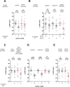
Figure 2.. The function of Or47b and Or47b-positive olfactory receptor neurons (ORNs) is essential for male-induced EHP shortening (MIES).
(A, C–E) ΔEHP of females of the indicated genotypes, incubated with or without naive males after mating. The female genotypes are as follows from left to right: (A) control (Or47b>TNTinactive), Or47b ORN silencing (Or47b>TNTactive); (C) Orco mutant (Orco1/Orco1), Orco rescue in Or47b ORNs of Orco mutant (Orco1/Orco1; Or47b>Orco); (D) control 1 (Or47b2/+), control 2 (Or47b3/+), Or47b mutant (Or47b2/Or47b3); (E) Or47b mutant (Or47b2/Or47b2), Or47b rescue (Or47b>Or47b; Or47b2/Or47b2). (B) Thermogenetic activation of Or47b-positive ORNs shortens EHP in females kept alone after mating. The female genotypes are as follows from left to right: control 1 (Or47b-Gal4/+), control 2 (UAS-dTRPA1/+), Or47b>dTRPA1 (Or47b-Gal4/UAS-dTRPA1). Mann-Whitney test (n.s. p>0.05; *p<0.05; **p<0.01; ****p<0.0001). The ΔEHP is calculated by subtracting the mean of the reference EHP of females kept alone after mating (‘-’ in A, C–E) or incubated at 21°C control conditions (B) from the EHP of individual females in comparison. Gray circles indicate the ΔEHP of individual females, and the mean ± SEM of data is presented. The gray circles with dashed borders indicate ΔEHP values that exceed the axis limits (>90 or <-90 min). Numbers below the horizontal bar represent the mean of the EHP differences between the indicated treatments. EHP, ejaculate holding period. Genotype and sample size are shown in Table 1.
Figure 2—figure supplement 1.
Figure 2—figure supplement 1.. The identification of trichoid and intermediate sensilla olfactory receptor neurons (ORNs) that are necessary for the production of male-induced EHP shortening (MIES).
ΔEHP of females of the indicated genotypes, incubated with or without naive males immediately after mating. The female genotypes are as follows from left to right:+>TNTactive, Or13a>TNTactive, Or19a>TNTactive, Or23a>TNTactive, Or43a>TNTactive, Or47b>TNTactive, Or65a>TNTactive, Or65b>TNTactive, Or65c>TNTactive, Or67d>TNTactive, Or83c>TNTactive, Or88a>TNTactive. Mann-Whitney test (n.s. p>0.05; *p<0.05; **p<0.01; ***p<0.001). Gray circles indicate the ΔEHP of individual females, and the mean ± SEM of data is presented. The ΔEHP is calculated by subtracting the mean of the reference EHP of females kept alone after mating (‘-’) from the EHP of individual females in comparison. Numbers below the horizontal bar represent the mean of the EHP differences between the indicated treatments. EHP, ejaculate holding period. Genotype and sample size are shown in Table 1.
Figure 3.
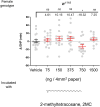
Figure 3.. 2-Methyltetracosane (2MC) can induce ejaculate holding period (EHP) shortening through Or47b.
(A–D) ΔEHP of females of the indicated genotypes, incubated in solvent vehicle or 2MC. Mated females were incubated with a piece of filter paper perfumed with either vehicle (-) or 750 ng 2MC (+). The female genotypes are as follows: (A) w1118, (B) Orco mutant (Orco1/Orco1), (C) Or47b mutant (Or47b2/Or47b2), (D) Gal4 control (Or47b-Gal4/+; Orco1/Orco1), UAS control (UAS-Orco/+; Orco1/Orco1), Orco rescue in Or47b olfactory receptor neurons (ORN) (Orco1/Orco1; Or47b-Gal4/UAS-Orco). (A–C) Mann-Whitney test (n.s. p>0.05; *p<0.05). (D) One-way analysis of variance (ANOVA) test with Fisher’s LSD multiple comparison (n.s. p>0.05; *p<0.05). Gray circles indicate the ΔEHP of individual females and the mean ± SEM of data is presented. The ΔEHP is calculated by subtracting the mean of the reference EHP of females incubated with vehicle-perfumed paper (the leftmost column in A–C) or the mean of the Gal4 control and UAS control female incubated with vehicle-perfumed paper (the two leftmost columns in D) from the EHP of individual females in comparison. Gray circles with dashed borders indicate ΔEHP values that exceed the axis limits (>90 or <-90 min). Numbers below the horizontal bar represent the mean of the EHP differences between the indicated treatments. Genotype and sample size are shown in Table 1.
Figure 3—figure supplement 1.
Figure 3—figure supplement 1.. Known odorant ligands for Or47b, methyl laurate and trans-palmitoleic acid, were unable to induce ejaculate holding period (EHP) shortening.
ΔEHP of w1118 females incubated with a piece of filter paper perfumed with solvent vehicle or with the indicated amounts of two known Or47b odorant ligands, methyl laurate (A) and trans-palmitoleic acid (B) immediately after mating. Mann-Whitney test (n.s. p>0.05). The ΔEHP is calculated by subtracting the mean of the reference EHP of females incubated with vehicle-perfumed paper (the leftmost column) from the EHP of individual females in comparison. Gray circles indicate the ΔEHP of individual females, and the mean ± SEM of data is presented. Numbers below the horizontal bar represent the mean of the EHP differences between vehicle and odorant treatments. Genotype and sample size are shown in Table 1.
Figure 3—figure supplement 2.
Figure 3—figure supplement 2.. Ejaculate holding period (EHP) shortening is induced by males with feminized oenocytes, females with masculinized oenocytes, and males of other closely related Drosophila species.
(A) ΔEHP of w1118 females incubated with males with feminized oenocytes (Oe Fem male; PromE(800)-Gal4/UAS-Tra) or virgin females with masculinized oenocytes (Oe Mas female; PromE(800)-Gal4/UAS-Tra-RNAi). (B) ΔEHP of w1118 females incubated with naive males of the indicated Drosophila species. D. mel (D. melanogaster), D. sim (D. simulans), D. sec (D. sechellia), D. ere (D. erecta), D. yak (D. yakuba). Mann-Whitney test (n.s. p>0.05; ****p<0.0001). The ΔEHP is calculated by subtracting the mean of the reference EHP of females kept alone after mating (the leftmost column) from the EHP of individual females in comparison. Gray circles indicate the ΔEHP of individual females, and the mean ± SEM of data is presented. Numbers below the horizontal bar represent the mean EHP differences between the indicated treatments. Genotype and sample size are shown in Table 1.
Figure 3—figure supplement 3.
Figure 3—figure supplement 3.. 2-Methyltetracosane (2MC) shortens ejaculate holding period (EHP) at a specific concentration.
ΔEHP of w1118 females incubated with a piece of filter paper perfumed with solvent vehicle or the indicated amounts of 2MC. Mann-Whitney test (n.s. p>0.05; *p<0.05). The ΔEHP is calculated by subtracting the mean of the reference EHP of females incubated with vehicle-perfumed paper (the leftmost column) from the EHP of individual females in comparison. Gray circles indicate the ΔEHP of individual females, and the mean ± SEM of data is presented. Numbers below the horizontal bar represent the mean of the EHP differences between vehicle and odorant treatments. Genotype and sample size are shown in Table 1.
Figure 4.
Figure 4.. 7-Tricosene (7-T) present in mated females and males reduces ejaculate holding period (EHP) via ppk23 neurons.
(A–D) ΔEHP of females of the indicated genotypes, incubated with mated females (A), a piece of filter paper perfumed with 150 ng 7-T (B), 200 ng 11-cis-vaccenyl acetate (cVA) (C), or naive males (D) after mating. The female genotypes are as follows: (A–C) w1118, (D) control (ppk23-Gal4/UAS-TNTinactive), ppk23 silencing (ppk23-Gal4/UAS-TNTactive). (A) Unpaired t-test. (B–D) Mann-Whitney test (n.s. p>0.05; *p<0.05). The ΔEHP is calculated by subtracting the mean of the reference EHP of females kept alone (‘-’ in A, D) or incubated with vehicle-perfumed paper (the leftmost column in B, C) from the EHP of individual females in comparison. Gray circles indicate the ΔEHP of individual females, and the mean ± SEM of data is presented. The gray circles with dashed borders indicate ΔEHP values that exceed the axis limits (>90 or <-90 min). Numbers below the horizontal bar represent the mean of the EHP differences between the indicated treatments. Genotype and sample size are shown in Table 1.
Figure 4—figure supplement 1.
Figure 4—figure supplement 1.. 7-Tricosene (7-T) induces ejaculate holding period (EHP) shortening at physiological concentrations, but DEG/ENaC channels expressed in ppk23 neurons are not required for male-induced EHP shortening (MIES).
(A, B) ΔEHP of w1118 females incubated with a piece of filter paper perfumed with solvent vehicle or the indicated amounts of 7-T (A), or 7-pentacosene (B) after mating. Incubation with a specific concentration of 7-T significantly shorten EHP, but 7-pentacosene does not. Unpaired t-test (n.s. p>0.05; *p<0.05). (C–E) ΔEHP of females of the indicated genotypes, incubated with or without naive males after mating. The female genotypes are as follows from left to right: (C) control 1 (w1118), control 2 (ppk23-/+), and ppk23- (ppk23-/ppk23-); (D) control 1 (w1118), control 2 (ppk28-/+), and ppk28- (ppk28-/ppk28-); (E) control 1 (w1118), control 2 (ppk29-/+), and ppk29- (ppk29-/ppk29-). Mann-Whitney test (n.s. p>0.05; *p<0.05; **p<0.01; ****p<0.0001). The ΔEHP is calculated by subtracting the mean of the reference EHP of females incubated with vehicle-perfumed paper (the leftmost column in A, B) or kept alone after mating (‘-’ in C–E) from the EHP of individual females in comparison. Gray circles indicate the ΔEHP of individual females, and the mean ± SEM of data is presented. Numbers below the horizontal bar represent the mean of the EHP differences between the indicated treatments. Genotype and sample size are shown in Table 1.
Figure 5.
Figure 5.. A subset of pC1 neurons, comprising pC1b and pC1c subtypes, regulates ejaculate holding period (EHP) and exhibits CRE-luciferase reporter activity in response to 2-methyltetracosane (2MC) and 7-tricosene (7-T).
(A–D) The optogenetic silencing of a pC1 neuron subset comprising pC1b and pC1c neurons (pC1b, c) increases EHP. Females of the indicated genotypes were cultured on food with or without all trans-retinal (ATR) after eclosion. The ΔEHP is calculated by subtracting the mean of the reference EHP of females cultured in control ATR - food (the leftmost column) from the EHP of individual females in comparison. The female genotypes are as follows: (A) pC1a,b,c>GtACR1 (pC1-S-Gal4/UAS-GtACR1), (B) pC1d,e>GtACR1 (pC1-A-Gal4/UAS-GtACR1), (C) pC1a>GtACR1 (pC1a-split-Gal4/UAS-GtACR1), and (D) pC1b,c>GtACR1 (Dh44-pC1-Gal4/UAS-GtACR1). Gray circles indicate the ΔEHP of individual females, and the mean ± SEM of data is presented. The gray circles with dashed borders indicate ΔEHP values that exceed the axis limits (>120 min). Mann-Whitney test (n.s. p>0.05; *p<0.05; ****p<0.0001). Numbers below the horizontal bar represent the mean of the EHP differences between the indicated treatments. (E, F) Relative CRE-luciferase reporter activity of pC1 neurons in females of the indicated genotypes, incubated with a piece of filter paper perfumed with solvent vehicle control or the indicated pheromones immediately after mating. The CRE-luciferase reporter activity of pC1 neurons of Or47b-deficient females (Or47b2/2 or Or47b3/3) was observed to increase in response to 7-T but not to 2MC. To calculate the relative luciferase activity, the average luminescence unit values of the female incubated with the vehicle are set to 100%. Mann-Whitney test (n.s. p>0.05; **p<0.01; ***p<0.001; ****p<0.0001). Gray circles indicate the relative luciferase activity (%) of individual females, and the mean ± SEM of data is presented. Genotype and sample size are shown in Table 1.
Figure 5—figure supplement 1.
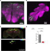
Figure 5—figure supplement 1.. Characterization of pC1a-split-Gal4.
(A, B) Z-projection confocal images of the brain (A) and VNC (B) of a female carrying pC1a-split-GAL4 and UAS-myrEGFP, stained with anti-EGFP (green) and anti-nc82 (magenta). Scale bars, 50 μm. In the brain, only the pC1a cells are labeled, but in the VNC, several cells are labeled in the abdominal ganglion. (C) An anatomical comparison between pC1a-split-GAL4 neurons (above; pC1a-ss) in the brain and a pC1a neuron (below; NeuPRINT body ID, 5813046951). The panel above shows the maximum intensity projection image (MIP) of an aligned confocal image of the brain from a female carrying pC1a-split-GAL4 and UAS-myrEGFP stained with anti-EGFP and anti-nc82. (D) Mating frequencies of pC1a>GtACR1 (pC1a-split-Gal4/UAS-GtACR1) females during optogenetic silencing, scored as the percentage of females that copulate within 1 hr. Females were cultured on food with or without all trans-retinal (ATR) prior to the mating assay. The optogenetic silencing of pC1a neurons with GtACR1 was observed to suppress mating receptivity almost completely. Chi-square test (****p<0.0001).
Figure 5—figure supplement 2.
Figure 5—figure supplement 2.. Incubation with 2-methyltetracosane (2MC) or 7-tricosene (7-T) increases cAMP levels in pC1 neurons.
The relative CRE-luciferase reporter activity of pC1 neurons in females incubated with a piece of filter paper perfumed with the indicated amounts of 2MC (A) and 7-T (B). It is noteworthy that the concentration range within which 2MC or 7-T increases cAMP levels in pC1 neurons is narrow. To calculate the relative luciferase activity, the average luminescence unit values of the female incubated with the vehicle are set to 100%. Gray circles indicate the relative luciferase activity (%) of individual females, and the mean ± SEM of data is presented. Mann-Whitney test (n.s. p>0.05; **p<0.01; ***p<0.001; ****p<0.0001). Genotype and sample size are shown in Table 1.
Figure 5—figure supplement 3.
Figure 5—figure supplement 3.. Incubation with 2-methyltetracosane (2MC) or 7-tricosene (7-T) increases cAMP levels in pC1a as well as pC1b, c neurons in virgin females.
The relative CRE-luciferase reporter activity of pC1 neurons in virgin females of the indicated genotypes, incubated with a piece of filter paper perfumed with the indicated odorants. To calculate the relative luciferase activity, the average luminescence unit values of the female incubated with the vehicle are set to 100%. Mann-Whitney test (n.s. p>0.05; **p<0.01; ***p<0.001; ****p<0.0001). Gray circles indicate the relative luciferase activity (%) of individual females, and the mean ± SEM of data is presented. Genotype and sample size are shown in Table 1.
Figure 6.
Figure 6.. Elevated cAMP levels in pC1 neurons reduce ejaculate holding period (EHP) and increase the responsiveness of pC1 neurons to male courtship cues, thereby promoting subsequent re-mating.
(A) The optogenetic production of cAMP in the pC1b, c neurons shortens EHP, whereas the same treatment in pC1a or pC1d, e neurons does not. ΔEHP is calculated by subtracting the mean of the reference EHP of females incubated in the control illumination (Dim light), which does not activate a photoactivatable adenylate cyclase (PhotoAC), from the EHP of individual females. Mann-Whitney test (n.s. p>0.05, ****p<0.0001). (B) The optogenetic production of cAMP transiently increases the excitability of pC1 neurons. Left, schematic of the experimental procedure. Right, peak ΔF/F in the LPC projections of pC1 neurons from freshly mated females in response to the pheromone 11-cis-vaccenyl acetate (cVA), before and after photoactivation of PhotoAC expressed in pC1 neurons. The calcium response was measured at specific time points after photoactivation: after 1 min (blue dots and box) or 10 min (purple dots and box) after activation. Repeated measures one-way ANOVA test with the Geisser-Greenhouse correction followed by Tukey’s multiple comparisons test (*p<0.05; ***p<0.001; ****p<0.0001). (C) Left, schematic of the experimental procedure. Right, re-mating rate of females during optogenetic cAMP production in pC1b, c neurons, scored as the percentage of females that copulate with a naive Canton-S (CS) male within 6 hr after the first mating. The female genotypes are as follows: Control (+/UAS-PhotoAC), pC1b,c>UAS-PhotoAC (Dh44-pC1-Gal4/UAS-PhotoAC). Chi-square test (*p<0.05). Genotype and sample size are shown in Table 1.
Figure 6—figure supplement 1.
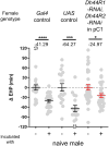
Figure 6—figure supplement 1.. The knockdown of Dh44R1 and Dh44R2 in pC1 neurons has a limited impact on male-induced EHP shortening (MIES).
ΔEHP of females of the indicated genotypes, incubated with or without naive males immediately after mating. The female genotypes are as follows from left to right: Gal4 control (UAS-Dcr2/+; GMR71G01-Gal4/+), UAS control (UAS-Dh44R1-RNAi/+; UAS-Dh44R2-RNAi/+), Dh44R1-RNAi, Dh44R2-RNAi in pC1 (UAS-Dcr2/+; GMR71G01-Gal4/Dh44R1-RNAi; Dh44R2-RNAi/+). Mann-Whitney test (*p<0.05; ***p<0.001; ****p<0.0001). The ΔEHP is calculated by subtracting the mean of the reference EHP of females kept alone after mating (‘-’) from the EHP of individual females in comparison. Gray circles indicate the ΔEHP of individual females, and the mean ± SEM of data is presented. The gray circles with dashed borders indicate ΔEHP values that exceed the axis limits (>120 or <-120 min). Numbers below the horizontal bar represent the mean of the EHP differences between the indicated treatments. EHP, ejaculate holding period. Genotype and sample size are shown in Table 1.
Figure 7.
Figure 7.. The presence of male odorants, which reflect changes in the social-sexual context, stimulates newly mated females to remove the male ejaculate and engage in subsequent re-mating.
Following the initial mating, a female that encounters a new courting male removes the male ejaculate after a shorter ejaculate holding period (EHP) than those that do not encounter new male partners. This phenomenon, referred to as male-induced EHP shortening (MIES) in this study, is followed by a second mating with the new partner. The production of MIES depends on the functions of the Or47b+ olfactory and ppk23+ gustatory neurons, which are activated by 2-methyltetracosane (2MC) and 7-T, respectively. These odorants increase cAMP levels in pC1b, c neurons, enhancing their responsiveness to male courtship cues and increasing mating receptivity. Consequently, 2MC and 7-T promote a second mating with a faster removal of the male ejaculate or mating plug.
Author response image 1.
Author response image 1.. The prolonged optogenetic activation of pC1b, c neurons increases EHP, mimicking silencing of pC1b, c neurons.
Females of the indicated genotypes were cultured on food with or without all-trans-retinal (ATR). The ΔEHP is calculated by subtracting the mean of the reference EHP of females cultured in control ATR- food from the EHP of individual females in comparison. The female genotypes are as follows: (A) 71G01-GAL4/UAS-CsChrimson, (B) pC1a-split-Gal4/UAS-CsChrimson, (C) pC1b,c-split-Gal4/UAS-CsChrimson, (D) pC1d-split-Gal4/UAS-CsChrimson, and (E) pC1e-split-Gal4/UAS-CsChrimson. Gray circles indicate the ΔEHP of individual females, and the mean ± SEM of data is presented. Mann-Whitney Test (n.s. p > 0.05; *p <0.05; ****p < 0.0001). Numbers below the horizontal bar represent the mean of the EHP differences between the indicated treatments.

Update of

  • doi: 10.1101/2023.11.28.568981
  • doi: 10.7554/eLife.96013.1
  • doi: 10.7554/eLife.96013.2

Similar articles

References

    1. Ali MZ, Bilgrami AL, Ahsan J. Drosophila melanogaster chemosensory pathways as potential targets to curb the insect menace. Insects. 2022;13:142. doi: 10.3390/insects13020142. - DOI - PMC - PubMed
    1. Anderson DJ. Circuit modules linking internal states and social behaviour in flies and mice. Nature Reviews. Neuroscience. 2016;17:692–704. doi: 10.1038/nrn.2016.125. - DOI - PubMed
    1. Antony C, Davis TL, Carlson DA, Pechine JM, Jallon JM. Compared behavioral responses of male Drosophila melanogaster (Canton S) to natural and synthetic aphrodisiacs. Journal of Chemical Ecology. 1985;11:1617–1629. doi: 10.1007/BF01012116. - DOI - PubMed
    1. Bartelt RJ, Schaner AM, Jackson LL. cis-Vaccenyl acetate as an aggregation pheromone in Drosophila melanogaster. Journal of Chemical Ecology. 1985;11:1747–1756. doi: 10.1007/BF01012124. - DOI - PubMed
    1. Becker SD, Hurst JL. In: Chemical Signals in Vertebrates 11. Hurst JL, Beynon RJ, Roberts SC, Wyatt TD, editors. New York, NY: Springer; 2008. Pregnancy block from a female perspective in; pp. 141–150. - DOI

LinkOut - more resources