. 2024 Sep 10;22(9):e3002791.
doi: 10.1371/journal.pbio.3002791. eCollection 2024 Sep.

PP1 phosphatase controls both daughter cell formation and amylopectin levels in Toxoplasma gondii

Affiliations

PP1 phosphatase controls both daughter cell formation and amylopectin levels in Toxoplasma gondii

Asma Sarah Khelifa et al. PLoS Biol. .

Abstract

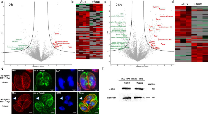
Virulence of apicomplexan parasites is based on their ability to divide rapidly to produce significant biomass. The regulation of their cell cycle is therefore key to their pathogenesis. Phosphorylation is a crucial posttranslational modification that regulates many aspects of the eukaryotic cell cycle. The phosphatase PP1 is known to play a major role in the phosphorylation balance in eukaryotes. We explored the role of TgPP1 during the cell cycle of the tachyzoite form of the apicomplexan parasite Toxoplasma gondii. Using a conditional mutant strain, we show that TgPP1 regulates many aspects of the cell cycle including the proper assembly of the daughter cells' inner membrane complex (IMC), the segregation of organelles, and nuclear division. Unexpectedly, depletion of TgPP1 also results in the accumulation of amylopectin, a storage polysaccharide that is usually found in the latent bradyzoite form of the parasite. Using transcriptomics and phospho-proteomics, we show that TgPP1 mainly acts through posttranslational mechanisms by dephosphorylating target proteins including IMC proteins. TgPP1 also dephosphorylates a protein bearing a starch-binding domain. Mutagenesis analysis reveals that the targeted phospho-sites are linked to the ability of the parasite to regulate amylopectin steady-state levels. Therefore, we show that TgPP1 has pleiotropic roles during the tachyzoite cell cycle regulation, but also regulates amylopectin accumulation.


Conflict of interest statement

The authors have declared that no competing interests exist.

Figures

Fig 1
Fig 1. TgPP1 is required for parasite growth and proliferation.
(a) Schematic representation of the construct used to generate iKD TgPP1 mutant parasites using the AID system that allows for the inducible degradation of the TgPP1 protein. The system involves introducing the AID domain into the gene of interest. In the presence of Auxin, the AID domain will be recognized by the Tir1 protein and the protein degraded by the proteasome. The HXGPRT-T2A-AID-2Ty cassette is inserted at the 5′ end of the gene using the CRISPR/Cas9 gene-editing strategy. (b) Confocal microscopy imaging of the AID-Ty iKD TgPP1 parasite in the absence of auxin labeled with anti-Ty (red, TgPP1) and TgAlba2 (green), a protein that localized to the cytoplasm. DAPI (blue) was used to stain the nucleus. The scale bar is indicated in the lower right corner of each image. Measurements of red and green fluorescence throughout length of the parasite is indicated in the upper right corner of the overlayed TgPP1 and TgAlba2 image. (c) Western blot of total protein extract from the iKD TgPP1strain in absence and presence of auxin (1 h and 2 h) displaying depletion of the TgPP1 protein after the addition of Auxin. Western blots were probed with anti-Ty antibodies to determine the presence of the TgPP1 protein (upper panel). Western blot was normalized using anti-TgActin antibodies (lower panel). (d) Growth assay of the Parental Tir1 and iKD TgPP1 strains in the absence and presence of Auxin for 48 h. The average number of parasites per vacuole was recorded. A Student’s t test was performed. *p < 0.05, **p < 0.01; mean ± SD (n = 3). (e) Plaque assay demonstrating proliferation of the Parental Tir 1 and iKD TgPP1 strain in the presence and absence of auxin. The data underlying this figure can be found in S1 Data. AID, auxin-inducible degron.
Fig 2
Fig 2. Depletion of TgPP1 results in a collapsed IMC and unsegregated nuclei.
(a) Confocal imaging of the Parental Tir1 and iKD TgPP1 strains labeled TgIMC3 (red) in the presence and absence of auxin treatment. DAPI was used to stain the nucleus. Scale bar (1 μm) is indicated in the lower right corner of each individual image. A nucleus without formed parasite body is indicated by a white arrow. (b) Expansion microscopy images of the iKD TgPP1 strain in absence and presence of auxin. The parasite IMC (IMC3, red) and cytoskeleton (acetylated tubulin, green) were labeled as well as the nucleus by DAPI (blue). A parasite without nucleus and a parasite bearing 2 nuclei are indicated by a white arrow. (c) Bar graph representing the percentage of Parental Tir1 and iKD TgPP1 vacuoles possessing a collapsed IMC by using anti-TgIMC3 antibodies for labeling the IMC in the absence and presence of auxin treatment for 48 h. A Student’s t test was carried out, ***p < 0.001; mean ± SD (n = 3). (d) Bar graph displaying the quantification of vacuole with nuclear segregation defects in the Parental Tir1 and iKD TgPP1 strain in the absence and presence of auxin treatment for 48 h. A Student’s t test was carried out, ***p < 0.001; mean ± SD (n = 3). (e) EM image demonstrating the structural morphology characteristics of the IMC and PM of the iKD TgPP1 mutant parasite in the absence of auxin treatment. In this case, both IMC and PM remain intact Magnified region is boxed. (f) EM image demonstrating the structural morphology of the IMC and PM of the iKD TgPP1 parasite after auxin treatment for 48 h. In this case, PM membrane remains intact, but the IMC is absent. (I) stand for IMC. Scale bar (5 μm) is demonstrated in the lower right region of each EM. Magnified regions are boxed. Region missing the IMC are indicated by black arrows. A region with both the IMC and PM is indicated by a white arrow. The data underlying this figure can be found in S1 Data. EM, electron microscopy; IMC, inner membrane complex; PM, plasma membrane.
Fig 3
Fig 3. TgPP1 depletion induce plastid and Golgi missegregation.
(a) Confocal imaging of the Parental Tir1 and iKD TgPP1 strains with labeled plastid (red) and Golgi (green) in the presence and absence of auxin treatment. DAPI was used to stain the nucleus. Scale bar (1 μm) is indicated in the lower right corner of each individual image. (b) Zoom images representing the normal segregation of Golgi (G, green) and plastid (P, red). A zoom image representing the abnormal segregation of Golgi (G, green) and plastid (P, red) is also shown on the right panel. DAPI was used to stain the nucleus. (c) Graph bar demonstrating the percentage of Parental Tir1 and iKD TgPP1 vacuoles possessing normal and abnormal plastid and Golgi segregation in the absence and presence of auxin treatment for 48 h. A Student’s t test was carried out. ****p < 0.0001; mean ± SD (n = 3). Blue bars represent normal plastid and Golgi segregation. Red bars represent abnormal plastid and Golgi segregation. The data underlying this figure can be found in S1 Data.
Fig 4
Fig 4. Absence of TgPP1 results in the accumulation of amylopectin granules.
(a) Confocal imaging of the Parental Tir1 and iKD TgPP1 strain stained with PAS (red) in the absence and presence of auxin for 48 h. DAPI was used to stain the nucleus. Scale bar (1 μm) is located in the far-right corner of each image. (b) EM scan of the iKD TgPP1 parasites in the absence (i) or presence (ii, 24 h and iii, 48 h) of auxin treatment depicting the presence of AG in the iKD mutant. Starch was labeled using periodic acid. Note the accumulation of AG in the tachyzoites in (ii) and (iii). AG, amylopectin granules; DG, dense granules; N, nucleus; PV, parasitophorous vacuole; HC, host cell. Scale bar (1 μm or 0.5 μm) is demonstrated in the lower left region of each EM scan. (c) Bar graph representing the percentage of PAS positive vacuoles in the Parental Tir1 and iKD TgPP1 strains in the absence and presence of auxin for 48 h. A Student’s t test was carried out. **p < 0.01, ***p < 0.001; mean ± SD (n = 3). (d) Quantification of the amount of amylopectin, as measured by biochemical assay, present within Parental Tir1 and iKD TgPP1 parasites in the absence and presence of auxin for 48 h. A Student’s t test was carried out. ***p < 0.001, ****p < 0.0001; mean ± SD (n = 3). The data underlying this figure can be found in S1 Data. EM, electron microscopy; PAS, periodic acid–Schiff.
Fig 5
Fig 5. TgPP1 depletion results in differentially phosphorylated proteins including a high number of IMC proteins.
(a) Volcano plot of the total phosphosites in the iKD TgPP1 mutant parasite after the treatment of auxin for 2 h resulting from phosphoproteomics analysis (n = 7,822). Selected proteins presenting hyperphosphorylated peptides in absence of TgPP1 are highlighted in red. Selected proteins presenting hypophosphorylated peptides are highlighted in green. See the full list in S2 Table. (b) Heat map of phosphosites which are differentially phosphorylated as a result of 2 h of auxin treatment in the iKD TgPP1 mutant. (c) Volcano plot of the total phosphosites in the iKD TgPP1 mutant parasite after the treatment of auxin for 24 h as demonstrated following phosphoproteomics analysis (n = 7,509). Selected proteins presenting hyperphosphorylated peptides in absence of TgPP1 are highlighted in red. Selected proteins presenting hypophosphorylated peptides are highlighted in green. See the full list in S2 Table. (d) Heat map displaying phosphosites which are differentially phosphorylated following the treatment of auxin for 24 h. (e) Expansion microscopy images of the iKD TgPP1 strain in absence and presence of auxin. The parasite IMC (IMC17, red) and cytoskeleton (acetylated tubulin, green) were labeled as well as the nucleus by DAPI (blue). A parasite with IMC formation defects is indicated by a white arrow. (f) Western blot image showing the unchanged level of expression of the TgIMC17 (IMC17-myc) protein in presence or absence of Auxin after 24 h. Sortilin was used as a loading control. IMC, inner membrane complex.
Fig 6
Fig 6. The phosphorylation status of the TGGT1_297720 protein affects amylopectin steady-state levels.
(a) Schematic representation of the trehalose synthase-phosphatase protein (TGGT1_297720) that consisting of CBM20, trehalose synthase (TPS), and the trehalose phosphatase (TPP) domains. Differentially phosphorylated sites after TgPP1 depletion at Serine 1054 and Serine 1073 are indicated by red arrow heads. (b) Confocal imaging of the mutated TGGT1_297720 proteins. The myc tagged TGGT1_297720 protein is labeled with anti-myc antibodies (red). DAPI was used to stain the nucleus. Scale bar (2 μm) is indicated in the lower right corner of each individual image. The identity of the mutation is indicated on the left side. (c) Bar graph representing the percentage of PAS positive vacuoles at a pH of 7.0 in the WT and mutant 1054A, 1054D, 1073A, and 1073D parasites. A Student’s t test was carried out. ns > 0.05; mean ± SD (n = 3). (d) Bar graph representing the percentage of PAS positive vacuoles at a pH of 8.2 in the WT and mutant 1054A, 1054D, 1073A, and 1073D parasites. A Student’s t test was carried out. ns > 0.05, **p < 0.01; mean ± SD (n = 3). The data underlying this figure can be found in S1 Data. PAS, periodic acid–Schiff; TPP, trehalose-6-phosphate phosphatase; TPS, trehalose-6-phosphate synthase.

Similar articles

References

    1. Gubbels M-J, Keroack CD, Dangoudoubiyam S, Worliczek HL, Paul AS, Bauwens C, et al.. Fussing About Fission: Defining Variety Among Mainstream and Exotic Apicomplexan Cell Division Modes. Front Cell Infect Microbiol. 2020;10:269. doi: 10.3389/fcimb.2020.00269 - DOI - PMC - PubMed
    1. Khelifa AS, Guillen Sanchez C, Lesage KM, Huot L, Mouveaux T, Pericard P, et al.. TgAP2IX-5 is a key transcriptional regulator of the asexual cell cycle division in Toxoplasma gondii. Nat Commun. 2021;12:116. doi: 10.1038/s41467-020-20216-x - DOI - PMC - PubMed
    1. Zarringhalam K, Ye S, Lou J, Rezvani Y, Gubbels M-J. Cell cycle-regulated ApiAP2s and parasite development: the Toxoplasma paradigm. Curr Opin Microbiol. 2023;76:102383. doi: 10.1016/j.mib.2023.102383 - DOI - PMC - PubMed
    1. O’Shaughnessy WJ, Dewangan PS, Paiz EA, Reese ML. Not your Mother’s MAPKs: Apicomplexan MAPK function in daughter cell budding. PLoS Pathog. 2022;18:e1010849. doi: 10.1371/journal.ppat.1010849 - DOI - PMC - PubMed
    1. Yang C, Arrizabalaga G. The serine/threonine phosphatases of apicomplexan parasites. Mol Microbiol. 2017;106:1–21. doi: 10.1111/mmi.13715 - DOI - PMC - PubMed

LinkOut - more resources