Clinical Trial
. 2024 Sep 10;15(1):7438.
doi: 10.1038/s41467-024-51575-4.

Interim safety and efficacy of gene therapy for RLBP1-associated retinal dystrophy: a phase 1/2 trial

Affiliations
Clinical Trial

Interim safety and efficacy of gene therapy for RLBP1-associated retinal dystrophy: a phase 1/2 trial

Anders Kvanta et al. Nat Commun. .

Abstract

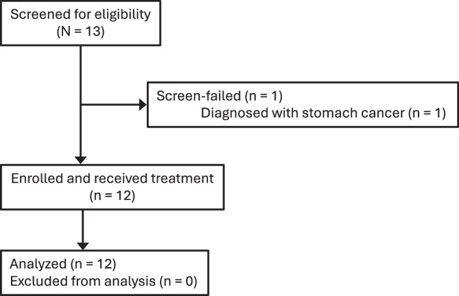
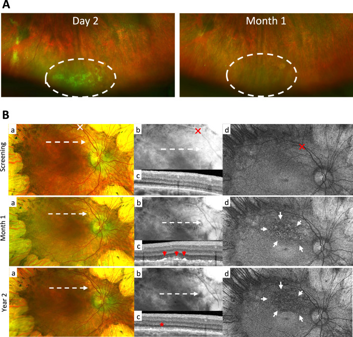
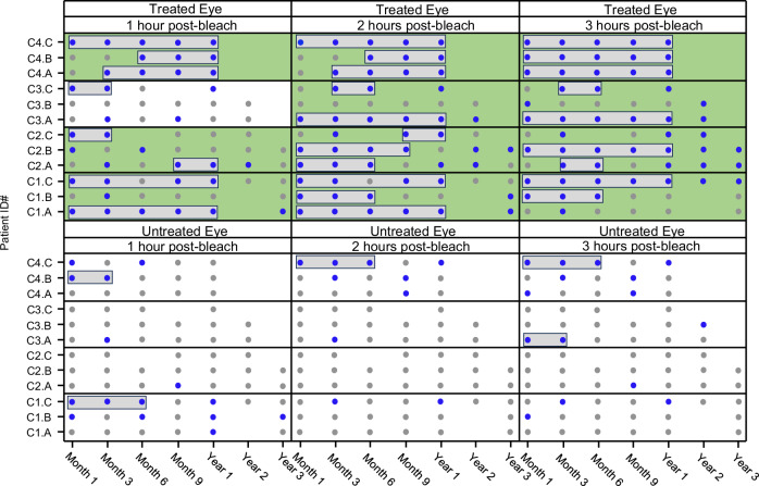
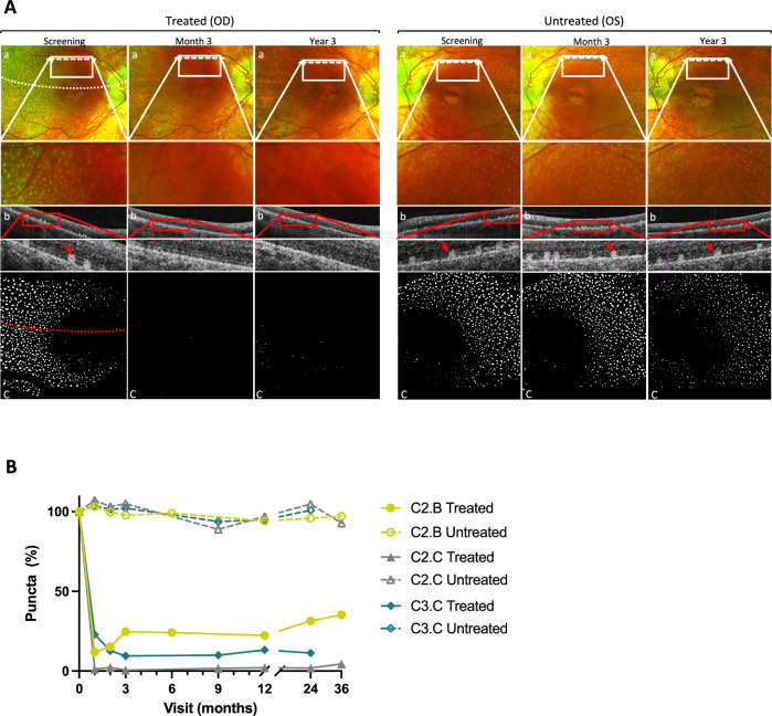
Gene therapy holds promise for treatment of inherited retinal dystrophies, a group of rare genetic disorders characterized by severe loss of vision. Here, we report up to 3-year pre-specified interim safety and efficacy results of an open-label first-in-human dose-escalation phase 1/2 gene therapy clinical trial in 12 patients with retinal dystrophy caused by biallelic mutations in the retinaldehyde-binding protein 1 (RLBP1) gene of the visual cycle. The primary endpoints were systemic and ocular safety and recovery of dark adaptation. Secondary endpoints included microperimetry, visual field sensitivity, dominant eye test and patient-reported outcomes. Subretinal delivery of an adeno-associated viral vector (AAV8-RLBP1) was well tolerated with dose-dependent intraocular inflammation which responded to corticosteroid treatment, and focal atrophy of the retinal pigment epithelium as the dose limiting toxicity. Dark adaptation kinetics, the primary efficacy endpoint, improved significantly in all dose-cohorts. Treatment with AAV8-RLBP1 resulted in the resolution of disease-related retinal deposits, suggestive of successful restoration of the visual cycle. In conclusion, to date, AAV8-RLBP1 has shown preliminary safety and efficacy in patients with RLBP1-associated retinal dystrophy. Trial number: NCT03374657.


Conflict of interest statement

N.R., K.H., C.W., N.J., C.B., K.S., M.L., and C.G. were employees of the sponsor of the trial, Novartis Pharma AG. The remaining authors declare no competing interests.

Figures

Fig. 1
Fig. 1. Diagram of patient flow.
This figure shows reasons for exclusion from the study and the number of patients included in the analyses.
Fig. 2
Fig. 2. Examples of inferior subretinal exudates and subretinal pigmented deposits.
A Inferior subretinal exudates (dashed area) in patient C2.B two days after treatment with AAV8-RLBP1. On day 29 post-treatment the exudates had resolved. B Subretinal pigmented deposits inside the superior vascular arcade shown on the color fundus image (a) and enhanced on the extracted red channel image (b) of patient C3.B 1-month post-treatment. On the corresponding SD-OCT scan (c) and FAF image (d), the deposits appear as hyperreflective dots at the RPE level (red arrows) and as hyperfluorescent dots (outlined by white arrows), respectively. Two years post-treatment, the deposits are no longer visible on either the color fundus photo or by SD-OCT. Instead, patchy loss of RPE (red star) and a hypofluorescent area are present (outlined by white arrows). The SD-OCT scan planes are shown on the fundus photos (dashed arrow). The retinotomy site of the subretinal bleb is marked (X).
Fig. 3
Fig. 3. Dark adaptation recovery kinetics.
Dark adaptation recovery for short wavelength stimulus (450 nm) outside of the pre-treatment prediction interval per timepoint, patient, visit, and cohort. Improvement (blue circles) versus non-improvement (gray circles) are shown for treated (upper panels) and untreated (lower panels) eyes at all available visits for all patients. Responders by timepoints are shown with gray boxes and efficacious cohorts by timepoints are shown with a green background. Treated eyes (p-values): C1 (1/2/3 h post-bleach), p = 0.104/0.08/0.104; C2 (1/2/3 h post-bleach), p = 0.104/0.08/0.104; C3 (1/2/3 hours post-bleach), p = 0.488/0.104/0.104; C4 (1/2/3 h post-bleach), p = 0.008/0.008/0.008. Untreated eyes (p-values): C1 (1/2/3 h post-bleach), p = 0.488/1/1; C2 (1/2/3 hours post-bleach), p = 1/1/1; C3 (1/2/3 h post-bleach), p = 1/1/0.488; C4 (1/2/3 h post-bleach), p = 0.488/0.488/0.488. Patient C1.A and C1.B missed the year 2 visit, and patient C2.C missed the month 6 visit due to the COVID-19 pandemic. Source data are provided as a Source Data file.
Fig. 4
Fig. 4. Regression of subretinal white puncta.
A Punctata albescens deposit in patient C2.C is present in both eyes on color fundus photos (a) at the screening visit. On the corresponding SD-OCT scan (b), the puncta appear as hyperreflective dots at the RPE level (red arrows). The SD-OCT scans are shown on the color fundus photos (dashed arrows in (a)). Three months through year 3 post-treatment, the puncta persisted in the untreated eyes but were no longer visible in the treated eye. The border of the subretinal bleb is shown on the screening visit images (dotted lines in (a) and (c)). Processed threshold en face images (c) highlight the difference in puncta between treated and untreated eyes. B The change in the number of punctata albescens deposits from the screening visit to the latest visit post-treatment is shown for the treated (solid lines) and untreated eye (hatched lines) of patients C2.B (circle), C2.C (triangle) and C3.C (diamond). The quantity of puncta was captured from digitally processed fundus images within a square area of 4 × 4 disc diameters as described in Methods. Source data are provided as a Source Data file.

References

    1. Bocquet, B. et al. Retinitis punctata albescens and RLBP1-allied phenotypes: phenotype-genotype correlation and natural history in the aim of gene therapy.Ophthalmol. Sci.1, 100052 (2021). 10.1016/j.xops.2021.100052 - DOI - PMC - PubMed
    1. Morimura, H., Berson, E. L. & Dryja, T. P. Recessive mutations in the RLBP1 gene encoding cellular retinaldehyde-binding protein in a form of retinitis punctata albescens. Invest. Ophthalmol. Vis. Sci.40, 1000–1004 (1999). - PubMed
    1. Burstedt, M. et al. Genotype-phenotype correlations in Bothnia dystrophy caused by RLBP1 gene sequence variations. Acta Ophthalmol.91, 437–444 (2013). 10.1111/j.1755-3768.2012.02431.x - DOI - PubMed
    1. Burstedt, M. et al. Retinal dystrophy associated with RLBP1 retinitis pigmentosa: A 5-year prospective natural history study. Invest. Ophthalmol. Vis. Sci.64, 42 (2023). 10.1167/iovs.64.13.42 - DOI - PMC - PubMed
    1. Burstedt, M. S. & Golovleva, I. Central retinal findings in Bothnia dystrophy caused by RLBP1 sequence variation. Arch. Ophthalmol.128, 989–995 (2010). 10.1001/archophthalmol.2010.167 - DOI - PubMed

Publication types

Substances

Associated data