. 2024 Sep 10;15(1):7865.
doi: 10.1038/s41467-024-51850-4.

LOXHD1 is indispensable for maintaining TMC1 auditory mechanosensitive channels at the site of force transmission

Affiliations

LOXHD1 is indispensable for maintaining TMC1 auditory mechanosensitive channels at the site of force transmission

Pei Wang et al. Nat Commun. .

Erratum in

Abstract

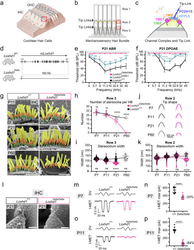
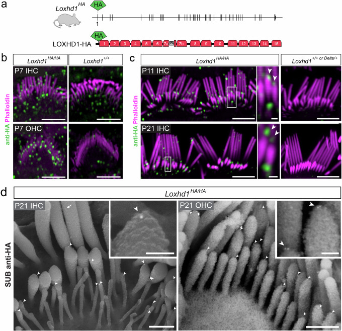
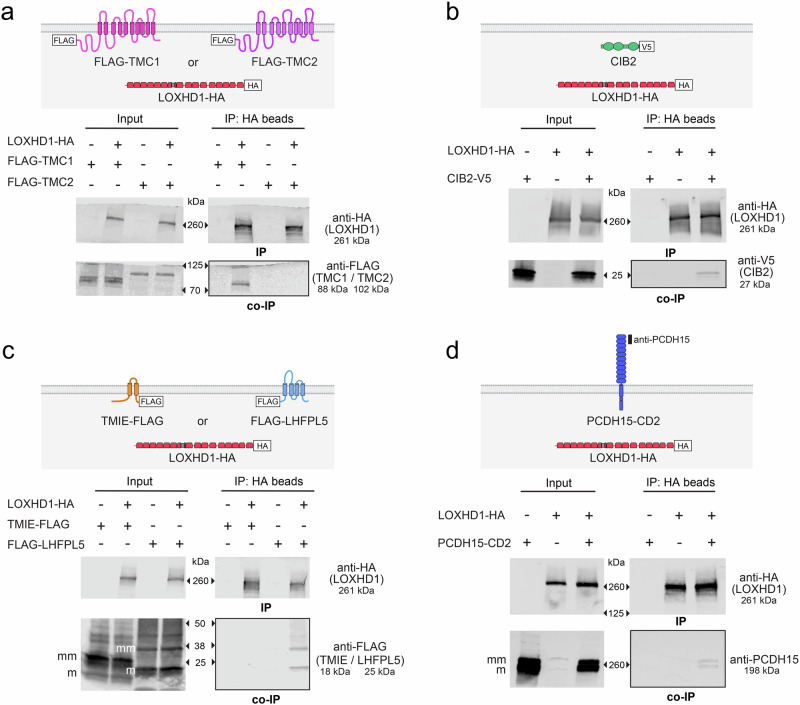
Hair cell bundles consist of stereocilia arranged in rows of increasing heights, connected by tip links that transmit sound-induced forces to shorter stereocilia tips. Auditory mechanotransduction channel complexes, composed of proteins TMC1/2, TMIE, CIB2, and LHFPL5, are located at the tips of shorter stereocilia. While most components can interact with the tip link in vitro, their ability to maintain the channel complexes at the tip link in vivo is uncertain. Return, using mouse models, we show that an additional component, LOXHD1, is essential for keeping TMC1-pore forming subunits at the tip link but is dispensable for TMC2. Using SUB-immunogold-SEM, we showed that TMC1 localizes near the tip link but mislocalizes without LOXHD1. LOXHD1 selectively interacts with TMC1, CIB2, LHFPL5, and tip-link protein PCDH15. Our results demonstrate that TMC1-driven mature auditory channels require LOXHD1 to stay connected to the tip link and remain functional, while TMC2-driven developmental channels do not.


Conflict of interest statement

The authors declare no competing interests.

Figures

Fig. 1
Fig. 1. The absence of LOXHD1 leads to hair bundle mechanotransduction deficits.
a Illustration of a cochlear sensory epithelium, which contains IHCs and OHCs that sense sound-induced forces with their apical hair bundles. b Illustration of a P21 IHC hair bundle comprising three stereocilia rows interconnected by tip links. c Cartoon representing the known auditory MET channel complex subunits located near the lower tip-link insertion point. d The Loxhd1Delta allele contains a large-scale genomic deletion at the Loxhd1 locus (see also Supplementary Fig. 1). e ABR and f DPOAE thresholds of P21 Loxhd1Delta mice across a 4–45 kHz frequency range. Data are represented as mean threshold ± SEM and were obtained from two independent blinded experiments (nanimal ≥ 4). Two-way ANOVA (age/genotype) followed by the Dunnett multiple comparisons test with adjusted p values, with wild-type means being the reference. g SEM of apical IHC Loxhd1Delta mice at P11, P21, and P60, with each stereocilia row being colored in post-production. Similar results were obtained in two independent experiments. Scale bars = 3 µm. h Number of row 3 stereocilia in apico-medial Loxhd1Delta/Delta IHCs. Means are connected with a line. Per group: P7–P21: ncells ≥ 9; P60: ncells ≥ 5; two-way ANOVA followed by the Šídák tests. i IHC row 3 stereocilia width. Means are connected with a line. Row 3: Per group: P7–P21: ncells ≥ 9, nstereocilia ≥ 143; P60: ncells ≥ 2, nstereocilia = 2 for Loxhd1Delta/Delta and nstereocilia = 61 for Loxhd1Delta/Delta. Two-way ANOVA followed by the Šídák tests. j IHC row 2 stereocilia tip shape. Superposition of tip traces from 12–20 row 2 stereocilia from 3–4 IHCs from 1–4 animals per genotype. k IHC row 2 stereocilia width. Row 2: Per group: P7-P21: ncells ≥ 9; nstereocilia ≥ 132; P60: ncells ≥ 5; nstereocilia ≥ 71. Micrographs obtained from two independent experiments. Two-way ANOVA followed by the Šídák tests. l Tip links (arrowheads) can be detected in P21 Loxhd1Delta/Delta apical IHCs. Similar results were obtained in two independent experiments. Scale bars = 100 nm. Mechanotransduction current (I-MET) traces from P7 (m) and P11 (o) IHC Loxhd1+/+ and Loxhd1Delta/Delta mechanically stimulated with a fluid jet and the maximum I-MET ± SD currents recorded (n, p). P7: ncells ≥ 10; P11: ncells ≥ 9 recorded over more than 12 independent experiments. Unpaired two-tailed t test: ****p < 0.0001. DV = driving voltage of the fluid jet piezoelectric stimulator. HB: hair bundle. For all experiments. ns (non significative) = p > 0.05; *p ≤ 0.05; **p ≤ 0.01; ***p ≤ 0.001; ****p ≤ 0.0001. The exact p values are presented in Supplementary Table 1. Source data are provided as a Source Data file.
Fig. 2
Fig. 2. LOXHD1 localizes at tips of transducing stereocilia of IHCs and OHCs.
a The Loxhd1HA allele comprises an HA-tag insertion in exon 1 before the first PLAT repeat. b, c Phalloidin labeling of the actin-rich stereocilia and anti-HA to detect LOXHD1 in Loxhd1HA/HA animals by Airyscan super-resolution microscopy. Specific HA staining is detected in Loxhd1HA/HA apical IHC and OHC hair bundles at P7 (b), P11, and P21 (c) but not in negative controls without HA. Scale bars = 4 µm. LOXHD1-HA can clearly be detected at the tip of row 2 stereocilia (arrowheads in insets) of apical P11 and P21 IHCs. Pictures from one of three replicate experiments. Scale bars = 4 µm, Inset scale bars = 0.4 µm. d SUB-immunogold-SEM anti-HA detects LOXHD1-associated gold beads on P21 IHC and OHC hair bundles. LOXHD1-gold beads (10 nm) were found at the tips of row 2 and 3 stereocilia (arrowhead) and more rarely in row 1 (arrow). High-magnification insets were taken from different hair bundles. Images from one of two replicate experiments, both yielding similar results. Scale bars = 500 nm, insets 100 nm. Source data are provided as a Source Data file.
Fig. 3
Fig. 3. TMC1 is progressively missing from row 2 stereocilia tips of IHCs in the absence of LOXHD1.
a TMC1 immunofluorescence localization (anti-HA on Tmc1HA/HA animals) at the tips of row 2 IHC stereocilia at P7, P11, and P21 showed a progressive reduction in Loxhd1Delta/Delta but not in Loxhd1Delta/+. Insets show high magnification at some row 2 tips. The last panels are negative control animals without a tag. Scale bars = 4 µm. b Percentage of row 2 tips with TMC1 puncta identified from 3D-volumes per genotype, represented as mean % ± SEM. (see Supplementary Fig. 2 and the Material and Methods section for more details). Per group: nmice ≥ 3, ncells ≥ 9. The experiment was replicated twice at P7 and three times at P11 and P21. One-way ANOVA followed by the Tukey multiple comparisons test. c TMC2 immunofluorescence localization (anti-MYC on Tmc2MYC/MYC animals) at the tips of row 2 IHC stereocilia at P7 and P11. Scale bars = 4 µm. d The percentage of row 2 tips with TMC2 puncta was not affected in Loxhd1Delta/Delta at P7 and P11. Represented as mean % ± SEM. The experiment was replicated twice at each age. One-way ANOVA followed by the Tukey multiple comparisons. e TMIE immunofluorescence localization (anti-HA on TmieHA/HA animals) at the tips of row 2 IHC stereocilia at P11. Scale bars = 4 µm. f  The percentage of row 2 tips with TMIE puncta was reduced in Loxhd1Delta/Delta at P11 but to a lesser extent than TMC1. Represented as mean % ± SEM. Per group: nmice = 4, ncells = 9. Unpaired two-tailed t test. The experiment was replicated twice. For all experiments. ns (non significative) = p > 0.05; *p ≤ 0.05; **p ≤ 0.01; ***p ≤ 0.001; ****p ≤ 0.0001. The exact p values are presented in Supplementary Table 1. Source data are provided as a Source Data file.
Fig. 4
Fig. 4. TMC1 is progressively mislocalized away from the first 100 nm of row 2 stereocilia tips of IHCs in the absence of LOXHD1.
a Sub-membranous (or SUB) immunogold-SEM anti-HA with a secondary antibody conjugated to 10 nm gold-beads localizes TMC1 at the tip of rows 2 and 3 stereocilia of P21 IHCs. Note that some row 3 stereocilia were disconnected from row 2, while gold-beads were attached to row 2 shafts at the presumptive upper tip link insertion position. Scale bar = 200 nm. b Distance between TMC1-gold beads to the stereocilia tip in rows 2 and 3 stereocilia measured from SUB-immunogold-SEM micrographs in Loxhd1+/+ IHCs. TMC1-gold beads were concentrated within 100 nm from the stereocilia tips (see the focused panel). Medians were indicated by a blue line. P21: ncells = 13; row 2: nstereocilia = 40; row 3: nstereocilia = 45. ce SUB-immunogold-SEM anti-HA detects TMC1-associated gold beads (arrowheads) at IHC row 2 tips of Loxhd1+/+ and Loxhd1Delta/Delta at P7, P11, and P21. Scale bar = 200 nm. The experiment was performed once at each age. Distance between TMC1-gold beads to the stereocilia tip in row 2 (f) and row 3 (g) stereocilia measured from SUB-immunogold-SEM micrographs in Loxhd1+/+ and Loxhd1Delta/Delta IHCs. Row 2: Per group: P7: ncells ≥ 18; nstereocilia ≥ 113; P11: ncells ≥ 16; nstereocilia ≥ 51; P21: ncells ≥ 13; nstereocilia ≥ 30. Row 3: P11: ncells ≥ 14; nstereocilia = 117 for Loxhd1+/+ and 12 for Loxhd1Delta/Delta; P21: nstereocilia = 45 for Loxhd1+/+ and 0 for Loxhd1Delta/Delta. Medians were indicated by a blue line. The experiment was performed once at each age. h, i Stereocilia classification based on the TMC1-gold distribution pattern (with at least one gold bead present regardless of its position, with at least one gold bead in the first 100 nm, with at least one gold bead below 100 nm, and without any gold beads). Row 2 (h): per group: P7: ncells ≥ 16; P11-P21: ncells ≥ 12; row 3 (i): per group: P11: ncells ≥ 13; P21: ncells ≥ 11. Represented as mean % ± SEM. Two-way ANOVA followed by Tukey’s multiple comparisons tests. For all experiments. ns (non significative) = p > 0.05; *p ≤ 0.05; ** p ≤ 0.01; ***p ≤ 0.001; ****p ≤ 0.0001. The exact p values are presented in Supplementary Table 1. Source data are provided as a Source Data file.
Fig. 5
Fig. 5. TMC1 localization in OHC stereocilia is affected by the Loxhd1Delta allele.
a TMC1 localization by immunofluorescence (anti-HA) in P7 OHC hair bundles (Phalloidin) along the tonotopical axis in Tmc1HA/HA with Loxhd1+/+, Loxhd1Delta/+, or Loxhd1Delta/Delta. TMC1+/+; Loxhd1Delta/+ was used as “non-HA” negative control. Scale bars = 4 µm. b Whole hair bundle TMC1-HA fluorescence intensity quantification on P7 apical, medial, and basal OHCs of Tmc1HA/HA with Loxhd1+/+, Loxhd1Delta/+, or Loxhd1Delta/Delta animals. Then intensity values were normalized to the mean fluorescence intensity of apical Tmc1HA/HA; Loxhd1+/+ OHCs. Per group: ncells ≥ 27. The experiment was replicated twice. Two-way ANOVA followed the Tukey test. c SUB-immunogold-SEM anti-HA detects TMC1-associated gold beads (10 nm, arrowheads) at OHC row 2 and 3 tips of Loxhd1+/+ and Loxhd1Delta/Delta P7 medial OHCs. The experiment was performed once. Scale bars = 200 nm. d Distance between TMC1-gold beads to the stereocilia tips in row 2 and 3 stereocilia measured from SUB-immunogold-SEM micrographs in Loxhd1+/+ and Loxhd1Delta/Delta P7 OHCs. Quantified OHCs were from pooled tonotopic positions distributed as follow: 35% apical, 27% middle, and 38% basal. Per group: P7 row 2: ncells ≥ 38; nstereocilia ≥ 113; row 3: ncells ≥ 39; nstereocilia ≥ 75; The experiment was performed once. e, f Stereocilia classification based on the TMC1-gold distribution pattern: with at least one gold bead present regardless of its position, with at least one gold bead in the first 100 nm, with at least one gold bead below 100 nm, and without any gold beads. Row 2 (e): per group: ncells ≥ 33; row 3 (f): per group: ncells ≥ 35; Represented as mean % ± SEM. Two-way ANOVA followed by Tukey’s multiple comparisons tests. For all experiments. ns (non significative) = p > 0.05; *p ≤ 0.05; **p ≤ 0.01; ***p ≤ 0.001; ****p ≤ 0.0001. The exact p values are presented in Supplementary Table 1. Source data are provided as a Source Data file.
Fig. 6
Fig. 6. The absence of LOXHD1 leads to BAIAP2L2 mislocalization.
a Anti-BAIAP2L2 immunofluorescence on P21 apical HCs shows a strong signal at transducing stereocilia tips which is absent in the mechanotransduction deficient IHC mutants TmieKO/KO and Tmc1KO/KO. In Loxhd1Delta/Delta, a strong BAIAP2L2 signal is detected in row 2 stereocilia but with a different distribution than control Loxhd1Delta/+ littermates. Boxes correspond to magnified panels. Scale bars = 2 µm. Representative results from experiments replicated once (TmieKO) or twice (Tmc1KO and Loxhd1Delta). b Stereocilia classification based on the BAIAP2L2 fluorescence signal distribution pattern: only at the tip, at both the tip and along the shaft, at the shaft, or absent from stereocilia. Data are represented as mean ± SEM. The experiment was replicated twice. Per group: ncells ≥ 8; Mann–Whitney test, two-tailed. c SUB-immunogold-SEM anti-BAIAP2L2 shows the enrichment of gold beads at the tip of rows 2 and 3 P21 IHC stereocilia in Loxhd1+/+, while gold beads were more distant from the tip in Loxhd1Delta/Delta. Arrowheads indicate the gold beads closest to the tips. Scale bars = 500 nm. Distance between BAIAP2L2-gold beads to tip of rows 2 (d) and 3 (e) stereocilia measured from SUB-immunogold-SEM micrographs in Loxhd1+/+ and Loxhd1Delta/Delta P21. IHCs corresponded to ~70% apical, ~12% medial, and ~18% basal. Per group: P21 row 2 (d) ncells ≥ 34; nstereocilia ≥ 168; row 3 (e) ncells ≥ 34; nstereocilia = 155 for Loxhd1+/+ and 4 for Loxhd1Delta/Delta; The experiment was replicated twice. Stereocilia classification based on the BAIAP2L2-gold distribution pattern: with at least one gold bead present regardless of its position, with at least one gold bead in the first 100 nm, with at least one gold bead below 100 nm, and without any gold beads. Represented as mean % ± SEM. Row 2 (f): per group: ncells ≥ 33; row 3 (g): per group: ncells = 32. Mann-Whitney test, two-tailed. For all experiments. ns (non significative) = p > 0.05; *p ≤ 0.05; **p ≤ 0.01; ***p ≤ 0.001; ****p ≤ 0.0001. The exact p values are presented in Supplementary Table 1. Source data are provided as a Source Data file.
Fig. 7
Fig. 7. LOXHD1 interacts in vitro with MET channel complex and tip link proteins.
ad Co-immunoprecipitation experiments from HEK293T cells overexpressing LOXHD1-HA with tagged channel complex or tip link proteins. FLAG-TMC1 was co-immunoprecipitated with LOXHD1-HA using anti-HA magnetic beads, but FLAG-TMC2 was not (a); CIB2-V5 (b), FLAG-LHFPL5 (c) and PCDH15 (d) were co-immunoprecipitated with LOXHD1-HA, while TMIE-FLAG was not (b). Each experiment was replicated at least thrice; (a) was replicated five times. Predicted protein sizes based on primary sequence: LOXHD1-HA: 261 kDa; TMC1-FLAG: 88 kDa; TMC2-FLAG: 102 kDa; CIB2-V5: 27 kDa; HA-CIB2: 23 kDa; TMIE-FLAG: 18 kDa; FLAG-LHFPL5: 25 kDa; PCDH15: 198 kDa. m: monomeric protein molecular size; mm: multimeric molecular size; IP: Immunoprecipitation. For corresponding full blots, see Supplementary Fig. 7.
Fig. 8
Fig. 8. LOXHD1 and TMC1 are part of protein complexes attached to the tip-link branches in vivo, and molecular interaction models between TMC1 and the tip link, with or without LOXHD1.
a In SUB experiments on P21 IHCs, some row 2 tips are detached from the rest of the stereocilia but still connected to the tip links (arrowheads and high magnification in the lower panels). Anti-HA gold-beads were specifically found at detached row 2 tips for LOXHD1-HA and TMC1-HA but not in the WT sample. Scale bars: 500 nm and 50 nm for the low and high mag pictures, respectively. b Quantification of the percentage of P21 IHC detached row 2 tips containing gold. Loxhd1HA/HA anti-HA: ncells = 17, ndetached tip with gold / total = 31/54; WT anti-HA: ncells = 5, ndetached tip with gold / total = 0/11; Tmc1HA/HA anti HA: ncells = 10, ndetached tip with gold / total = 34/44; WT anti-BAIAP2L2: ncells = 14, ndetached tip with gold / total = 1/42. The experiment was performed once. c Cartoon representing the mature auditory mechanotransduction protein complex in the wild-type (WT). d Model summarizing the molecular connections between TMC1 and PCDH15. e Cartoon representing the mature auditory mechanotransduction protein complexes in the absence of LOXHD1: initially, a population of TMC1 channels still functions while a second population, progressively increasing in proportion, is physically disconnected from the tip-link and cannot receive sound-induced forces. f Model summarizing the molecular connections between TMC1 and PCDH15 persisting upon the absence of LOXHD1: some TMC1/LHFPL5/TMIE proteins are initially sufficient to maintain some TMC1 channels at the tip link. However, with time, more and more TMC1 is found at a distance from the PCDH15, with LHFPL5 remaining attached to PCDH15 along with some TMIE molecules. A subset of TMIE proteins is predicted to remained attached to the mislocalized TMC1 molecules. The predicted localization of CIB2 remains hypothetical. However, considering that CIB2 can interact with BAIAP2L2 and is necessary for proper localization of BAIAP2L2 in hair bundles, and given that we observed BAIAP2L2 mislocalization similar to TMC1 at P21, it is probable that CIB2 is also associated with the mislocalized TMC1 molecules and absent from the stereocilia tip. Predicted positions are indicated with transparent colors. Source data are provided as a Source Data file.

Update of

References

    1. Coste, B. et al. Piezo proteins are pore-forming subunits of mechanically activated channels. Nature483, 176–181 (2012) - PMC - PubMed
    1. Ranade, S. S. et al. Piezo2 is the major transducer of mechanical forces for touch sensation in mice. Nature516, 121–125 (2014). - PMC - PubMed
    1. Kawashima, Y. et al. Mechanotransduction in mouse inner ear hair cells requires transmembrane channel–like genes. J. Clin. Investig.121, 4796–4809 (2011). - PMC - PubMed
    1. Pan, B. et al. TMC1 and TMC2 are components of the mechanotransduction channel in hair cells of the mammalian inner ear. Neuron79, 504–515 (2013). - PMC - PubMed
    1. Jia, Y. et al. TMC1 and TMC2 Proteins Are Pore-Forming Subunits of Mechanosensitive Ion Channels. Neuron105, 310–321. (2020). - PubMed

Publication types

LinkOut - more resources