. 2024 Sep 10;14(1):21155.
doi: 10.1038/s41598-024-71695-7.

Ortho-positronium lifetime for soft-tissue classification

Affiliations

Ortho-positronium lifetime for soft-tissue classification

Ashish V Avachat et al. Sci Rep. .

Abstract

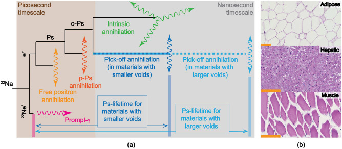
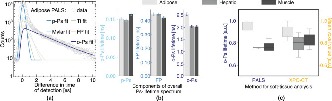
The objective of this work is to showcase the ortho-positronium lifetime as a probe for soft-tissue characterization. We employed positron annihilation lifetime spectroscopy to experimentally measure the three components of the positron annihilation lifetime-para-positronium (p-Ps), positron, and ortho-positronium (o-Ps)-for three types of porcine, non-fixated soft tissues ex vivo: adipose, hepatic, and muscle. Then, we benchmarked our measurements with X-ray phase-contrast imaging, which is the current state-of-the-art for soft-tissue analysis. We found that the o-Ps lifetime in adipose tissues (2.54 ± 0.12 ns) was approximately 20% longer than in hepatic (2.04 ± 0.09 ns) and muscle (2.03 ± 0.12 ns) tissues. In addition, the separation between the measurements for adipose tissue and the other tissues was better from o-Ps lifetime measurement than from X-ray phase-contrast imaging. This experimental study proved that the o-Ps lifetime is a viable non-invasive probe for characterizing and classifying the different soft tissues. Specifically, o-Ps lifetime as a soft-tissue characterization probe had a strong sensitivity to the lipid content that can be potentially implemented in commercial positron emission tomography scanners that feature list-mode data acquisition.

Keywords: PALS; Positronium annihilation lifetime spectroscopy; Soft tissue analysis; X-ray phase-contrast imaging.


Conflict of interest statement

The authors declare no competing interests.

Figures

Figure 1
Figure 1
(a) Illustration of the physics of positronium transport. 22Na undergoes β+ decay (positron and neutrino emissions) and turns into an excited 22Ne, which in turn decays by emitting a prompt-γ (1274.6 keV gamma emission). Hereafter, the emitted positron is referred to as free positron (FP). FP can either directly annihilate by 2γ emission or can form a positronium (Ps); Ps can take a form of a para-positronium (p-Ps) or an ortho-positronium (o-Ps); p-Ps directly annihilates by 2γ emission; and o-Ps can either annihilate by 2γ emission (pick-off annihilation) or 3γ emission. (b) Histological photomicrographs of example soft tissues. Hematoxylin and eosin (H &E) stained histological photomicrographs for adipose, hepatic, and muscle tissues. Scale bars represent 100 μm.
Figure 2
Figure 2
Results of PALS analysis and its benchmarking with XPC-CT. (a) The five components Ps-lifetime—p-Ps, fixed component for titanium (Ti) foil encapsulation of the source (248 ps), fixed component for Mylar foil that was used to wrap the tissue samples (382 ps), free-positron (FP), and o-Ps lifetimes—can be decomposed from the measured PALS spectrum (example PALS spectrum and its decomposed components for adipose tissues in a). (b) The comparison of three non-fixed components of the PALS spectrum for the three soft tissue types with 5 repeats for each type (n = 5) showed that o-Ps was the most sensitive out of the three components for discerning the subtle changes between the different types of soft-tissue. Single standard-deviation error bars are added to the plot. (c) Comparison of PALS as a method for soft-tissue analysis with the current state-of-the-art, XPC-CT. PALS probe measurement—o-Ps lifetime—showed less variations across the samples than the mean voxel value measurement through XPC-CT. The adipose tissue was significantly more discernible from the hepatic and the muscle tissues using mean o-Ps lifetime compared to using mean voxel values in XPC-CT. (For the box-and-whiskers plot in (c), the PALS measurements and XPC-CT mean voxel values were normalized to their respective maximums for presentation purposes.)
Figure 3
Figure 3
Methods for PALS analysis and its benchmarking with XPC-CT. (a) The PALS measurement setup: two scintillation detectors with tissue samples in the middle irradiated by the 22Na source. (b) 22Na source was placed between two tissue samples that were kept cooled by a Peltier-cell based system. The tissue samples were wrapped with Mylar foils of 2.5 μm thickness to avoid these tissue samples from touching the 22Na source. (c) An example tomographic slice of XPC-CT images. The blue, green, and red boxes represent a 2D slice of the 3D regions-of-interest corresponding to the adipose, hepatic, and muscle tissues, respectively, in the given example XPC-CT slice. (d) Sample fast pulse acquired by the organic scintillation detector, (1) its attenuated version by a factor F, (2) the original pulse delayed by Δ ns and inverted, and the (3) bipolar pulse obtained by subtracting (2) from the original pulse. The zero-crossing point corresponds to the pulse time stamp. (e) Energy spectrum and its average energy for 70 kVp X-ray beam from the liquid-metal-jet-based X-ray source used by XPC-CT system.

References

    1. Gidley, D. W., Peng, H. G. & Vallery, R. S. Positron annihilation as a method to characterize porous materials. Annu. Rev. Mater. Res.36, 49–79. 10.1146/annurev.matsci.36.111904.135144 (2006).10.1146/annurev.matsci.36.111904.135144 - DOI
    1. Tao, S. J. Positronium annihilation in molecular substances. J. Chem. Phys.56, 5499–5510. 10.1063/1.1677067 (1972).10.1063/1.1677067 - DOI
    1. Goworek, T. Positronium as a probe of small free volumes in crystals, polymers and porous media. Annales UMCS, Chemia69, 1–110. 10.2478/umcschem-2013-0012 (2015).10.2478/umcschem-2013-0012 - DOI
    1. DeBenedetti, S. New atoms - positronium and mesonic atoms. Il Nuovo Cimento4, 1209–1270. 10.1007/BF02744346 (1956).10.1007/BF02744346 - DOI
    1. Ore, A. & Powell, J. L. Three-photon annihilation of an electron-positron pair (1949).

LinkOut - more resources