. 2024 Sep 11;22(1):193.
doi: 10.1186/s12915-024-01990-2.

Hypoxia and Foxn1 alter the proteomic signature of dermal fibroblasts to redirect scarless wound healing to scar-forming skin wound healing in Foxn1-/- mice

Affiliations

Hypoxia and Foxn1 alter the proteomic signature of dermal fibroblasts to redirect scarless wound healing to scar-forming skin wound healing in Foxn1-/- mice

Barbara Gawronska-Kozak et al. BMC Biol. .

Abstract

Background: Foxn1-/- deficient mice are a rare model of regenerative skin wound healing among mammals. In wounded skin, the transcription factor Foxn1 interacting with hypoxia-regulated factors affects re-epithelialization, epithelial-mesenchymal transition (EMT) and dermal white adipose tissue (dWAT) reestablishment and is thus a factor regulating scar-forming/reparative healing. Here, we hypothesized that transcriptional crosstalk between Foxn1 and Hif-1α controls the switch from scarless (regenerative) to scar-present (reparative) skin wound healing. To verify this hypothesis, we examined (i) the effect of hypoxia/normoxia and Foxn1 signalling on the proteomic signature of Foxn1-/- (regenerative) dermal fibroblasts (DFs) and then (ii) explored the effect of Hif-1α or Foxn1/Hif-1α introduced by a lentiviral (LV) delivery vector to injured skin of regenerative Foxn1-/- mice with particular attention to the remodelling phase of healing.

Results: We showed that hypoxic conditions and Foxn1 stimulation modified the proteome of Foxn1-/- DFs. Hypoxic conditions upregulated DF protein profiles, particularly those related to extracellular matrix (ECM) composition: plasminogen activator inhibitor-1 (Pai-1), Sdc4, Plod2, Plod1, Lox, Loxl2, Itga2, Vldlr, Ftl1, Vegfa, Hmox1, Fth1, and F3. We found that Pai-1 was stimulated by hypoxic conditions in regenerative Foxn1-/- DFs but was released by DFs to the culture media exclusively upon hypoxia and Foxn1 stimulation. We also found higher levels of Pai-1 protein in DFs isolated from Foxn1+/+ mice (reparative/scar-forming) than in DFs isolated from Foxn1-/- (regenerative/scarless) mice and triggered by injury increase in Foxn1 and Pai-1 protein in the skin of mice with active Foxn1 (Foxn1+/+ mice). Then, we demonstrated that the introduction of Foxn1 and Hif-1α via lentiviral injection into the wounded skin of regenerative Foxn1-/- mice activates reparative/scar-forming healing by increasing the wounded skin area and decreasing hyaluronic acid deposition and the collagen type III to I ratio. We also identified a stimulatory effect of LV-Foxn1 + LV-Hif-1α injection in the wounded skin of Foxn1-/- mice on Pai-1 protein levels.

Conclusions: The present data highlight the effect of hypoxia and Foxn1 on the protein profile and functionality of regenerative Foxn1-/- DFs and demonstrate that the introduction of Foxn1 and Hif-1α into the wounded skin of regenerative Foxn1-/- mice activates reparative/scar-forming healing.

Keywords: Dermal fibroblasts; Foxn1; Hypoxia; Pai-1; Regeneration; Skin wound healing.


Conflict of interest statement

The authors declare that they have no competing interests.

Figures

Fig. 1
Fig. 1
Scheme of the proteomic experiment. DFs collected from the Foxn1−/− (CBy. Cg-Foxn1 < nu > /cmdb) mice were cocultured with keratinocytes collected from Foxn1.+/+ (C57BL/6 J) mice. Keratinocytes seeded in inserts were transduced with adenovirus carrying Foxn1 (Ad-Foxn1) or control (Ad-GFP). Keratinocytes and DFs were cocultured under hypoxic (1% O2) or normoxic (21% O2) conditions (n = 3, 2 animals per experiment, total n = 6 animals). After 24 h of coculture, DFs and DFCM were collected separately and subjected to detailed mass spectrometry analysis (LC‒MS/MS)
Fig. 2
Fig. 2
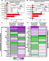
Classification of DF proteins regulated upon hypoxia (1% O2) vs. normoxia (21% O2) that were cocultured with Ad-Foxn1 (A)- or Ad-GFP (B)-transduced keratinocytes for 24 h according to GO using PANTHER and enriched with the g: Profiler database. A, B Diagrams illustrate detected proteins in terms of “molecular function” (with detailed proteins included in the catalytic activity class). C, D Heatmap of DF proteins linked to ECM organization (C) or the proteins interacting with ECM (D) enriched by hypoxia separately for coculture with Ad-Foxn1 or Ad-GFP keratinocytes. The scale bar in the heatmap indicates protein fold changes ranging from + 12 to − 1
Fig. 3
Fig. 3
Classification of proteins identified in DFCM regulated upon hypoxia (1% O2) vs. normoxia (21% O2) for keratinocytes transduced with Ad-Foxn1 (A) or Ad-GFP (B). Diagrams illustrate detected proteins in terms of the “Pathway” category with detailed proteins included in the plasminogen activating cascade. C Interactive network of proteins up- (Pai-1, Mmp-13, Mmp-8, Vegfa, Bsg) or downregulated (Ccl7 and Adamts5) in the DFCM from DFs cocultured with Ad-Foxn1 keratinocytes
Fig. 4
Fig. 4
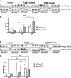
Pai-1 (A,B) and Vegfa (C,D) protein levels in cultured DFs isolated from the skin of Foxn1+/+ or Foxn1.−/− mice. Western blots for control, Tgfβ1- or Tgfβ3-treated DFs (A and C) followed by densitometric analyses (B and D). Values are the lsmean ± SE; asterisks indicate significant differences (*p < 0.05; ** p < 0.01, *** p < 0.001)
Fig. 5
Fig. 5
Oxygen availability (normoxia vs. hypoxia) modulates DFs: proliferation (A), metabolic activity (B) and migration (C). Foxn1.−/− DFs cultured under hypoxic or normoxic conditions with KCM (keratinocyte conditioned media) from Ad-Foxn1- or Ad-GFP-transduced keratinocytes were analysed with a BrdU incorporation assay followed by flow cytometry analysis (A; n = 4), MTT metabolic activity assay (B; n = 4) or scratch migratory assay (C; n = 4). Values are the lsmean ± SE; asterisks indicate significant differences between normoxic or hypoxic condition transduction (**p < 0.01)
Fig. 6
Fig. 6
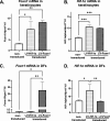
Transduction efficiency of LVs carrying Hif-1α-eGFP or Foxn1-mCherry in cell culture model. qRT-PCR analysis of Foxn1 (A, C) or Hif-1α (B, D) mRNA expression in keratinocytes (A, B) or DFs (C, D). Asterisks indicate significant differences (* p < 0.05; ** p < 0.01; *** p < 0.001). Data represent the mean ± SD
Fig. 7
Fig. 7
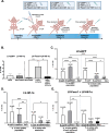
Transduction efficiency of LVs carrying eGFP (control), Hif-1α-eGFP or Foxn1-mCherry + Hif-1α-eGFP injected into the wounded skin of Foxn1−/− mice at postwounding day 1 analysed by flow cytometry at day 14 or by immunohistochemistry for mCherry (LV-Foxn1-mCherry) at postwounding day 6. A Scheme of experiment. B Percentage of cells positive for eGFP or mCherry isolated from the injured skin of LV-eGFP (control)-, LV-Hif-1α-eGFP- or LV-Hif-1α-eGFP + LV-Foxn1-mCherry-injected Foxn1−/− mice. C–E Percentage of E-cadherin (E-cad +), vimentin (Vim +) or E-cadherin + vimentin double-positive cells (E-cad/Vim +) within eGFP-positive (LV-eGFP or LV-Hif-1α) or mCherry + eGFP-positive (LV-Hif-1α + LV-Foxn1) populations in comparison to the whole cell population. F Immunohistochemical detection of mCherry (LV-Foxn1-mCherry) localization in the skin of Foxn1.−/− mice. Asterisks indicate significant differences (* p < 0.05; ** p < 0.01; *** p < 0.001; **** p < 0.0001). Data represent the mean ± SD. E-epidermis, D-dermis, arrowheads indicate positivity in epidermis, arrows point out positivity in dermis (F). Scale bar (F) 50 μm, (inset; 20 μm)
Fig. 8
Fig. 8
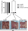
LV-Foxn1 + LV-Hif-1α introduction into the postwounded skin (at day 1) of Foxn1−/− mice delays skin wound healing (at day 14). Morphometrical analysis of the percentage of the initial (100%; day 0) postwounded surface wound areas measured at postwounded day 14 (d14) (n = 12 measurements per time point). Values are the lsmean ± SE; asterisks indicate significant differences between the LV-eGFP- and LV-Hif-1α + LV-Foxn1-treated Foxn1.−/− mice at postwounding day 14 (*p < 0.05)
Fig. 9
Fig. 9
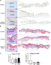
Distinctive accumulation of hyaluronic acid (A) and collagen (B) and expression of collagen 1a2 and collagen 3a1 mRNA (C) in injured skin of LV-eGFP (control)-, LV-Hif-1α- or LV-Hif-1α + LV-Foxn1-treated Foxn1−/− mice at postwounding day 6 (A) and day 14 (B,C). A Representative histological sections stained with alcian blue (marker for hyaluronic acid) and immunostained for EpCAM (epithelial cell adhesion molecule; marker of epidermis) and B Masson trichrome (marker for collagen) collected from the skin of Foxn1−/− mice. Scale bar A 50 µm, B 100 µm
Fig. 10
Fig. 10
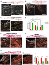
Evaluation of collagen type I vs. collagen type III content in stained picrosirius red histological skin sections collected from LV-eGFP (control)-, LV-Hif-1α- or LV-Hif-1α + LV-Foxn1-treated Foxn1−/− mice at day 6 (A–D) and day 14 (E–G and I) after injury. Representative histological sections of wounded (A–C; E–G; n = 3 per LV treatment) and unwounded skin (H; n = 3) were analysed. Collagen fibres: yellow‒red (collagen type I) and green (collagen type III) were evaluated as a Relative Object Count [%] at postwounding days 6 (D) and 14 (I). Scale bar (A-G) 200 µm, insets (A–G) 50 µm and (H) 50 µm; wb – wound bed
Fig. 11
Fig. 11
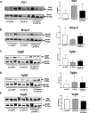
LV-Hif-1α and LV-Foxn1 + LV-Hif-1α regulate ECM content in the wounded skin of Foxn1−/− mice. Western blot and densitometric analyses of Pai-1 (A), Mmp-9 (B), Tgfβ1 (C), Tgfβ3 (D) and Vegfa (E) proteins in the skin of LV-eGFP (control), LV-Hif-1α- or LV-Foxn1 + LV-Hif-1α-treated Foxn1.−/− mice at postwounding day 14 (n = 3 per treatment group)
Fig. 12
Fig. 12
Pai-1 (A,B) and Foxn1 (C) protein levels in the intact (day 0) and wounded (days 1, 3, 7, 14) skin of Foxn1−/− (A,B) and Foxn1.+/+ (A–C) mice. Western blots (A, C) followed by densitometric analyses (B). Values are the lsmean ± SE; asterisks indicate significant differences (*p < 0.05)
Fig. 13
Fig. 13
Scheme of the proposed Foxn1 → Hif-1α → Pai-1 pathway in reparative scar-forming skin wound healing in Foxn1+/+ mice

Similar articles

Cited by

References

    1. Gurtner GC, Werner S, Barrandon Y, Longaker MT. Wound repair and regeneration. Nature. 2008;453(7193):314–21. Epub 2008/05/16. 10.1038/nature07039 - DOI - PubMed
    1. Gawronska-Kozak B, Bogacki M, Rim JS, Monroe WT, Manuel JA. Scarless skin repair in immunodeficient mice. Wound Repair Regen. 2006;14:265–76. 10.1111/j.1743-6109.2006.00121.x - DOI - PubMed
    1. Gawronska-Kozak B. Scarless skin wound healing in FOXN1 deficient (nude) mice is associated with distinctive matrix metalloproteinase expression. Matrix Biol. 2011;30(4):290–300. Epub 2011/05/05. 10.1016/j.matbio.2011.04.004 - DOI - PMC - PubMed
    1. Machcinska S, Kopcewicz M, Bukowska J, Walendzik K, Gawronska-Kozak B. Impairment of the Hif-1alpha regulatory pathway in Foxn1-deficient (Foxn1(-/-) ) mice affects the skin wound healing process. FASEB J. 2021;35(2):e21289 Epub 2021/01/22. 10.1096/fj.202001907R - DOI - PubMed
    1. Corbeaux T, Hess I, Swann JB, Kanzler B, Haas-Assenbaum A, Boehm T. Thymopoiesis in mice depends on a Foxn1-positive thymic epithelial cell lineage. Proc Natl Acad Sci U S A. 2010;107(38):16613–8. Epub 2010/09/09. 10.1073/pnas.1004623107 - DOI - PMC - PubMed

LinkOut - more resources