. 2024 Sep 10;24(1):312.
doi: 10.1186/s12935-024-03490-3.

Exosome-mediated transfer of lncRNA RP3-340B19.3 promotes the progression of breast cancer by sponging miR-4510/MORC4 axis

Affiliations

Exosome-mediated transfer of lncRNA RP3-340B19.3 promotes the progression of breast cancer by sponging miR-4510/MORC4 axis

Bo Wang et al. Cancer Cell Int. .

Abstract

Background: This study aims to explore the molecular mechanism of lncRNA RP3-340B19.3 on breast cancer cell proliferation and metastasis and clinical significance of lncRNA RP3-340B19.3 for breast cancer.

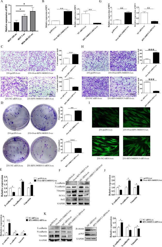
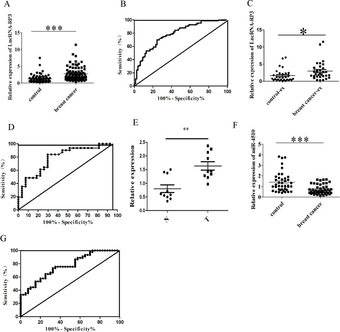
Methods: The subcellular localization of lncRNA RP3-340B19.3 was identified using RNA fluorescence in situ hybridization (FISH). The expression of lncRNA RP3-340B19.3 in breast cancer cells, breast cancer tissues, as well as the serum and serum exosomes of breast cancer patients, was measured through quantitative RT-PCR. In the in vitro setting, we conducted experiments to observe the effects of RP3-340B19.3 on both cell migration and proliferation. This was achieved through the utilization of transwell migration assays as well as clone formation assays. Meanwhile, transwell migration assays and clone formation assays were used to observe the effects of MDA-MB-231-exosomes enriched in RP3-340B19.3 on breast cancer microenvironment cells MCF7 and BMMSCs. Additionally, western blotting techniques were used to assess the expression levels of proteins associated with essential cellular processes such as proliferation, apoptosis, and metastasis. In vivo, the impact of RP3-340B19.3 knockdown on tumour weight and volume was observed within a nude mice model. We aimed to delve into the intricate molecular mechanisms involving RP3-340B19.3 by using bioinformatics analysis, dual luciferase reporter gene experiments and western blotting. Moreover, the potential correlations between RP3-340B19.3 expression and various clinical pathological characteristics were analyzed.

Results: Our investigation revealed that RP3-340B19.3 was expressed in both the cytoplasm and nucleus, with a noteworthy increase in breast cancer cells. Notably, we found that RP3-340B19.3 exerted a promoting influence on the proliferation and migration of breast cancer cells, both in vitro and in vivo. MDA-MB-231-exosomes enriched in RP3-340B19.3 promoted the proliferation and migration of MCF7 and BMMSCs in vitro. Mechanistically, RP3-340B19.3 demonstrated the capability to modulate the expression of MORC4 by forming a complex with miR-4510. This interaction subsequently triggered the activation of the NF-κB and Wnt-β-catenin signaling pathways. Furthermore, our study highlighted the potential diagnostic utility of RP3-340B19.3. We discovered its presence in the serum and exosomes of breast cancer patients, showing promising efficacy as a diagnostic marker. Notably, the diagnostic potential of RP3-340B19.3 was particularly significant in relation to distinguishing between different pathological types of breast cancer and correlating with tumour diameter.

Conclusion: Our findings establish that RP3-340B19.3 plays a pivotal role in driving the proliferation and metastasis of breast cancer. Additionally, exosomes enriched in RP3-340B19.3 could influence MCF7 and BMMSCs in tumour microenvironment, promoting the progression of breast cancer. This discovery positions RP3-340B19.3 as a prospective novel candidate for a tumour marker, offering substantial potential in the realms of breast cancer diagnosis and treatment strategies.

Keywords: Breast cancer; Exosome; Metastasis; Proliferation; lncRNA RP3-340B19.3.


Conflict of interest statement

The authors declare no competing interests.

Figures

Fig. 1
Fig. 1
RP3-340B19.3 promotes proliferation and migration of breast cancer in vitro. (A) Gene chip screening and bioinformatics prediction were used to select lnc RP3-340B19.3 as the subject of investigation. C, control MDA-MB-231; L, LPS treated MDA-MB-231. The concentration of LPS was 10 µg/ml. (B) The expression of RP3-340B19.3 in LPS-stimulated MDA-MB-231 breast cancer cells was examined by quantitative RT-PCR. The concentration of LPS was 10 µg/ml. **P < 0.01. (C) The expression of RP3-340B19.3 in MDA-MB-231 and MCF7 was examined by quantitative RT-PCR. *P < 0.05. (D) The expression of RP3-340B19.3 in MCF10A, MCF7 and MDA-MB-231 was examined by FISH. (E) To determine the cellular localization of RP3-340B19.3, the fractionation experiment was performed using digitonin. The level of GAPDH and U6 were checked for verification of cytoplasmic and nuclear extracts, respectively. The levels of RP3-340B19.3 mRNA were determined by RT-qPCR. (F) The migratory ability of RP3-340B19.3-overexpressing and RP3-340B19.3-knockdown MDA-MB-231 cells were evaluated by transwell migration assay. ***P < 0.001. (G) The proliferation ability of RP3-340B19.3-overexpressing and RP3-340B19.3-knockdown MDA-MB-231 cells were evaluated by cell colony assay. ***P < 0.001. (H) The migratory ability of RP3-340B19.3-overexpressing and RP3-340B19.3-knockdown MCF7 cells were evaluated by using transwell migration assay. ***P < 0.001. (I) The proliferation ability of RP3-340B19.3-overexpressing and RP3-340B19.3-knockdown MCF7 cells were evaluated by cell colony assay. ***P < 0.001. (J) The proliferation ability of RP3-340B19.3-overexpressing and RP3-340B19.3-knockdown MCF7 and MDA-MB-231 cells were evaluated by CCK8. ***P < 0.001, **P < 0.01, *P < 0.05. (K) The expression of EMT related genes in RP3-340B19.3-overexpressing and RP3-340B19.3-knockdown cells was examined by using quantitative RT-PCR. ***P < 0.001, **P < 0.01, *P < 0.05. (L) The expression of EMT related proteins, cleaved caspase3, PARP, PCNA and Bcl2 in RP3-340B19.3-overexpressing and RP3-340B19.3-knockdown cells was examined by using western blot. (M) The apoptosis rate of MCF7 cells pretreated with pcDNA, RP3-340B19.3 overexpression plasmids, NC siRNA, and RP3-340B19.3 siRNA. **P < 0.01. (N) The expression of wnt-β-catenin related genes in RP3-340B19.3-overexpressing and RP3-340B19.3-knockdown cells was examined by using quantitative RT-PCR. **P < 0.01, *P < 0.05. (O) The expression of β-catenin, P-P65 in RP3-340B19.3-overexpressing and RP3-340B19.3-knockdown cells was examined by using western blot
Fig. 2
Fig. 2
RP3-340B19.3 promotes the proliferation and migration of breast cancer in vivo. (A), (B) and (C) The macroscopic appearance, volume and weight of subcutaneous tumours in mice(n = 6/group) transplanted with NC siRNA and RP3-340B19.3 siRNA transfected MDA-MB-231 cells. **P < 0.01, *P < 0.05. (D), (E) and (F) The macroscopic appearance, volume and weight of subcutaneous tumours in mice(n = 6/group) transplanted with NC siRNA and RP3-340B19.3 siRNA transfected MCF7 cells. **P < 0.01, *P < 0.05. (G) and (H) The expression of RP3-340B19.3 in the tumour tissues in mice transplanted with NC siRNA and RP3-340B19.3 siRNA transfected MDA-MB-231 and MCF7 cells was examined by using quantitative RT-PCR. **P < 0.01. (I) and (J) The expression of EMT related genes in mice with NC siRNA and RP3-340B19.3 siRNA transfected MDA-MB-231 and MCF7 cells was examined by using quantitative RT-PCR. ***P < 0.001, **P < 0.01, *P < 0.05. (K) and (L) The expression of RP3-340B19.3 in the tumour tissues in mice transplanted with NC siRNA and RP3-340B19.3 siRNA transfected MDA-MB-231 cells was examined by using quantitative RT-PCR. **P < 0.01. (H) The expression of EMT related genes in mice with NC siRNA and RP3-340B19.3 siRNA transfected MDA-MB-231 cells was examined by using quantitative RT-PCR. ***P < 0.001, **P < 0.01. (K) and (L) The expression of EMT related proteins and PCNA in mice transplanted with NC siRNA and RP3-340B19.3 siRNA transfected MDA-MB-231 and MCF7 cells was determined by using immunohistochemistry
Fig. 3
Fig. 3
Role of RP3-340B19.3 in exosomes in tumour microenvironment. (A) The expression of RP3-340B19.3 in MCF-10-ex, MCF7-ex and MDA-MB-231-ex was examined by using quantitative RT-PCR. *P < 0.05. (B) The expression of RP3-340B19.3 in MCF7 cells treated with 231-pcDNA-ex, 231-Over-RP3-340B19.3-ex, 231-NC-siRNA-ex and 231-RP3-340B19.3-siRNA-ex was examined by using quantitative RT-PCR. **P < 0.01. (C) The effect of 231-Over-RP3-340B19.3-ex and 231-RP3-340B19.3-siRNA-ex on the migration of MCF7. ***P < 0.001. (D) The effect of 231-Over-RP3-340B19.3-ex and 231-RP3-340B19.3-siRNA-ex on the proliferation of MCF7. ***P < 0.001, **P < 0.01. (E) The effect of 231-Over-RP3-340B19.3-ex and 231-RP3-340B19.3-siRNA-ex on the expression of EMT related genes in MCF7 was examined by using quantitative RT-PCR. *P < 0.05. (F) The effect of 231-Over-RP3-340B19.3-ex and 231-RP3-340B19.3-siRNA-ex on the expression of EMT related proteins, PCNA and Bcl2 in MCF7 was examined by using western blot. (G) The expression of RP3-340B19.3 in BMMSCs treated with 231-pcDNA-ex, 231-Over-RP3-340B19.3-ex, 231-NC-siRNA-ex and 231-RP3-340B19.3-siRNA-ex was examined by using quantitative RT-PCR. **P < 0.01. (H) The effect of 231-Over-RP3-340B19.3-ex and 231-RP3-340B19.3-siRNA-ex on the migration of BMMSCs. ***P < 0.001. (I) The expression of α-SMA in BMMSCs treated with 231-pcDNA-ex, 231-Over-RP3-340B19.3-ex, 231-NC-siRNA-ex and 231-RP3-340B19.3-siRNA-ex was examined by immunofluorescence. (J) and (K) The effect of 231-Over-RP3-340B19.3-ex and 231-RP3-340B19.3-siRNA-ex on the expression of EMT related genes in BMMSCs was examined by using quantitative RT-PCR and western blot. *P < 0.05. β-catenin and p-P65 was examined by using western blot
Fig. 4
Fig. 4
RP3-340B19.3 targets and inhibits miR-4510. (A) The predicted binding sites in the seed region of RP3-340B19.3 and miR-4510. (B) The effect of miR-4510 on the stability of RP3-340B19.3 was determined by using a luciferase reporter gene assay. *P < 0.05. (C) and (D)The expression of miR-4510 in breast cancer cells transfected with pcDNA, RP3-340B19.3 overexpression plasmids, NC siRNA, and RP3-340B19.3 siRNA was examined by qRT-PCR. **P < 0.01, *P < 0.05. (E) and (G) The migratory ability of NC mimics, miR-4510-mimics, NC inhibitor and miR-4510 inhibitor transfected cells was evaluated by using transwell migration assay. ***P < 0.001. (F) and (H) Cell colony formation in NC mimics, miR-4510-mimics, NC inhibitor and miR-4510 inhibitor transfected cells. ***P < 0.001. (I) The expression of EMT related genes in NC mimics, miR-4510-mimics, NC inhibitor and miR-4510 inhibitor transfected cells was examined by qRT-PCR. ***P < 0.001, **P < 0.01, *P < 0.05. (J) The expression of EMT related proteins, PCNA and Bcl2 in NC mimics, miR-4510-mimics, NC inhibitor and miR-4510 inhibitor transfected cells was examined by western blot
Fig. 5
Fig. 5
miR-4510 targets and inhibits MORC4. (A) The predicted binding sites in the seed region of miR-4510 and the 3’UTR of MORC4 mRNA. (B) The effect of miR-4510 on the stability of MORC4 mRNA was determined by using a luciferase reporter gene assay. **P < 0.01. (C) and (D) The expression of MORC4 in breast cancer cells transfected with MNC, and 4510 mimics was examined by qRT-PCR and western blot. **P < 0.01. (E) The expression of MORC4 in mice transplanted with MDA-MB-231 cells pretreated with NC siRNA and RP3-340B19.3 siRNA was determined by immunohistochemistry. (F) The expression of MORC4 in MCF10A, MCF7 and MDA-MB-231 was examined by qRT-PCR. **P < 0.01. (G) The mRNA levels of MORC4 in breast cancer tissues and adjacent normal tissues. **P < 0.01. N, adjacent normal tissues; T, breast cancer tissues. (H) The protein levels of MORC4 in breast cancer tissues and adjacent normal tissues was determined by using immunohistochemistry. (I) and (J) The expression of MORC4 in breast cancer cells transfected with NC siRNA and MORC4 siRNA was examined by qRT-PCR and western blot. (K) The expression of EMT related proteins in NC siRNA, MORC4 siRNA transfected cells was examined by western blot. (L) The migratory ability of NC siRNA and MORC4 siRNA transfected cells was evaluated by transwell migration assay. **P < 0.01, *P < 0.05. (M) Cell colony formation in NC siRNA and MORC4 siRNA. **P < 0.01
Fig. 6
Fig. 6
Clinical detection analysis of RP3-340B19.3 in serum and its exosomes. (A) The expression of RP3-340B19.3 in the serum of 66 patients with breast cancer and that of 66 healthy volunteers. ***P < 0.001. (B) ROC curves for the diagnostic values of RP3-340B19.3 in breast cancer. (C) The expression of exosomal RP3-340B19.3 in the serum of 27 patients with breast cancer and that of 27 healthy volunteers. ***P < 0.001. (D) ROC curves for the diagnostic values of serum CA153 and exosomal RP3-340B19.3 in breast cancer. (E) The expression of RP3-340B19.3 in 10 breast cancer tissues and 10 adjacent normal tissues was determined by quantitative RT-PCR. *P < 0.05. N, adjacent normal tissues; T, breast cancer tissues. (F) The expression of miR-4510 in the serum of 35 patients with breast cancer and that of 35 healthy volunteers. ***P < 0.001. (G) ROC curves for the diagnostic values of miR-4510 in breast cancer

References

    1. Siegel RL, Miller KD, Fuchs HE, Jemal A. Cancer statistics, 2022. CA Cancer J Clin. 2022;72(1):7–33. 10.3322/caac.21708 - DOI - PubMed
    1. Jokar N, Velikyan I, Ahmadzadehfar H, Rekabpour SJ, Jafari E, Ting HH, et al. Theranostic Approach in breast Cancer: a treasured tailor for future oncology. Clin Nucl Med. 2021;46(8):e410–20. 10.1097/RLU.0000000000003678 - DOI - PubMed
    1. Hutchinson L. Breast cancer: challenges, controversies, breakthroughs. Nat Rev Clin Oncol. 2010;7(12):669–70. 10.1038/nrclinonc.2010.192 - DOI - PubMed
    1. Ghafouri-Fard SM, Taheri. Long non-coding RNA signature in gastric cancer. Exp Mol Pathol. 2020;113:104365. 10.1016/j.yexmp.2019.104365 - DOI - PubMed
    1. Chi Y, Wang JP, Yu WD, Yang JC. Long non-coding RNA in the pathogenesis of cancers. Cells. 2019;8(9):1015. 10.3390/cells8091015 - DOI - PMC - PubMed

LinkOut - more resources