. 2024 Sep 11;15(1):286.
doi: 10.1186/s13287-024-03908-9.

Connexin 25 maintains self-renewal and functions of airway basal cells for airway regeneration

Affiliations

Connexin 25 maintains self-renewal and functions of airway basal cells for airway regeneration

Jingyuan Zhang et al. Stem Cell Res Ther. .

Abstract

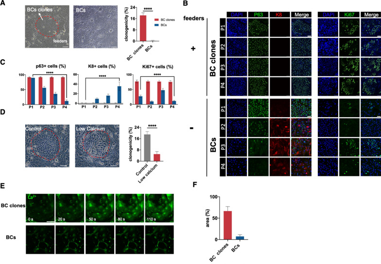
Background: The formation of stem cell clones enables close contact of stem cells inside. The gap junctions in such clone spheres establish a microenvironment that allows frequent intercellular communication to maintain self-renewal and functions of stem cells. Nevertheless, the essential gap junction protein for molecular signaling in clones is poorly known.

Methods: Primary human airway basal cells (hBCs) were isolated from brushing samples through bronchoscopy and then cultured. A tightly focused femtosecond laser was used to excite the local Ca2+ in an individual cell to initiate an internal Ca2+ wave in a clone to screen gap junction proteins. Immunoflourescence staining and clonogenicity assay were used to evaluate self-renewal and functions. RNA and protein levels were assessed by PCR and Western blot. Air-liquid interface assay was conducted to evaluate the differentiation potential. A Naphthalene injury mouse model was used to assess the regeneration potential.

Results: Herein, we identify Connexin 25 (Cx25) dominates intercellular Ca2+ communications in clones of hBCs in vitro to maintain the self-renewal and pluripotency of them. The self-renewal and in vitro differentiation functions and in vivo regeneration potential of hBCs in an airway damage model are both regulated by Cx25. The abnormal expression of Cx25 is validated in several diseases including IPF, Covid-19 and bronchiectasis.

Conclusion: Cx25 is essential for hBC clones in maintaining self-renewal and functions of hBCs via gap junctions.

Keywords: Airway basal cell; Calcium; Connexin; Self-renewal.


Conflict of interest statement

The authors declare no competing interests.

Figures

Fig. 1
Fig. 1
hBC clones maintain self-renewal and pluripotent during long-term in vitro culture. A hBC clones and hBCs cultured without feeders. Right panel: the ratio of clones in BC clones and BCs cultured without feeders on Day 4–5 post-seeding (n = 5 independent experiments). Red dashed lines: the outline of clones. Bar: 100 μm. B Immunofluorescent microscopy images of p63, K8 and Ki67 in hBC clones and hBCs without feeders in 4 continous passengers. Bar: 50 μm. C The quantified ratio of positive hBCs with above markers (n = 13 images from 4 independent experiments). D hBCs cultured with control medium and low calcium medium. Right panel: the ratio of clones in BCs cultures with control medium and low calcium medium on Day 4–5 post-seeding. Red dashed lines: the outline of clones. Bar: 200 μm. E Intercellular Ca2+ waves in hBC clones and hBCs treated with TG. Bar: 100 μm. F The quantified area of Ca2+ waves propagation (n = 4 independent trials). Data information: Data are presented as means ± SD. ****p < 0.0001 (Unpaired T-test for two groups and one-way ANOVA for multiple groups)
Fig. 2
Fig. 2
Intercellular Ca2+ waves propagate through gap junction in hBC clones. A The schematic diagram of photostimulation to an invidual target hBC in clones and hBCs without feeders by a tightly-focused femtosecond laser to initiate Ca2+ waves. B Ca2+ waves propagation in hBC clones and hBCs without feeders after photostimulation. C The quantified propagation area and speed of Ca2+ waves (n = 24). D Intercellular Ca2+ waves in hBC clones in presence of octanol, PPADS, 2-APB and Ca2+ free buffer respectively. E The quantified propagation area and speed of Ca2+ waves in groups treated with octanol, PPADS, 2-APB and Ca2+ free buffer respectively (n = 20). Bar: 50 μm. Data information: Data are presented as means ± SD. *p < 0.05, **p < 0.01, ***p < 0.001, ****p < 0.0001 (Unpaired T-test for two groups and one-way ANOVA for multiple groups)
Fig. 3
Fig. 3
Cx25 is essential to intercellular Ca2+ propagation in hBC clones. A RNA expression level of connexins in hBC clones and hBCs without feeders. B Protein expression level of Cx31.9, Cx32, Cx59 and Cx25 in hBC clones and hBCs without feeders (n = 3 independent experiments). C Ca2+ waves initiated by photostimulation in BCs with Cx31.9, Cx32, Cx59 and Cx25 transiently KD respectively (n = 20). D,E The quantified amplitude (C) and propagation area (D) of Ca2+ waves in those hBCs. F The protein expression level of Cx25 in stable Cx25-KD and Cx32-KD hBCs (n = 3 independent experiments). G Ca2+ waves in stable Cx25-KD, Cx32-KD, and Vehicle hBCs (n = 22). H The quantified propagation area and speed of Ca2+ waves in Cx25-KD, Cx32-KD, and Vehicle hBCs. Bar: 25 μm. Data information: Data are presented as means ± SD. *p < 0.05, **p < 0.01, ***p < 0.001, ****p < 0.0001 (Unpaired T-test for two groups and one-way ANOVA for multiple groups)
Fig. 4
Fig. 4
Cx25 dominates the self-renewal of in vitro cultured BC clones. A Immunofluorescent microscopy images of Cx25 Vehicle hBCs and Cx25-KD hBCs. Bar: 25 μm. B Clones in Cx25-KD hBCs and Vehicle hBCs. Right panel: the ratio of clones in Cx25-KD hBCs and Vehicle hBCs on Day 4–5 post-seeding. (n = 5 independent experiments). Red dashed lines: the outline of clones. Bar: 50 μm. C Immunofluorescent staining of p63, K8 and Ki67 in Cx25-KD hBCs and Vehicle hBCs. Bar: 100 μm. D The quantified ratio of positive hBCs with above markers (n = 13 images from 4 independent experiments). E Protein level of Wnt, Hippo and Hedgehog pathway in Cx25-KD BCs and Vehicle BCs (n = 3 independent experiments). F Protein level of Cx25 in cultured BCs from normal people, COPD, bronchiectasis and BALF of IPF patients (n = 3 independent experiments). Data information: Data are presented as means ± SD. *p < 0.05, **p < 0.01, ***p < 0.001, ****p < 0.0001 (Unpaired T-test for two groups)
Fig. 5
Fig. 5
Cx25 overexpression restores self-renewal and proliferation ability in hBCs. A Immunofluorescent microscopy images of Cx25-OE hBCs and Vehicle-OE hBCs. Bar: 25 μm. B The protein expression level of Cx25 in stable Cx25-OE and Vehciel-OE hBCs (n = 3 independent experiments). C Clones in Cx25-OE hBCs and Vehicle-OE hBCs. Right panel: the ratio of clones in Cx25-OE hBCs and Vehicle-OE hBCs on Day 5-7 post-seeding (n = 5 independent experiments). Red dashed lines: the outline of clones. Bar: 50 μm. D Ca2+ waves in stable Cx25-OE and Vehicle-OE hBCs. E The quantified propagation area and speed of Ca2+ waves (n = 19). F Immunofluorescent staining of p63, K8 and Ki67 in Cx25-OE hBCs and Vehicle-OE hBCs. Bar: 100 μm. G The quantified ratio of positive hBCs with above markers (n = 10 images from 4 independent experiments). Data information: Data are presented as means ± SD. *p < 0.05, **p < 0.01, ***p < 0.001, ****p < 0.0001 (Unpaired T-test for two groups)
Fig. 6
Fig. 6
Cx25 dominates the functions of in vitro cultured hBC clones. A Images of Cx25-KD hBCs and Vehicle hBCs with ALI and the H&E staining after 20-day in vitro culture for differentiation. Red arrows: Ciliated cells. Blue arrows: Goblet cells. Bar: 100 μm. B Immunofluorescent staining of hBC marker P63, ciliated cell marker acetylated tubulin (Ace-Tub) and goblet cell marker Muc5AC of those 20-day cultured hBCs. Bar: 50 μm. C The quantified ratio of hBCs, ciliated cells and goblet cells in those 20-day cultured hBCs (n = 24 images from 5 independent experiments). D The schematic diagram of the test planting ability of Cx25-KD hBCs and Vehicle hBCs in naphthalene-treatment mice. E & F H&E staining and immunofluorescent staining of the airways tissue after transplantation at Day 7 and Day 14 respectively. Right panel: thequantification of cells planted in the lung. (n = 15 images from 3 mice in each group). Bar: 100 μm. Data information: Data are presented as means ± SD. *p < 0.05, **p < 0.01, ***p < 0.001, ****p < 0.0001 (Unpaired T-test for two groups)

Similar articles

References

    1. Hough SR, Thornton M, Mason E, Mar JC, Wells CA, Pera MF. Single-cell gene expression profiles define self-renewing, pluripotent, and lineage primed states of human pluripotent stem cells. Stem Cell Rep. 2014;2(6):881–95. 10.1016/j.stemcr.2014.04.014. 10.1016/j.stemcr.2014.04.014 - DOI - PMC - PubMed
    1. Warmflash A, Sorre B, Etoc F, Siggia ED, Brivanlou AH. A method to recapitulate early embryonic spatial patterning in human embryonic stem cells. Nat Methods. 2014;11(8):847–54. 10.1038/nmeth.3016. 10.1038/nmeth.3016 - DOI - PMC - PubMed
    1. Yu L, Li J, Hong J, et al. Low cell-matrix adhesion reveals two subtypes of human pluripotent stem cells. Stem Cell Rep. 2018;11(1):142–56. 10.1016/j.stemcr.2018.06.003 - DOI - PMC - PubMed
    1. Joshi R, Fuller B, Mosadegh B, Tavana H. Stem cell colony interspacing effect on differentiation to neural cells. J Tissue Eng Regen Med. 2018;12(10):2041–54. 10.1002/term.2739 - DOI - PMC - PubMed
    1. Nallet-Staub F, Yin X, Gilbert C, et al. Cell density sensing alters TGF-β signaling in a cell-type-specific manner, independent from hippo pathway activation. Dev Cell. 2015;32(5):640–51. 10.1016/j.devcel.2015.01.011. 10.1016/j.devcel.2015.01.011 - DOI - PMC - PubMed

LinkOut - more resources