. 2024 Nov 6;32(11):4021-4044.
doi: 10.1016/j.ymthe.2024.09.005. Epub 2024 Sep 10.

Targeting Rap1b signaling cascades with CDNF: Mitigating platelet activation, plasma oxylipins and reperfusion injury in stroke

Affiliations

Targeting Rap1b signaling cascades with CDNF: Mitigating platelet activation, plasma oxylipins and reperfusion injury in stroke

Jui-Sheng Wu et al. Mol Ther. .

Abstract

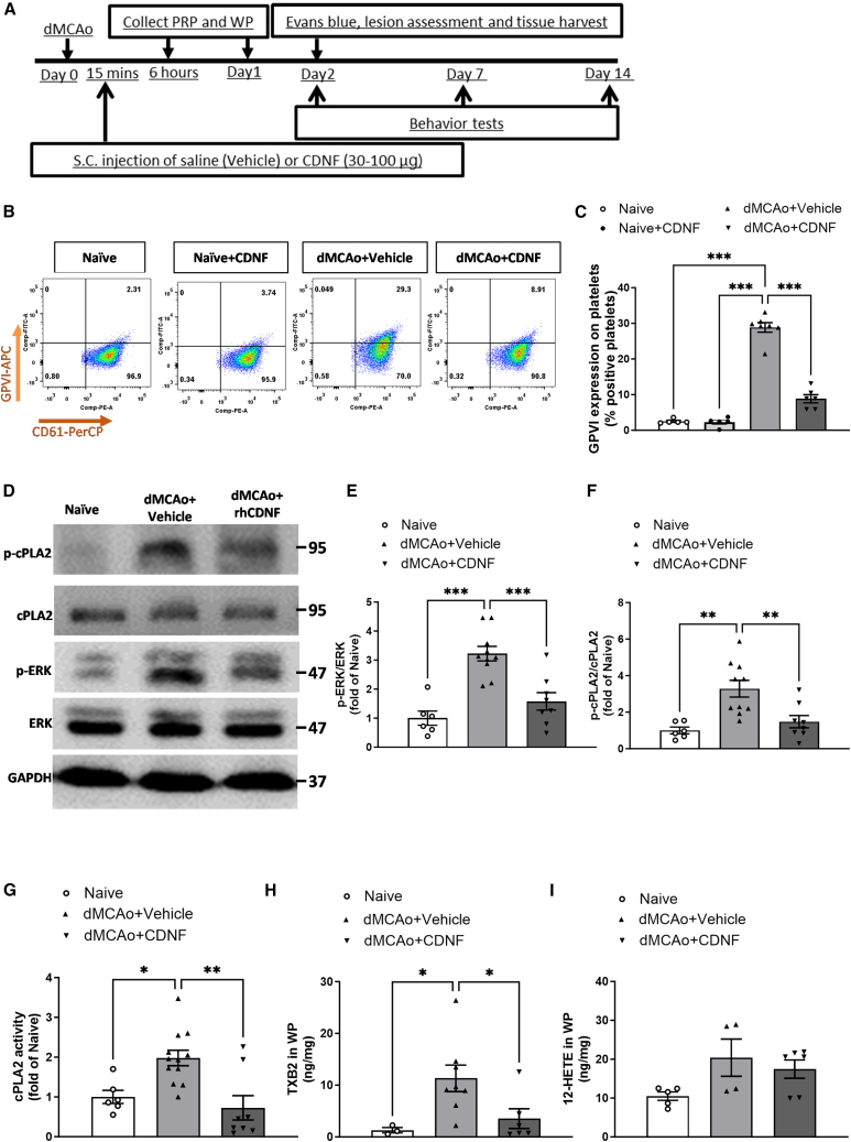
Cerebral reperfusion injury in stroke, stemming from interconnected thrombotic and inflammatory signatures, often involves platelet activation, aggregation and its interaction with various immune cells, contributing to microvascular dysfunction. However, the regulatory mechanisms behind this platelet activation and the resulting inflammation are not well understood, complicating the development of effective stroke therapies. Utilizing animal models and platelets from hemorrhagic stroke patients, our research demonstrates that human cerebral dopamine neurotrophic factor (CDNF) acts as an endogenous antagonist, mitigating platelet aggregation and associated neuroinflammation. CDNF moderates mitochondrial membrane potential, reactive oxygen species production, and intracellular calcium in activated platelets by interfering with GTP binding to Rap1b, thereby reducing Rap1b activation and downregulating the Rap1b-MAPK-PLA2 signaling pathway, which decreases release of the pro-inflammatory mediator thromboxane A2. In addition, CDNF reduces the inflammatory response in BV2 microglial cells co-cultured with activated platelets. Consistent with ex vivo findings, subcutaneous administration of CDNF in a rat model of ischemic stroke significantly reduces platelet activation, aggregation, lipid mediator production, infarct volume, and neurological deficits. In summary, our study highlights CDNF as a promising therapeutic target for mitigating platelet-induced inflammation and enhancing recovery in stroke. Harnessing the CDNF pathway may offer a novel therapeutic strategy for stroke intervention.

Keywords: CDNF; Rap1b; cerebral dopamine neurotrophic factor; dMCAo; distal middle cerebral artery occlusion; ischemic stroke; oxylipin metabolism; platelet activation.


Conflict of interest statement

Declaration of interests M.S. is one of inventors of the CDNF-related patent (7452969), which is owned by the Herantis Pharma Company (Espoo, Finland).

Figures

None
Graphical abstract
Figure 1
Figure 1
PRP derived from hemorrhagic stroke patients exhibits lower levels of CDNF, but higher aggregation responses compared with healthy controls Extracellularly added CDNF suppressed certain agonist-induced platelet aggregation in PRP. (A) A flow diagram illustrates the experimental design process used in this study. (B) CDNF concentrations in PRP were measured using ELISA in healthy controls, traumatic hemorrhage, and hemorrhagic stroke patients. ∗∗∗p < 0.0001 in comparison with the health donor. Data were analyzed by one-way ANOVA followed by Bonferroni corrections. (C–E) Representative the aggregation rates of healthy donor, traumatic hemorrhagic, and stroke patients’ PRP stimulated with various doses of ADP (C), TRAP6 (D), or collagen (E). Data were analyzed one-way ANOVA + post hoc Bonferroni test, ∗p < 0.05, ∗∗p < 0.01, ∗∗∗p < 0.0001 in comparison with the healthy controls. (F and G) Changes in platelet glycoprotein VI (GPVI) expression were observed in PRP obtained from healthy donors or stroke patients by flow cytometry. (G) Quantification of GPVI+/CD61+ expression in PRP (n > 6, each group) were analyzed as two-tailed Student’s t test. ∗p < 0.05 in comparison with the healthy donors. (H–K) Aggregation responses of PRP obtained from healthy controls and patients with traumatic brain hemorrhage or hemorrhagic stroke treated with different agonists. Collagen- (J)- and AA- (K) induced stroke patient’s PRP aggregation were significantly suppressed by CDNF treatment in an agonist dose-dependent manner. One-way ANOVA + original FDR method of Benjamini and Hochberg, ∗p < 0.05 in comparison with the PBS group. Mean ± SEM is shown.
Figure 2
Figure 2
Extracellularly delivered CDNF hinders the decrease in mitochondrial membrane potential (TREM), reduces ROS production, and inhibits Ca2+ efflux in arachidonic acid-treated platelets (A–D) Changes in the level of mitochondrial membrane potential (TREM) of platelet stimulated by AA or TXA2 analog, U46619. Decreased TREM florescence was shown in AA- or TXA2-treated washed platelets. Pre-treatment of CDNF could maintain mitochondria membrane potential in human platelets in the presence of AA (0.6 mM) or TXA2 (30 μM). ∗p < 0.05, ∗∗p < 0.01, ∗∗∗p < 0.0001 by one-way ANOVA followed by Bonferroni corrections. Mean ± SEM is shown. Lucigenin (bis-N-methylacridinium nitrate) is the most used chemiluminescent probe for the detection of superoxide ROS in the cells and tissue. Pre-treatment of CDNF could significantly decrease O2 production estimated by lucigenin method in human washed platelets in presence of AA. (E and F) Data were analyzed as two-tailed Student’s t test. ∗p < 0.05 (n = 6). Plate reader Fluo-4-loaded platelets were stimulated in black 96-well plates (FLUOStar Plate Reader; excitation, 485 nm; emission, 520 nm) to determine the concentration of cytoplasmic calcium. (G and H) Shown are (G) mean florescent intensity (Ft/F0) and (H) statistical analysis of platelets with responses reaching the threshold for detection relative to amplitude (F/F0) in response to 1 U/mL thrombin. Data were analyzed as two-tailed Student’s t test. ∗p < 0.05 (n > 40). When platelets were stimulated with the thrombin, these platelets had high Flur-4 fluorescence. However, pre-administration of CDNF could significantly reduce Fluo-4 florescent amplitude of the thrombin-treated washed platelets.
Figure 3
Figure 3
Exogenous CDNF interacts with Rap1b to interfere with GTP-bound Rap1b active form in AA-stimulated human platelets (A) Through immunoprecipitation and western blot analysis, it was shown that adding CDNF antibody to human platelets treated with CDNF exhibited greater efficacy in capturing CDNF compared with IgG. The CDNF served as the positive control in the experiment. (B) The protein interactome networks show CDNF and CDNF-interacted proteins. The representative protein interaction network was generated using IPA software from CDNF-treated human platelets in the presence of AA. (C) Through co-immunoprecipitation (coIP) and western blot analysis, the findings provided evidence of the interaction between CDNF and Rap1b in human platelets treated with AA and supplemented with CDNF. Consistent with the previous study, we also found that CDNF could interact with GRP78 (Bip). (D) The immunoblots show active Rap1b (Rap1-GTP) isolated by the pull-down assay with GST-RalGDS-RBD and the level of total Rap1b present in the lysates used for the Rap1b activation assays. A representative western blot was conducted to visualize Rap1b-GTP levels in human platelets under different conditions: untreated (alone), treated with CDNF, stimulated with AA alone, or treated with a combination of AA and CDNF. The addition of GTPγS and GDP was utilized as a positive control and negative control, respectively for assessment. (E) Quantitation of Rap1b activation as a ratio of Rap1b to GAPDH. Two-way ANOVA + post hoc Bonferroni test, ∗∗p < 0.01, ∗∗∗p < 0.0001 in comparison with the AA group. As shown in (F) and (G), CDNF could suppress accumulation of GTP-bound Rap1b in the GTPrS treatment with concentrations of 0.01, 0.05 mM (GTPrS 0.01 mM: ∗p < 0.05 vs. vehicle and vehicle + CDNF; GTPrS 0.05 mM: ∗p < 0.05 vs. vehicle and vehicle + CDNF), but not 0.1 mM, indicating that CDNF treatment could suppress Rap1b activation in washed platelets exposed to GTP in a concentration-dependent manner. TCL, total cell lysates.
Figure 4
Figure 4
CDNF inhibits the AA-induced phosphorylation of cPLA2 and ERK, resulting in the suppression of cPLA2 activation and thromboxane B2 production, but not 12-HETE synthesis in platelets (A–D) Fluorescently labeled CDNF is detected in phalloidin-stained washed human platelets (A and B). Alexa Fluro 647-labeled-CDNF was co-localized in Rap1b-expressed cells (B and C). A yellowish color in the merge (D) indicates co-localization of CDNF expression in phalloidin and Rap1b-double-positive platelets. Scale bars, 2 μm. The effect of CDNF on AA-induced cPLA2 and ERK phosphorylation in platelets was investigated. Washed human platelets were stimulated with AA in the presence or absence of CDNF. (E–G) The western blot images presented here display the changes in phospho-cPLA2, cPLA2, phospho-ERK, ERK, and CDNF levels in platelets subjected to treatment with CDNF or PBS, in the presence or absence of AA. The quantification of phosphorylation levels for ERK (F) and cPLA2 (G) was represented as a fold change relative to the control group. ∗p < 0.05, ∗∗p < 0.01 indicates comparison with the AA group with two-way ANOVA and Tukey’s post hoc test. The data represent mean ± SEM. (H) The activity of cPLA2 was measured using the cPLA2 ELISA assay, revealing that CDNF effectively inhibited the AA-induced increase in cPLA2 activity of washed platelets. (I) Thromboxane B2 levels were quantified using the thromboxane B2 ELISA assay, and the results showed that CDNF effectively reduced the AA-induced increase in platelet thromboxane B2 production. (J) The addition of extracellular CDNF did not influence the AA-induced increase in platelet production of 12-HETE. ∗p < 0.05, ∗∗p < 0.01, ∗∗∗p < 0.001 by Tukey’s multiple comparisons test, following one-way ANOVA.
Figure 5
Figure 5
Pretreatment of CDNF attenuated the p-JNK and p-p38 signaling pathway, thereby suppressing the upregulation of iNOS, COX-2, IL-6, TNF-α, and IL-1β in BV2 microglia cells co-cultured with platelets from stroke patients (A–D) BV2, a microglia cell line, was treated with PBS, normal human washed platelets, or washed platelets obtained from patients with hemorrhagic stroke. Western blot assays were employed to determine the expression levels of COX-2 (B), IL-1β (C), and the upstream regulator p-JNK (D), with GAPDH serving as the internal control. The findings revealed that washed platelets from hemorrhagic stroke patients further boost inflammatory responses in BV2 cells, as evidenced by higher levels of COX-2, IL-1b, and p-JNK compared with cells treated with PBS or washed platelets from healthy donors. Data are shown as mean ± SEM (n > 3; ∗p < 0.05, ∗∗p < 0.01 by Tukey’s multiple comparisons test, following one-way ANOVA). (E–L) The addition of washed platelets from hemorrhagic stroke into BV2 microglial cells for 6 h elicited upregulated expressions of iNOS, IL-6, COX-2, IL-1β, TNF-α, p-JNK, and p-P38. When CDNF-treated washed platelets from hemorrhagic stroke patients were co-cultured with BV2 microglial cells, a dose-dependent inhibitory effect was observed on the expression of pro-inflammatory cytokines and their upstream regulators. The quantitative analysis of all the obtained results present as follows (F–L). ∗p < 0.05, ∗∗p < 0.01 indicates comparison with HS WP group with one-way ANOVA and Tukey’s post hoc test. The data represent mean ± SEM. HD WP, healthy donor washed platelets; HS WP, hemorrhagic stroke washed platelets. CDNF 0.5, 0.5 μg/mL; CDNF 1, 1 μg/mL; CDNF 2, 2 μg/mL.
Figure 6
Figure 6
Post-stroke systemic administration of CDNF reduced GPVI expression in PRP and downregulated ERK-cPLA2-TXA2 signaling transduction in circulating platelets (A) Timetable of experiment. The rats underwent dMCAo surgery, and they were divided into two groups randomly. Fifteen minutes after reperfusion, the animals received an s.c. injection of either CDNF or PBS (vehicle). PRP or washed platelet samples were collected at 6 h and 1 day post-surgery. Behavioral functions were evaluated on days 2, 7, and 14 post-stroke. Rats were sacrificed for analysis at different time points. (B and C) By using flow cytometry, it was observed that CDNF treatment suppressed the elevated expression of GPVI+/CD61+ in PRP at 6 h after dMCAo. (C) Quantitation of GPVI+/CD61+ expression in PRP derived from naive, CDNF alone, dMCAo + vehicle and dMCAo + CDNF groups. n = 5–6 per group. ∗∗∗p < 0.001 by Tukey’s multiple comparisons test, following one-way ANOVA. (D–F) Western blot analysis revealed that systemic administration of CDNF suppressed the upregulated expressions of p-ERK (E) and p-cPLA2 (F) in washed platelets at 6 h post-stroke. cPLA2 or ERK was used as an internal control for normalization. The statistical analysis of the results is presented as a fold change relative to the naive group. ∗∗p < 0.01, ∗∗∗p < 0.001 indicates comparison with the dMCAo + vehicle group by Dunnett’s multiple comparisons test, following one-way ANOVA. (G–I) The activity of p-cPLA2 as well as production of TXB2 and 12-HETE in washed platelets at 6 h after dMCAo were measured using ELISA. The findings revealed that CDNF treatment suppressed stroke-upregulated cPLA2 activity (G) and the production of TXB2 by platelets (H), while it had no effect on the synthesis of platelet 12-HETE (I). ∗p < 0.05, ∗∗p < 0.001, Holm-Šidák multiple comparisons test, following one-way ANOVA. The mean ± SEM of three independent experiments is shown.
Figure 7
Figure 7
The levels of oxylipins in plasma increase 5 h after ischemic stroke in a rat model and s.c. administration of CDNF reduces the levels down to the baseline (A) Timetable of the experiment. The rats were divided into four groups randomly (n = 4–6 animals/group). Two control groups without stroke received an s.c. injection of either CDNF or saline (vehicle). Two stroke groups underwent dMCAo surgery with 60 min occlusion and 4 h after reperfusion the animals received an s.c. injection of either CDNF or saline (vehicle). Rat plasma was collected 1 h after injection. (B) Significantly increased oxylipins after dMCAo surgery and decreased after CDNF treatment: 9-HETE, (±)9-hydroxy-5Z,7E,11Z,14Z-eicosatetraenoic acid; 9-HODE, (±)9-hydroxy-10E,12Z-octadecadienoic acid; 9-KODE, 9-oxo-10E,12Z-octadecadienoic acid; 11-HETE, 11-hydroxy-5Z,8Z,11E,14Z-eicosatetraenoic acid; 12-HEPE, (±)12-hydroxy-5Z,8Z,10E,14Z,17Z-eicosapentaenoic acid; 12-HETE, (±)12-hydroxy-5Z,8Z,10E,14Z-eicosatetraenoic acid; 12-HHTrE, 12S-hydroxy-5Z,8E,10E-heptadecatrienoic acid; 12-KETE, 12-oxo-5Z,8Z,10E,14Z-eicosatetraenoic acid; 12(13)-EpOME(cis), (±)12,13-epoxy-9Z-octadecenoic acid; 13-HODE, (±)13-hydroxy-9Z,11E-octadecadienoic acid; 13-HOTrE, 13S-hydroxy-9Z,11E,15Z-octadecatrienoic acid; 15-HETE, (±)15-hydroxy-5Z,8Z,11Z,13E-eicosatetraenoic acid; PGD2, 9α,15S-dihydroxy-11-oxo-prosta-5Z,13E-dien-1-oic acid; PGE2, 9-oxo-11α,15S-dihydroxy-prosta-5Z,13E-dien-1-oic acid; PGF, 9α,11α,15S-trihydroxy-prosta-5Z,13E-dien-1-oic acid; TXB2, 9α,11,15S-trihydroxythromba-5Z,13E-dien-1-oic acid. ∗p < 0.05, ∗∗p < 0.01, ∗∗∗p < 0.001 by Tukey’s multiple comparisons test, following one-way ANOVA. Mean ± SEM is shown.
Figure 8
Figure 8
Systemic administration of CDNF decreased infarction volume, accelerated neurobehavioral recovery, attenuated neuroinflammation, and preserved BBB integrity and junction protein expressions in the lesioned cortex after dMCAo (A) Representative images of brain sections stained with TTC showing infarction area. (B) Quantification of infarction volume using TTC staining after 48 h of reperfusion. n = 8–10 per group; ∗p < 0.05 vs. dMCAo + vehicle; Student’s t test was used for the analysis of statistical significance. (C) Modified neurological severity scores (mNSS) were examined at 2 days post-dMCAo n = 8–10 per group. ∗∗p < 0.01 vs. dMCAo + vehicle; Student’s t test was used for the analysis of statistical significance. (D) Forepaw use bias of the rats was assessed in the cylinder test at 2, 7, and 14 days after dMCAo. n = 5–6 per group. ∗∗p < 0.01 by Fisher’s LSD test, following two-way ANOVA (effect of treatment: F(1,28) = 11.77, p < 0.0019). (E) Horizontal distance traveled for 30 min on days 2, 7, and 14 post-dMCAo. ∗p < 0.05 by original FDR method of Benjamini and Hochberg test, following two-way ANOVA (effect of treatment: F(2,31) = 11.97, p < 0.0001). (F and G) Body asymmetry test and Bederson’s score were analyzed on days 2, 7, and 14 after dMCAo, ∗∗p < 0.01, ∗∗∗p < 0.001 indicate comparison with vehicle with Bonferroni’s post hoc test following two-way ANOVA. (H) Western blot bands of iNOS, COX-2, IL-1β, TNF-α, IL-10, CD163, CD36, and GAPDH at 2 days after dMCAo. (I) Representative brain coronal sections (2 mm thickness) show Evans blue extravasation on day 2 post-dMCAo. (J and K) Comparison of dye concentrations in the ipsilateral (J) and contralateral (K) cortex between dMCAo + vehicle and dMCAo + CDNF groups. Dye concentration is presented as μg/g of tissue weight and calculated from a standard curve obtained from known amounts of dye. ∗p < 0.05, paired t test. Mean ± SEM is shown. (L) Western blotting showed the levels of tight junction proteins ZO-1, claudin-5, LRP-1, and 92 kDa type IV collagenase, MMP-9, within the peri-infarct cortical area. (M) At 2 days post-stroke, the decreased levels of iNOS, COX-2, IL-1β, TNF-α, and MMP-9, but increased levels of IL-10, CD163, CD36, ZO-1, Claudin-5, and LRP-1 were detected in the ischemic cortex of CDNF treatment group. ∗p < 0.05, ∗∗p < 0.01, ∗∗∗p < 0.001 by multiple unpaired t test. Mean ± SEM is shown.

References

    1. Donkor E.S. Stroke in the 21(st) Century: A Snapshot of the Burden, Epidemiology, and Quality of Life. Stroke Res. Treat. 2018;2018:3238165. doi: 10.1155/2018/3238165. - DOI - PMC - PubMed
    1. De Meyer S.F., Denorme F., Langhauser F., Geuss E., Fluri F., Kleinschnitz C. Thromboinflammation in Stroke Brain Damage. Stroke. 2016;47:1165–1172. doi: 10.1161/STROKEAHA.115.011238. - DOI - PubMed
    1. De Meyer S.F., Langhauser F., Haupeltshofer S., Kleinschnitz C., Casas A.I. Thromboinflammation in Brain Ischemia: Recent Updates and Future Perspectives. Stroke. 2022;53:1487–1499. doi: 10.1161/STROKEAHA.122.038733. - DOI - PubMed
    1. Jurcau A., Simion A. Neuroinflammation in Cerebral Ischemia and Ischemia/Reperfusion Injuries: From Pathophysiology to Therapeutic Strategies. Int. J. Mol. Sci. 2021;23:14. doi: 10.3390/ijms23010014. - DOI - PMC - PubMed
    1. Stoll G., Nieswandt B. Thrombo-inflammation in acute ischaemic stroke - implications for treatment. Nat. Rev. Neurol. 2019;15:473–481. doi: 10.1038/s41582-019-0221-1. - DOI - PubMed

Substances

LinkOut - more resources