[Preprint]. 2024 Aug 29:2024.08.28.610074.
doi: 10.1101/2024.08.28.610074.

O-glycosylation contributes to mammalian glycoRNA biogenesis

Affiliations

O-glycosylation contributes to mammalian glycoRNA biogenesis

Jennifer Porat et al. bioRxiv. .

Abstract

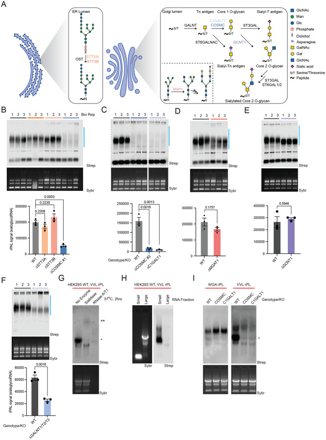
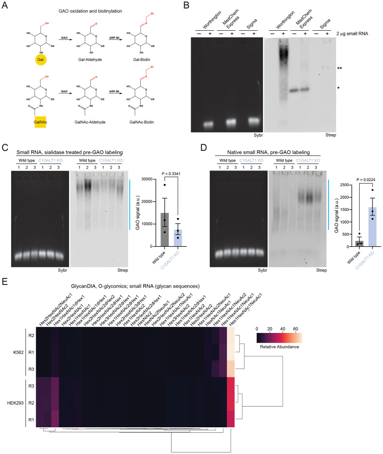
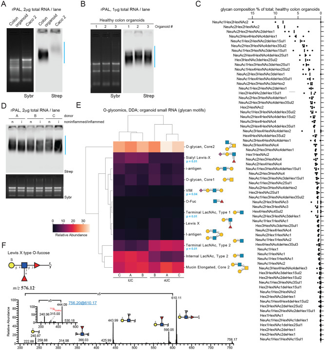
There is an increasing appreciation for the role of cell surface glycans in modulating interactions with extracellular ligands and participating in intercellular communication. We recently reported the existence of sialoglycoRNAs, where mammalian small RNAs are covalently linked to N-glycans through the modified base acp3U and trafficked to the cell surface. However, little is currently known about the role for O-glycosylation, another major class of carbohydrate polymer modifications. Here, we use parallel genetic, enzymatic, and mass spectrometry approaches to demonstrate that O-linked glycan biosynthesis is responsible for the majority of sialoglycoRNA levels. By examining the O-glycans associated with RNA from cell lines and colon organoids we find known and previously unreported O-linked glycan structures. Further, we find that O-linked glycans released from small RNA from organoids derived from ulcerative colitis patients exhibit higher levels of sialylation than glycans from healthy organoids. Together, our work provides flexible tools to interrogate O-linked glycoRNAs (O-glycoRNA) and suggests that they may be modulated in human disease.


Conflict of interest statement

DECLARATION OF INTERESTS R.A.F. is a stockholder of ORNA Therapeutics. R.A.F. is a board of directors member and stockholder of Chronus Health and Blue Planet Systems. The other authors declare no competing interests.

Figures

Figure 1.
Figure 1.. SialoglycoRNAs are dependent O-glycan biosynthetic enzymes.
A. Schematic of N-glycan biosynthesis (Endoplasmic Reticulum, Golgi inset) and O-glycan biosynthesis, with the associated glycosyltransferases responsible for each maturation step. B. RNA periodate oxidation and aldehyde ligation (rPAL) northern blot of total RNA from wild type HEK293, STT3A Knockout (KO), STT3B KO, and COSMC KO. An in-gel Sybr stain of total RNA and detection of biotinylated sialoglycoRNA on the membrane (Strep) are shown, along with a quantification of sialoglycoRNA signal (n= 3 biological replicates, mean±SEM, unpaired student’s t-test). C. rPAL northern as in (B), here with WT vs. COSMC KO clone #2 vs. C1GALT1 KO. D. rPAL northern as in (B), here with WT vs. MGAT1 KO. E. rPAL northern as in (B), here with WT vs. GCNT1 KO. F. rPAL northern as in (B), here with WT vs. GALNT1/GALNT2/GALNT3 KO. G. RNA proximity labeling (rPL) of live HEK293 cells using the O-glycan binding lectin VVL (binding GalNAc), followed by in vitro digestion of extracted total RNA with sialidase or RNase. *lower molecular weight (MW) signal, ** ultra-high molecular weight signal are noted. H. rPL as in (G), here fractionating small and large RNAs. I. rPL as in (G), here labeling in proximity of VVL (binding a terminal GalNAc) or the N-glycan binding lectin WGA (binding GlcNac and sialic acid) on WT, COSMC KO, or C1GALT1 KO HEK293 cells.
Figure 2.
Figure 2.. Galactose oxidase (GAO) labels glycoRNA.
A. Reaction schematic of the oxidation of galactose (Gal) or N-acetylgalactosamine (GalNAc), followed by ligation of biotin (ARP-Bt) to the newly generated aldehyde. B. GAO northern with and without wild type HEK293 small RNA labeled with GAO purchased from Worthington, MedChem Express, or Sigma. *RNA-independent labeling using MedChem Express GAO, **RNA-dependent labeling using Sigma GAO. An in-gel Sybr stain of small RNA and detection of biotinylated glycoRNA on the membrane (Strep) are shown. C. GAO northern as in (B), with wild type and C1GALT1 KO HEK293 small RNA pre-treated with sialidase. A quantification of GAO signal is shown (n= 3 biological replicates, mean±SEM, unpaired student’s t-test). D. GAO northern as in (C), with native (non sialidase-treated) wild type and C1GALT1 KO HEK293 small RNA. E. Heatmap of O-glycan structures released from K562 and HEK293 small RNA and identified by Data Independent acquisition (DIA). Glycan structures are indicated (n= 3 biological replicates).
Figure 3.
Figure 3.. Mass spectrometry defines the O-glycan profile of glycoRNAs.
A. rPAL northern blot of total RNA from a colon organoid derived from healthy cells and colon cancer cells (Caco 2). An in-gel Sybr stain of total RNA and detection of biotinylated sialoglycoRNA on the membrane (Strep) are shown. B. rPAL northern as in (A), here with total RNA from colon organoids derived from healthy patients (n= 3 biological replicates). C. Glycan composition percent (for all compositions with a percent greater than 0) of O-glycans released from RNA from healthy organoids and identified by Data-Dependent Acquisition (DDA) (n= 3 biological replicates). D. rPAL northern as in (A), here with total RNA from colon organoids derived from non-inflamed (n) or inflamed (i) cells (n= 3 biological replicates). E. Heatmap of O-glycans identified by DDA in small RNA from organoids derived from inflamed Ulcerative Colitis (iUC) and non-inflamed Ulcerative Colitis (nUC) cells (two-tailed Welch’s t-test corrected by the Benjamini-Hochberg procedure, n= 3 biological replicates). F. MS/MS spectrum of a novel sulfated O-fucose, SGalβ1-4(Fucα1-3)GlcNAcβ1-3Fucα (m/z 576).

References

    1. Flynn R.A., Pedram K., Malaker S.A., Batista P.J., Smith B.A.H., Johnson A.G., George B.M., Majzoub K., Villalta P.W., Carette J.E., et al. (2021). Small RNAs are modified with N-glycans and displayed on the surface of living cells. Cell 184, 3109–3124.e22. 10.1016/j.cell.2021.04.023. - DOI - PMC - PubMed
    1. Bi M., Zhang Z., Wang T., Liang H., and Tian Z. (2023). A draft of human N-glycans of glycoRNA. bioRxiv, 2023.09.18.558371. 10.1101/2023.09.18.558371. - DOI
    1. Zhang N., Tang W., Torres L., Wang X., Ajaj Y., Zhu L., Luan Y., Zhou H., Wang Y., Zhang D., et al. (2024). Cell surface RNAs control neutrophil recruitment. Cell 187, 846–860.e17. 10.1016/j.cell.2023.12.033. - DOI - PMC - PubMed
    1. Ma Y., Guo W., Mou Q., Shao X., Lyu M., Garcia V., Kong L., Lewis W., Ward C., Yang Z., et al. (2023). Spatial imaging of glycoRNA in single cells with ARPLA. Nature Biotechnology. 10.1038/s41587-023-01801-z. - DOI - PMC - PubMed
    1. Xie Y., Chai P., Till N.A., Hemberger H., Lebedenko C.G., Porat J., Watkins C.P., Caldwell R.M., George B.M., Perr J., et al. (2024). The modified RNA base acp3U is an attachment site for N-glycans in glycoRNA. Cell. 10.1016/j.cell.2024.07.044. - DOI - PMC - PubMed

Publication types

LinkOut - more resources