. 2024 Sep 5:19:9091-9107.
doi: 10.2147/IJN.S447645. eCollection 2024.

Silver Nanoparticles (AgNPs) Uptake by Caveolae-Dependent Endocytosis is Responsible for Their Selective Effect Towards Castration Resistant Prostate Cancer

Affiliations

Silver Nanoparticles (AgNPs) Uptake by Caveolae-Dependent Endocytosis is Responsible for Their Selective Effect Towards Castration Resistant Prostate Cancer

Mariana Morais et al. Int J Nanomedicine. .

Abstract

Purpose: Castration Resistant Prostate Cancer (CRPC) is characterized by poor prognosis and limited therapeutic options. AgNPs functionalized with glucose (G-AgNPs) were observed cytotoxic to CRPC cell lines (PC-3 and Du-145) and not LNCaP. This study aims to evaluate AgNPs and G-AgNPs' uptake mechanisms in these cells and understand their role in the selective effect against CRPC cells.

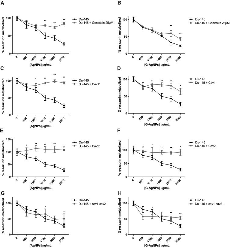
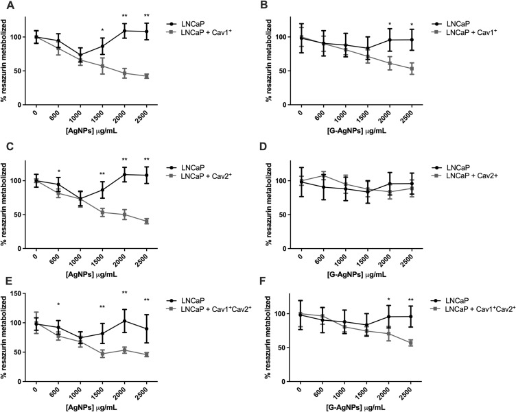
Methods: Uptake of AgNPs and G-AgNPs was assessed through transmission electron microscopy (TEM). A microRNA (miRNAs) analysis approach was used to uncover the main molecular differences responsible for the endocytic mechanisms' regulation. Caveolin (Cav) 1 and 2 mRNA and protein levels were assessed in the three cell lines. Caveolae-dependent endocytosis was inhibited with genistein or siCav1- and siCav2- in PC-3 and Du-145 and resazurin assay was used to evaluate viability after AgNPs and G-AgNPs administration. Caveolae-dependent endocytosis was induced with Cav1+ and Cav2+ plasmids in LNCaP, resazurin assay was used to evaluate viability after AgNPs and G-AgNPs administration and TEM to assess their location.

Results: AgNPs and G-AgNPs were not uptaked by LNCaP. miRNA analysis revealed 37 upregulated and 90 downregulated miRNAs. Functional enrichment analysis of miRNAs' targets resulted in enrichment of terms related to endocytosis and caveolae. We observed that Cav1 and Cav2 are not expressed in LNCaP. Inhibiting caveolae-dependent endocytosis in Du-145 and PC-3 led to a significative reduction of cytotoxic capacity of AgNPs and G-AgNPs and induction of caveolae-dependent endocytosis in LNCaP lead to a significative increase as well as their uptake by cells.

Conclusion: This study shows the potential of these AgNPs as a new therapeutic approach directed to CRPC patients, uncovers caveolae-dependent endocytosis as the uptake mechanism of these AgNPs and highlights deregulation of Cav1 and Cav2 expression as a key difference in hormone sensitive and resistant PCa cells which may be responsible for drug resistance.

Keywords: caveolins; endocytosis; prostate cancer resistant to castration; silver nanoparticles; uptake mechanism.


Conflict of interest statement

The authors report no conflicts of interest in this work.

Figures

Figure 1
Figure 1
TEM images of LNCaP cells treated with 2000μg/mL of AgNPs (A–C) or G-AgNPs (D–F). From each sample, three pictures were taken with different ampliations. Thus, scale bars in A, B and D are 2μm, in C and E are 1μm, and in F are 0.5 μm. NPs are the black dots presented in the figures.
Figure 2
Figure 2
Molecular profile comparison of PC-3 and Du-145 with LNCaP cells. (A) Volcano plots of the differentially expressed miRNAs when comparing PC-3 and Du-145 cells with LNCaP cells. The y-axis indicates the Log10 of the p values and the x-axis is the Log2 transformed ratio of the expression between the experimental groups. (B) PPI network of the 40 proteins derived from the target genes of the upregulated miRNAs performed using Cytoscape. (C) PPI network of the 45 proteins derived from the target genes of the downregulated miRNAs performed using Cytoscape. (D) Compartments, GO, KEGG and Reactome analysis of the 40 selected genes derived from the upregulated miRNAs. The functional enrichment analysis was made with the STRINGapp from Cytoscape. (E) Compartments, GO, KEGG and Reactome analysis of the 45 selected genes derived from the downregulated miRNAs. The functional enrichment analysis was made with the STRINGapp from Cytoscape.
Figure 3
Figure 3
(A) Variation of the relative expression levels of Cav1 and Cav2 in LNCaP, PC-3 and Du-145 cells (mean ± SE); (B) Protein levels of Cav1 and Cav2 in LNCaP, PC-3 and Du-145 cells according to Western Blot analysis; (C) Relative quantification of Cav1 expression relative to B-Actin in LNCaP, PC-3 and Du-145; (D) Relative quantification of Cav2 expression relative to B-Actin in LNCaP, PC-3 and Du-145; **p<0.001 (t-student test).
Figure 4
Figure 4
Comparison of cell viability, by Resazurin Assay, upon treatment with AgNPs and G-AgNPs for 24 h in PC-3 cell line with or without previous inhibition using Genistein 20μM (A and B), using siCav1 (Cav−) (C and D), using siCav2 (Cav−) (E and F), and both siCav1 and siCav2 (Cav−Cav−) (G and H). Genistein was added for 1 h at concentration of 20μM; siCav1 and siCav2 was added for 24h at concentration of 40nM in combination with 1μL of lipofectamine or 1.5μL, when added simultaneously. Both were washed off after incubation and nanoparticles were added. Results are expressed as percentage of control (-●-: -genistein/-AgNPs; -■-: +genistein/-AgNPs cells), as mean ± SEM. *p<0.05, ** p<0.001 (5 replicates, sample T test, Kruskal Wallis tests depending on whether the results followed or not a normal distribution).
Figure 5
Figure 5
Comparison of cell viability, by Resazurin Assay, upon treatment with AgNPs and G-AgNPs for 24 h in Du-145 cell line with or without previous inhibition using Genistein (A and B), using siCav1 (Cav1) (C and D), using siCav2 (Cav2) (E and F), and both siCav1 and siCav2 (Cav1Cav2) (G and H). Genistein was added for 2 h at concentration of 25μM; siCav1 and siCav2 was added for 24h at concentration of 40nM in combination with 1μL of lipofectamine or 1.5μL, when added simultaneously. Both were washed off after incubation and nanoparticles were added. Results are expressed as percentage of control (-●-: -genistein/-AgNPs; -■-: +genistein/-AgNPs cells), as mean ± SEM. *p<0.05, **p<0.001 (5 replicates, sample T test, Kruskal Wallis tests depending on whether the results followed or not a normal distribution).
Figure 6
Figure 6
Comparison of cell viability, by Resazurin Assay, upon treatment with AgNPs and G-AgNPs for 24 h in LNCaP cell line with or without previous induction of Cav1 (Cav1+) (A and B), Cav2 (Cav2+) (C and D), and both Cav1 and Cav2 (Cav1+Cav2+) (E and F). Results are expressed as percentage of control (-●-: -plasmid/-AgNPs; -■-: +plasmid/-AgNPs cells), as mean ± SEM. *p<0.05, **p<0.001 (5 replicates, sample T test, Kruskal Wallis tests depending on whether the results followed or not a normal distribution).
Figure 7
Figure 7
TEM images of LNCaP cells transfected with Cav1+ (AC and JL), Cav2+ (DF and MO), Cav1+Cav2+ (G–I and PR) and treated with 2000μg/mL of AgNPs (AI) or G-AgNPs (J–%). From each sample, three pictures were taken with different ampliations. Thus, scale bars in (A, D, G and H) are 2μm, in (J, K and M) are 1μm, and in (BF), (L, N, O, Q and R) are 0.5 μm.

Similar articles

Cited by

References

    1. Rezayatmand H, Razmkhah M, Razeghian-Jahromi I. Drug resistance in cancer therapy: the Pandora’s Box of cancer stem cells. Stem Cell Res Ther. 2022;13(1):181. doi:10.1186/s13287-022-02856-6 - DOI - PMC - PubMed
    1. Parker C, Castro E, Fizazi K, et al. Prostate cancer: ESMO Clinical Practice Guidelines for diagnosis, treatment and follow-up. Ann Oncol. 2020;31(9):1119–1134. doi:10.1016/j.annonc.2020.06.011 - DOI - PubMed
    1. Gandhi J, Afridi A, Vatsia S, et al. The molecular biology of prostate cancer: current understanding and clinical implications. Prostate Cancer Prostatic Dis. 2018;21(1):22–36. doi:10.1038/s41391-017-0023-8 - DOI - PubMed
    1. Crowley F, Sterpi M, Buckley C, Margetich L, Handa S, Dovey Z. A review of the pathophysiological mechanisms underlying castration-resistant prostate cancer. Res Rep Urol. 2021;13:457–472. doi:10.2147/rru.S264722 - DOI - PMC - PubMed
    1. Dong L, Zieren RC, Xue W, de Reijke TM, Pienta KJ. Metastatic prostate cancer remains incurable, why? Asian J Urol. 2019;6(1):26–41. doi:10.1016/j.ajur.2018.11.005 - DOI - PMC - PubMed

MeSH terms

LinkOut - more resources