. 2024 Aug 27:12:1449156.
doi: 10.3389/fcell.2024.1449156. eCollection 2024.

Effect of AMH on primordial follicle populations in mouse ovaries and human pre-pubertal ovarian xenografts during doxorubicin treatment

Affiliations

Effect of AMH on primordial follicle populations in mouse ovaries and human pre-pubertal ovarian xenografts during doxorubicin treatment

Xi Wei et al. Front Cell Dev Biol. .

Abstract

Introduction: Survival rates of the childhood cancer patients are improving, however cancer treatments such as chemotherapy may lead to infertility due to loss of the primordial follicle (PMF) reserve. Doxorubicin (DXR) is a gonadotoxic chemotherapy agent commonly used in childhood cancers. Anti-Müllerian Hormone (AMH) has been reported to have a protective effect on the mouse ovarian reserve against DXR in vivo. However, whether AMH can prevent PMF loss in conjunction with DXR in human ovarian tissue in vivo has not been determined.

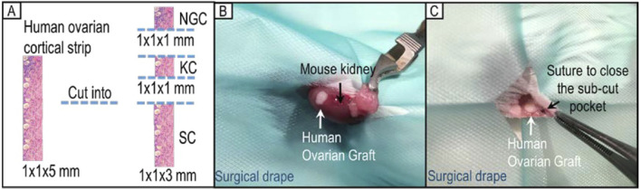
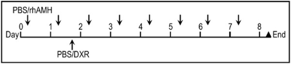
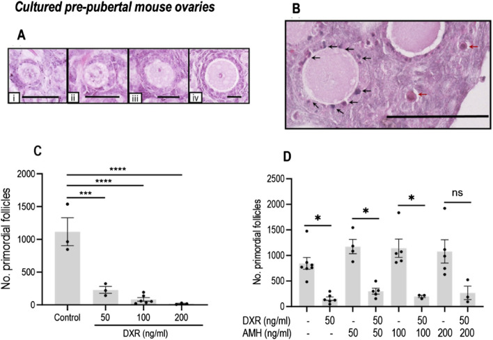
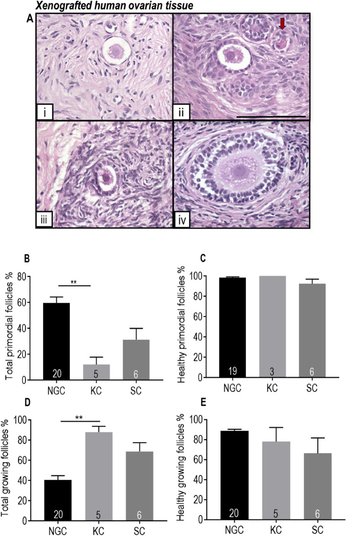
Methods: In order to investigate this, we first established an optimum dose of DXR that induced PMF loss in cultured mouse ovaries and investigated the efficacy of AMH on reducing DXR-induced PMF loss in mice in vitro. Second, we investigated the effects of DXR on pre-pubertal human ovarian tissue and the ability of AMH to prevent DXR-induced damage comparing using a mouse xenograft model with different transplantation sites.

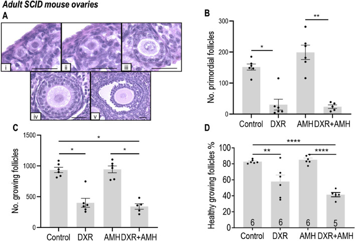
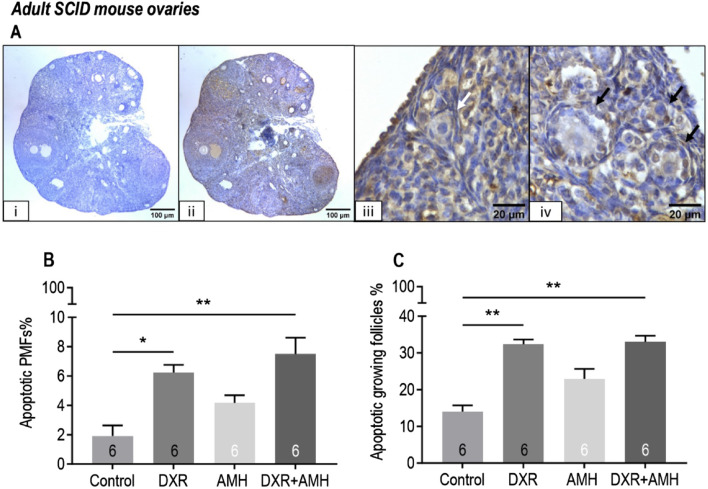
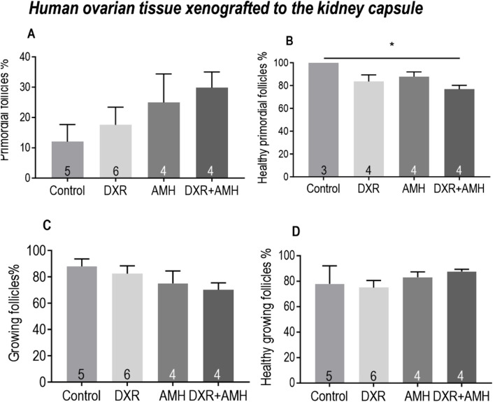
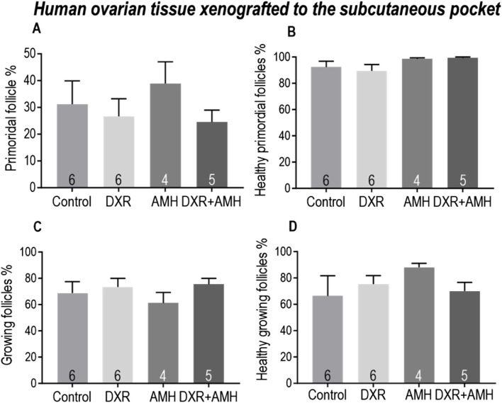
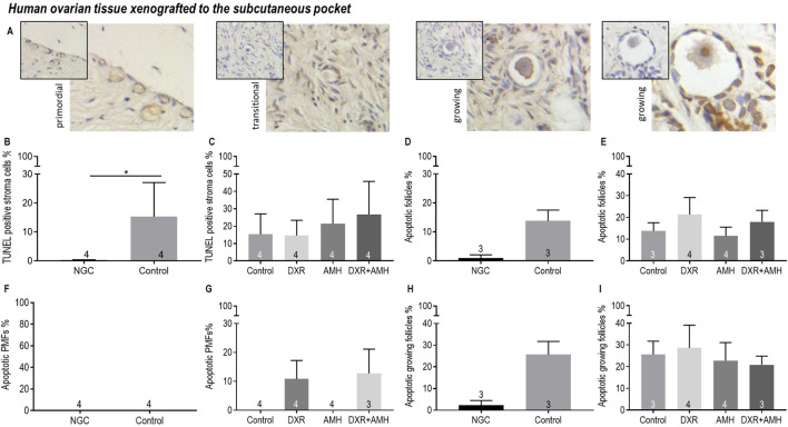
Results: Mouse ovaries treated with DXR in vitro and in vivo had reduced PMF populations and damaged follicle health. We did not observe effect of DXR-induced PMF loss or damage to follicle/stromal health in human ovarian cortex, this might have been due to an insufficient dose or duration of DXR. Although AMH does not prevent DXR-induced PMF loss in pre-pubertal and adult mouse ovaries, in mouse ovaries treated with higher concentration of AMH in vitro, DXR did not cause a significant loss in PMFs. This is the first study to illustrate an effect of AMH on DXR-induced PMF loss on pre-pubertal mouse ovaries. However, more experiments with higher doses of AMH and larger sample size are needed to confirm this finding.

Discussion: We did not observe that AMH could prevent DXR-induced PMF loss in mouse ovaries in vivo. Further studies are warranted to investigate whether AMH has a protective effect against DXR in xenotransplanted human ovarian tissue. Thus, to obtain robust evidence about the potential of AMH in fertility preservation during chemotherapy treatment, alternative AMH administration strategies need to be explored alongside DXR administration to fully interrogate the effect of DXR and AMH on human xenografted tissues.

Keywords: anti-Müllerian hormone; doxorubicin; fertility preservation; follicle; human ovarian xenograft; ovarian tissue; ovary; primordial follicles.


Conflict of interest statement

The authors declare that the research was conducted in the absence of any commercial or financial relationships that could be construed as a potential conflict of interest.

Figures

FIGURE 1
FIGURE 1
Human ovarian cortical strip division and allocation for xenotransplantation. (A) Each strip of ovarian cortical tissue was divided into three pieces for use (NGC: non-grafted control group; KC: kidney capsule; SC: subcutaneous pocket). (B) Representative image of xenotransplantation surgery placing one piece of tissue under the kidney capsule of a SCID mouse. (C) Representative image of xenotransplantation surgery grafting one piece of human ovarian tissue in a subcutaneous pocket of the same SCID mouse.
FIGURE 2
FIGURE 2
Treatment regimen for administration of anti-Müllerian hormone (AMH) and/or doxorubicin (DXR) to SCID mice (severe combined immunodeficient) containing human ovarian transplants. Ten days after human ovarian tissue was xenotransplanted into SCID mice, animals were intraperitoneally (IP) injected daily with PBS or rhAMH for 8 days with an additional single IP injection of PBS or DXR on the second day.
FIGURE 3
FIGURE 3
Analysis of mouse ovarian follicles in cultured pre-pubertal ovaries treated with DXR and AMH stained with H and E. (A) Representative images of morphologically healthy follicles (scale bar = 25 μm): i: Primordial follicles. ii-iv: Growing follicles (ii: Transitional; iii: Primary; iv: Secondary). (B) Unhealthy follicles (scale bar = 100 μm): black arrows denote pyknotic granulosa cells, red arrows denote atretic oocytes. (C) Total number of morphologically healthy primordial follicles in mouse ovaries treated with different concentrations of DXR. Values are presented as Mean ± S.E.M. Control n = 3; 50 ng/mL DXR n = 3; 100 ng/mL DXR n = 5; 200 ng/mL DXR n = 3. (D) Number of morphologically healthy primordial follicles in mouse ovaries cultured with different concentrations of rhAMH and with or without exposure to DXR. Values represent Mean ± S.E.M ns = not significant, *P < 0.05, ***P < 0.001, ****P < 0.0001.
FIGURE 4
FIGURE 4
Analysis of mouse ovarian follicles in SCID mice carrying human ovary xenografts treated with AMH and/or DXR stained with H and E. (A) Representative images of mouse ovarian follicles (scale bar = 25 μm). (I) Primordial follicles. ii-v: Growing follicles (ii: Transitional; iii: Primary; iv: Secondary; v: Antral). (B) Number of morphologically healthy primordial follicles in mouse ovaries after 8 days of treatment with PBS, DXR, AMH or co-treatment (DXR + AMH). (C) Number of growing follicle numbers in mouse ovaries from the Control, DXR, AMH, and DXR + AMH groups at the end of the experiment. (D) Proportion of morphologically healthy growing follicles in mouse ovaries from different groups. The data are presented as Mean ± S.E.M. Number in the base of the column is the sample number. *P < 0.05, **P < 0.01, ****P < 0.0001.
FIGURE 5
FIGURE 5
Analysis of mouse apoptotic follicles in SCID mice carrying human ovarian xenografts treated with AMH and/or DXR stained with TUNEL. (A) Representative images of TUNEL assay. (I) Negative control ovarian section (scale bar = 100 µm), ii: TUNEL-labelled ovarian section (scale bar = 100 µm), iii: TUNEL-negative primordial follicle (white arrow, scale bar = 20 µm), iv: TUNEL-positive growing follicles (black arrows, scale bar = 20 µm). (B) Proportion of apoptotic (TUNEL-positive) primordial follicles (PMFs) in mouse ovaries after 8 days of treatment with PBS (Control), DXR, AMH or co-treatment (DXR + AMH). (C) Proportion of apoptotic growing follicles in mouse ovaries after 8 days of treatment with PBS (Control), DXR, AMH or co-treatment (DXR + AMH). *P < 0.05, **P < 0.01.
FIGURE 6
FIGURE 6
Comparison of human ovarian follicles in xenografted tissue retrieved from the mouse kidney capsule and a subcutaneous pocket in the control group with non-grafted tissue stained with H and E. (A) Representative images of follicles at different stages in pre-pubertal human ovarian tissue. i Primordial follicle. ii-iv: Growing follicles (ii: Transitional follicle. iii: Primary follicle. iv: Secondary follicle). Red arrow denotes an unhealthy follicle. Scale bar represents 100 μm. (B) Proportion of follicles that were primordial. (C) Proportion of primordial follicles that were morphologically healthy. (D) Proportion of growing follicles. (E) Proportion of morphologically healthy growing follicles. NGC: Non-grafted group. KC: Kidney capsule. SC: Subcutaneous pocket. The data are presented as Mean ± S.E.M. Number in the base of the column is the sample number. **P < 0.01. Total follicle numbers (Mean ± S.E.M.): NGC 335.1 ± 55.3; KC control 76.7 ± 24.62; SC control 278.6 ± 137.6.
FIGURE 7
FIGURE 7
Analysis of follicles in human ovarian tissue xenografts retrieved from beneath the kidney capsule of mice treated with AMH and/or DXR stained with H and E. (A) Proportion of follicles that were primordial follicles in each treatment group. (B) Proportion of primordial follicles that were morphologically healthy (The sample number is different here. Since two pieces of ovarian tissue in the Control and DXR groups contained only growing follicles without primordial follicles, the ‘n’ in Control and DXR groups in Figure 7B is two fewer than that in Figure 7A. (C) Proportion of follicles that were growing. (D) Proportion of growing follicles that were morphologically healthy. The data are presented as Mean ± S.E.M. Number in the base of the column is the sample number. *P < 0.05. **P < 0.01. Total follicle numbers (Mean ± S.E.M.): Control 76.7 ± 24.62; AMH 147 ± 48.88; DXR 135.7 ± 42.19; DXR + AMH 131.1 ± 51.75.
FIGURE 8
FIGURE 8
Analysis of follicles in human ovarian tissue xenografts retrieved from a subcutaneous pocket of mice treated with AMH and/or DXR stained with H and E. (A) Proportion of follicles that were primordial follicles in each treatment group. (B) Proportion of primordial follicles that were morphologically healthy. (C) Proportion of follicles that were growing. (D) Proportion of growing follicles that were morphologically healthy. The data are presented as Mean ± S.E.M. Number in the base of the column is the sample number. *P < 0.05. Total follicle numbers (Mean ± S.E.M.): Control 278.6 ± 137.6; AMH 511.5 ± 157.5; DXR 330.2 ± 91.73; DXR + AMH 220.4 ± 105.3.
FIGURE 9
FIGURE 9
Analysis of apoptotic stroma cells, primordial follicles and growing follicles in non-transplanted human ovarian tissue and xenografts retrieved from a subcutaneous pocket of mice treated with AMH and/or DXR stained with TUNEL. (A) Representative images of follicles in human ovarian xenografts stained with TUNEL. (B) Proportion of stroma cells that were TUNEL-positive in the non-grafted control ovarian tissue (NGC) and the Control transplanted group. (C) Proportion of stroma cells that were TUNEL-positive in tissue retrieved from the control and treated groups. (D) Proportion of follicles that were classed as apoptotic in the non-grafted control ovarian tissue (NGC) and the Control transplanted group. (E) Proportion of follicles that were classed as apoptotic in tissue retrieved from the control and treated groups. (F) Proportion of primordial follicles (PMFs) that were classed as apoptotic in the non-grafted control ovarian tissue (NGC) and the Control transplanted group. (G) Proportion of PMF that were classed as apoptotic in tissue retrieved from the control and treated groups. (H) Proportion of growing follicles (PMF) that were classed as apoptotic in the non-grafted control ovarian tissue (NGC) and the Control transplanted group. (I) Proportion of growing follicles that were classed as apoptotic in tissue retrieved from the control and treated groups. The data are presented as Mean ± S.E.M. Number in the base of the column is the sample number. *P < 0.05.

Similar articles

Cited by

References

    1. Abercrombie M. (1946). Estimation of nuclear population from microtome sections. Anat. Rec. 94, 239–247. 10.1002/ar.1090940210 - DOI - PubMed
    1. Barpe D. R., Rosa D. D., Froehlich P. E. (2010). Pharmacokinetic evaluation of doxorubicin plasma levels in normal and overweight patients with breast cancer and simulation of dose adjustment by different indexes of body mass. Eur. J. Pharm. Sci. 41 (3-4), 458–463. 10.1016/j.ejps.2010.07.015 - DOI - PubMed
    1. Bertoldo M. J., Bernard J., Duffard N., Tsikis G., Alves S., Calais L., et al. (2016). Inhibitors of c-Jun phosphorylation impede ovine primordial follicle activation. Mol. Hum. Reprod. 22 (5), 338–349. 10.1093/molehr/gaw012 - DOI - PubMed
    1. Blanchard M. G., Josso N. (1974). Source of the anti-Müllerian hormone synthesized by the fetal testis: Müllerian-inhibiting activity of fetal bovine Sertoli cells in tissue culture. Pediatr. Res. 8 (12), 968–971. 10.1203/00006450-197412000-00011 - DOI - PubMed
    1. Braw-Tal R. (2002). The initiation of follicle growth: the oocyte or the somatic cells? Mol. Cell Endocrinol. 187 (1-2), 11–18. 10.1016/s0303-7207(01)00699-2 - DOI - PubMed

LinkOut - more resources