Review
. 2024 Nov 1;42(11):1848-1861.
doi: 10.1097/HJH.0000000000003829. Epub 2024 Sep 11.

Recent updates on the influence of iron and magnesium on vascular, renal, and adipose inflammation and possible consequences for hypertension

Affiliations
Review

Recent updates on the influence of iron and magnesium on vascular, renal, and adipose inflammation and possible consequences for hypertension

Benjamin J Connolly et al. J Hypertens. .

Abstract

The inflammatory status of the kidneys, vasculature, and perivascular adipose tissue (PVAT) has a significant influence on blood pressure and hypertension. Numerous micronutrients play an influential role in hypertension-driving inflammatory processes, and recent reports have provided bases for potential targeted modulation of these micronutrients to reduce hypertension. Iron overload in adipose tissue macrophages and adipocytes engenders an inflammatory environment and may contribute to impaired anticontractile signalling, and thus a treatment such as chelation therapy may hold a key to reducing blood pressure. Similarly, magnesium intake has proven to greatly influence inflammatory signalling and concurrent hypertension in both healthy animals and in a model for chronic kidney disease, demonstrating its potential clinical utility. These findings highlight the importance of further research to determine the efficacy of micronutrient-targeted treatments for the amelioration of hypertension and their potential translation into clinical application.


Conflict of interest statement

There are no conflicts of interest.

Figures

FIGURE 1
FIGURE 1
Over-saturation of lipid stores in obesity contributes to vascular inflammation, which leads to hypertension. Adipose tissue can only store a finite volume of lipids – when this volume is exceeded, lipids begin to accumulate in high concentrations in the circulation as free fatty acids (FFAs). These circulating FFAs interact with toll-like receptors (TLRs) -2 and -4 on proximal macrophages, leading to their activation. When activated, these macrophages cause surrounding vascular cells to express nuclear factor κβ (NF-κβ), inducing an inflammatory state in the vasculature. Once an inflammatory state has arisen in the blood vessel wall, the cell adhesion molecules E- and P-selectin are up-regulated, causing leukocytes to bind and roll along the inner surface of the vessel wall before extravasating into the surrounding tissue. Here, they release pro-inflammatory and pro-fibrotic cytokines, causing necrosis and tissue remodelling respectively. This causes the blood vessel wall to stiffen, resulting in reduced arterial compliance and increased blood pressure. Figure created with BioRender.com.
FIGURE 2
FIGURE 2
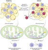
The inflammation-driving changes in cellular and mitochondrial iron regulation in adipose tissue macrophages (ATMs) in obese conditions. In healthy tissue, macrophages specially adapted for efficient iron processing (MFehi) display high expression of genes involved in iron import, storage metabolism and export. This allows for heathy homeostasis of iron. In obesity, hepcidin expression and inflammation increase, causing alterations in the expression of genes involved in iron processing and the recruitment of macrophages which cannot process iron efficiently (MFelo). As a result, iron uptake is increased, while its storage, metabolism and export are reduced. Iron homeostasis becomes significantly impaired, causing adipocytes to store excess labile iron. This iron can accumulate in the mitochondria where it binds to complexes I, II and III of the electron transport chain, increasing its activity and stimulating excess reactive oxygen species (ROS) production. The increased presence of ROS then stimulates the polarisation of M1 macrophages, which possess a pro-inflammatory phenotype. Figure created with BioRender.com.
FIGURE 3
FIGURE 3
The proposed mechanism in which mitochondrial iron (miFe) depletion may contribute to reduced blood pressure (BP). Where miFe is depleted in adipose tissue macrophages (ATMs), reactive oxygen species (ROS) production is low, and the ATMs exhibit an anti-inflammatory profile. The anti-inflammatory interleukins (ILs) -4, -10 and -13 are released, promoting an anti-inflammatory state in surrounding adipocytes. This allows them to express beneficial levels of adiponectin, which ultimately stimulates nitric oxide (NO) release by vascular endothelial cells, leading to vasorelaxation and reduced blood pressure. However, where there is an overload of miFe in ATMs, pro-inflammatory molecules are released by both ATMs and adipocytes due to increased ROS, namely interleukins (ILs) -6 and -1β, toll-like receptor (TLR)-4 and chemokine (C-C motif) ligand (CCL)2. There is also a reduction in the production of the anti-inflammatory ILs -4 and -10, as well as adiponectin, by ATMs and macrophages. This reduction in adiponectin means NO production by endothelial cells is greatly reduced, resulting in vasoconstriction and increased blood pressure. Figure created with BioRender.com.
FIGURE 4
FIGURE 4
The activation of CD8+ T-cells as a result of a high-salt diet and the subsequent hypertension. Monocytes and dendritic cells are two types of antigen-presenting cell (APC). In the presence of high dietary salt, the Nod-like receptor family pyrin domain-containing 3 (NLRP3) inflammasome is upregulated in these cells, and the production of isolevuglandins (isoLGs) as a result of fatty acid peroxidation by reactive oxygen species (ROS) is increased. NLRP3 upregulation leads to increased pro-inflammatory cytokine release (interleukins (ILs) -1β and -18), whilst isoLGs generate pyrrole adducts in the APCs. These adducts are likely presented as neoantigens to CD8+ T-cells, activating them. Once activated, T-cells promote inflammation and dysfunction in the vasculature and kidney, leading to increased blood pressure (through mechanisms detailed in Fig. 1). Based on evidence presented by Pitzer Mutchler et al.[8], magnesium (Mg) likely inhibits NLRP3 inflammasome activity and isoLG formation, leading to reduced inflammation and subsequent increased blood pressure, although its mechanism of action is not currently confirmed. Figure created with BioRender.com.
FIGURE 5
FIGURE 5
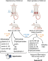
The effects of magnesium (Mg) supplementation on 5/6-nephrectomised rats. Rats underwent surgical removal of a full kidney and 2/3 of the other to generate a model for chronic kidney disease, in which renal failure and symptoms of metabolic syndrome are present. 5/6-nephrectomised rats fed a diet consisting of a standard Mg intake (0.1%) displayed increases in inflammation, ROS activity and endothelial dysfunction (ED) through increased expression of the inflammatory cytokines nuclear factor κβ (NF-κβ), interleukins (ILs) -6 and -1β, as well as lipid peroxides and endothelin-1. Reductions in glutathione peroxidase (GPx), an antioxidant, and nitric oxide (NO) a vasodilator, were also present. Blood pressure itself was increased compared to control sham-operated rats fed a standard Mg diet. On the other hand, 5/6-nephrectomised rats that consumed significantly more Mg displayed levels of these parameters tested by López-Baltanás et al.[9] similar to that of sham-operated controls fed a standard Mg diet. GPx expression, was still slightly reduced in Nx-rats, despite being fed a high-Mg diet. Figure created with BioRender.com.

References

    1. The World Health Organisation 2023. Hypertension. Available at: https://www.who.int/news-room/fact-sheets/detail/hypertension [Accessed 10 November 2023].
    1. Arima H, Barzi F, Chalmers J. Mortality patterns in hypertension. J Hypertens 2011; 29: (Suppl 1): S3–S7. - PubMed
    1. Elliot WJ. Systemic hypertension. Curr Probl Cardiol 2007; 32:201–259. - PubMed
    1. Oparil S, Zaman MA, Calhoun DA. Pathogenesis of hypertension. Ann Intern Med 2003; 139:761–776. - PubMed
    1. Bataillard A, Freiche JC, Vincent M, Sassard J, Touraine JL. Antihypertensive effect of neonatal thymectomy in the genetically hypertensive LH rat. Thymus 1986; 8:321–330. - PubMed