. 2024 Oct;300(10):107748.
doi: 10.1016/j.jbc.2024.107748. Epub 2024 Sep 12.

Impaired endocytosis and accumulation in early endosomal compartments defines herpes simplex virus-mediated disruption of the nonclassical MHC class I-related molecule MR1

Affiliations

Impaired endocytosis and accumulation in early endosomal compartments defines herpes simplex virus-mediated disruption of the nonclassical MHC class I-related molecule MR1

Carolyn Samer et al. J Biol Chem. 2024 Oct.

Abstract

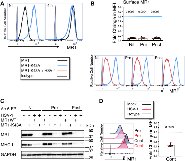
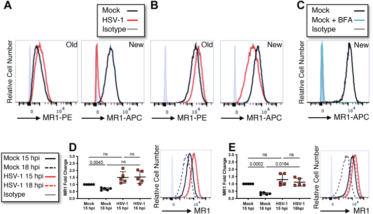
Presentation of metabolites by the major histocompatibility complex class I-related protein 1 (MR1) molecule to mucosal-associated invariant T cells is impaired during herpes simplex virus type 1 (HSV-1) and type 2 (HSV-2) infections. This is surprising given these viruses do not directly synthesise MR1 ligands. We have previously identified several HSV proteins responsible for rapidly downregulating the intracellular pool of immature MR1, effectively inhibiting new surface antigen presentation, while preexisting ligand-bound mature MR1 is unexpectedly upregulated by HSV-1. Using flow cytometry, immunoblotting, and high-throughput fluorescence microscopy, we demonstrate that the endocytosis of surface MR1 is impaired during HSV infection and that internalized molecules accumulate in EEA1-labeled early endosomes, avoiding degradation. We establish that the short MR1 cytoplasmic tail is not required for HSV-1-mediated downregulation of immature molecules; however it may play a role in the retention of mature molecules on the surface and in early endosomes. We also determine that the HSV-1 US3 protein, the shorter US3.5 kinase and the full-length HSV-2 homolog, all predominantly target mature surface rather than total MR1 levels. We propose that the downregulation of intracellular and cell surface MR1 molecules by US3 and other HSV proteins is an immune-evasive countermeasure to minimize the effect of impaired MR1 endocytosis, which might otherwise render infected cells susceptible to MR1-mediated killing by mucosal-associated invariant T cells.

Keywords: HSV; MR1; herpesvirus; immunosuppression; receptor endocytosis; viral immunology.


Conflict of interest statement

Conflict of interest The authors declare that they have no conflicts of interest with the contents of this article.

Figures

Figure 1
Figure 1
Upregulation of surface and early endosomal MR1 during HSV-1 and HSV-2 infection. ARPE-19 MR1 cells were pretreated with Ac-6-FP (5 μM) for 6 h and then infected with HSV-1 (strain F, MOI 5), HSV-2 (strain 186, MOI 3), or mock-infected in parallel. Cells were stained at 15 h p.i. with WGA (plasma membrane) and then permeabilized and stained for MR1 (clone 26.5), and calreticulin (ER), GM130 (Golgi apparatus), EEA1 (early endosomes), or LAMP1 (late endosomes/lysosomes). Cells were then stained with fluorophore-conjugated secondary antibodies and DAPI (nucleus). A, representative images of DAPI + WGA, MR1, EEA1, and merged images, with white arrows highlighting examples of colocation in enlarged image (box) on right. The scale bar represents 50 μm or 20 μm in the enlarged images on the right. Cell segmentation map for each field of view (FOV) was completed with ilastik software after training with a representative image set. ImageJ software was then used for quantification of fold change (FC) in MR1 MFI intensity relative to mock (B, C) or median cellular MR1 (D, E) within each FOV after overlaying the Ilastik-generated map. Violin plots depict fold change in MFI in mock (white), HSV-1 (red), and HSV-2 (blue) infected cells to either mock (C) or median cellular MR1 (E), with median and quartiles shown as horizontal lines. Statistical significance was calculated with one-way ANOVA with multiple comparisons, with p values < 0.05 annotated. Analysis of at least 14 FOV in two independent experiments. Ac-6-FP, acetyl-6-formylpterin; DAPI, 4′,6-diamidino-2-phenylindole; ER, endoplasmic reticulum; h p.i., hour post infection; HSV-1, herpes simplex virus type 1; HSV-2, herpes simplex virus type 2; MOI, multiplicity of infection; MR1, major histocompatibility complex class I–related protein 1; WGA, wheat germ agglutinin.
Figure 2
Figure 2
HSV-1 mediated loss of total and surface MR1 is not dependent on the cytoplasmic tail.A, ARPE-19 MR1 (black) or ARPE-19 ΔP305 (blue) cells transduced to express MR1 lacking the final C-terminal 15 amino acids were left untreated (nil) or treated with Ac-6-FP (5 μM) for 4 h prior to harvesting. Cells were stained for surface MR1 or isotype control (gray) and analyzed by flow cytometry. Histograms representative of two independent experiments. B and C, ARPE-19 ΔP305 cells were mock (blue) or HSV-1 (strain F, MOI 5, red) infected in parallel. Cells were left untreated (nil) or treated with Ac-6-FP (5 μM) 24 h prior to infection (pre) or 14 h p.i. (post). B, cells were harvested at 18 h p.i. when they were stained for surface MR1 or isotype control (gray) and analyzed by flow cytometry. Fold change of mean fluorescence intensity in infected cells relative to mock-infected cells (dotted blue line) with matching ligand treatment is graphed with SD. Statistical significance was calculated by paired Student’s t test, with p values < 0.05 annotated. Analysis of four independent experiments. C, cell lysates were harvested at 18 h p.i. and separated by gel electrophoresis before immunoblotting for MR1, MHC-I, and GAPDH. Ac-6-FP, acetyl-6-formylpterin; h p.i., hour post infection; HSV-1, herpes simplex virus type 1; MHC-I, MHC class I; MOI, multiplicity of infection; MR1, major histocompatibility complex class I–related protein 1.
Figure 3
Figure 3
Surface MR1 is downregulated during HSV-1 infection under conditions of continuous trafficking.A, ARPE-19 MR1 (black) or ARPE-19 MR1-K43A (blue) cells transduced to express MR1 auto folding mutant were left untreated (nil) or treated with Ac-6-FP (5 μM) for 4 h prior to harvesting. Cells were stained for surface MR1 or isotype control (gray) and analyzed by flow cytometry. Histograms representative of two independent experiments. B, ARPE-19 MR1-K43A cells were mock (blue) or HSV-1 (strain F, MOI 5, red) infected in parallel. Cells were left untreated (nil) or treated with Ac-6-FP (5 μM) 24 h prior to infection (pre) or 14 h p.i. (post). Cells were harvested at 18 h p.i. when they were stained for surface MR1 or isotype control (gray) and analyzed by flow cytometry. Fold change in mean fluorescence intensity of infected cells relative to mock-infected cells (dotted blue line) with matching ligand treatment is graphed with SD. Statistical significance was calculated by paired Student’s t test, with p values < 0.05 annotated. Analysis of three independent experiments. C, ARPE-19 MR1 or MR1-K43A cells were mock or HSV-1 (strain F) infected in parallel. Cells were left untreated or treated with Ac-6-FP (5 μM) 24 h prior to infection (pre) or 14 h p.i. (post) before harvesting at 18 h p.i. Cell lysates were separated by gel electrophoresis and before immunoblotting for MR1, MHC-I, GAPDH, and GFP. D, ARPE-19 MR1 cells were mock or HSV-1 (strain F) infected in parallel. Cells were left treated with Ac-6-FP (5 μM) 24 h prior to infection (pre) and optionally treated again immediately post infection (cont) before harvesting at 18 h p.i., when they were stained for surface MR1 or isotype control (gray) and analyzed by flow cytometry. Fold change of infected cells (red) relative to mock-infected cells (dotted line) is graphed with SD. Statistical significance was calculated by paired Student’s t test with p values < 0.05 annotated. Analysis of three independent experiments. Ac-6-FP, acetyl-6-formylpterin; HSV-1, herpes simplex virus type 1; MOI, multiplicity of infection; MR1, major histocompatibility complex class I–related protein 1; p.i., hour post infection.
Figure 4
Figure 4
HSV-1 and HSV-2 US3 protein expression modulation of surface MR1 precedes loss of total MR1.A, ARPE-19 MR1 cells were infected with RAd-Ctrl, RAd-H1-US3, RAd-H1-US3.5, RAd-H2-US3, or mock-infected. Cells were harvested at 44 h p.i. and lysates were separated by gel electrophoresis before immunoblotting for FLAG (to detect US3 expression), MR1, MHC-I, and GAPDH. B, ARPE-19 MR1-GFP cells were infected with RAd-Ctrl adenovirus or RAd-Ctrl modified to encode HSV-1 US3 (RAd-H1-US3), US3.5 gene (RAd-H1-US3.5), or HSV-2 US3 gene (RAd-H2-US3). Cells were treated with Ac-6-FP (5 μM) for 4 h prior to harvesting at 30 or 44 h p.i. Cells were stained for surface MR1 and analyzed by flow cytometry. C, ARPE-19 MR1-GFP cells were infected with RAd-Ctrl adenovirus (black), RAd-H1-US3 (red), RAd-H1-US3.5 (orange), or RAd-H2-US3 (blue). Cells were left untreated (nil) or treated with Ac-6-FP (5 μM) for 4 h (post) prior to harvesting at 44 h p.i. Cells were stained for surface MR1, MHC-I, or matching isotype control (gray) and analyzed by flow cytometry. Fold change in MFI of live-infected cells relative to RAd-Ctrl with matching ligand treatment is graphed with SD. Statistical significance was calculated by paired Student’s t test, with p values < 0.05 annotated. Analysis of four independent experiments. D, 293T cells were transfected with pSY10 (parental plasmid), pSY10-US3 (HSV-1 US3), or pSY10-US3kd (kinase dead US3, K220A). After adding ligand (Ac-6-FP) at 24 h, cells were harvested at 28 h and stained for surface MR1 and MHC-I and analyzed by flow cytometry. Fold change in MFI of GFP+ cells relative to mean pSY10 MFI is graphed with SD. Statistical significance was calculated by one-way ANOVA with multiple comparisons, with p values < 0.05 annotated. Analysis of four independent transfections for each plasmid. Ac-6-FP, acetyl-6-formylpterin; h p.i., hour post infection; HSV-1, herpes simplex virus type 1; HSV-2, herpes simplex virus type 2; MFI, mean fluorescence intensity; MHC-I, MHC class I; MR1, major histocompatibility complex class I–related protein 1.
Figure 5
Figure 5
Endocytosis of MR1 is reduced during HSV-1 infection.A and B, ARPE-19 MR1 cells were mock or HSV-1 (strain F) infected in parallel. Cells were treated with Ac-6-FP (5 μM) 24 h prior to infection (B) or left untreated (A). Existing surface MR1 (old) was captured by staining for surface MR1 (anti-MR1-PE or isotype control in gray) during the final 30 min of the 1-h period of viral adsorption. Newly trafficked surface MR1 (new) was detected at 16 h p.i., when cells were again stained for surface MR1 (anti-MR1-APC or isotype control in gray). C, ARPE-19 MR1 cells were stained for existing surface MR1 (anti-MR1-PE), treated with Ac-6-FP (5 μM), and optionally treated with brefeldin A. After 16 h, cells were harvested and stained for surface MR1 (new, anti-MR1-APC, or isotype control in gray). Histogram representative of two independent experiments. D, ARPE-19 MR1 cells were mock or HSV-1 (strain F) infected in parallel. Cells were treated with Ac-6-FP (5 μM) 6 h prior to infection. At 15 h p.i., cells were stained with anti-MR1-APC and then harvested immediately or after a further 3 h. E, ARPE-19 MR1 cells were mock or HSV-1 (strain F) infected in parallel. Cells were treated with Ac-6-FP (5 μM) 6 h prior to infection. At 15 h p.i., cells were stained with anti-MR1-biotin and then harvested immediately or after a further 3 h. Cells were stained with streptavidin-APC at time of harvest. D and E, Cells were analyzed by flow cytometry with analysis based on mean fluorescence intensity fold change to mock infected at 15 h p.i. Statistical significance was calculated by one-way ANOVA with multiple comparisons, with p values < 0.05 annotated. Analysis of five independent experiments. Ac-6-FP, acetyl-6-formylpterin; APC, allophycocyanin; h p.i., hour post infection; HSV-1, herpes simplex virus type 1; MR1, major histocompatibility complex class I–related protein 1.
Figure 6
Figure 6
The fate of MR1 molecules during HSV infection. HSV infection depletes the pool of immature MR1 molecules in the endoplasmic reticulum, limiting trafficking through the Golgi apparatus in response to ligand availability. Infection impairs endocytosis of ligand-bound mature molecules on the plasma membrane, although this is offset by HSV targeting of intracellular and surface MR1 molecules for degradation. After endocytosis MR1 preferentially accumulates in early endosomal compartments, but not late endosomes/lysosomes. HSV, herpes simplex virus; MR1, major histocompatibility complex class I–related protein 1.

References

    1. Keller A.N., Eckle S.B.G., Xu W., Liu L., Hughes V.A., Mak J.Y.W., et al. Drugs and drug-like molecules can modulate the function of mucosal-associated invariant T cells. Nat. Immunol. 2017;18:402–411. - PubMed
    1. Kjer-Nielsen L., Patel O., Corbett A.J., Le Nours J., Meehan B., Liu L.G., et al. MR1 presents microbial vitamin B metabolites to MAIT cells. Nature. 2012;491:717–723. - PubMed
    1. Gold M.C., Cerri S., Smyk-Pearson S., Cansler M.E., Vogt T.M., Delepine J., et al. Human mucosal associated invariant T cells detect bacterially infected cells. PLoS Biol. 2010;8 - PMC - PubMed
    1. Gherardin N.A., Keller A.N., Woolley R.E., Le Nours J., Ritchie D.S., Neeson P.J., et al. Diversity of T Cells restricted by the MHC class I-related molecule MR1 facilitates differential antigen recognition. Immunity. 2016;44:32–45. - PubMed
    1. Tsukamoto K., Deakin J.E., Graves J.A.M., Hashimoto K. Exceptionally high conservation of the MHC class I-related gene, MR1, among mammals. Immunogenetics. 2013;65:115–124. - PubMed

MeSH terms

Substances

LinkOut - more resources