. 2024:703:29-49.
doi: 10.1016/bs.mie.2024.05.019. Epub 2024 Jun 21.

Spectroscopic definition of ferrous active sites in non-heme iron enzymes

Affiliations

Spectroscopic definition of ferrous active sites in non-heme iron enzymes

Edward I Solomon et al. Methods Enzymol. 2024.

Abstract

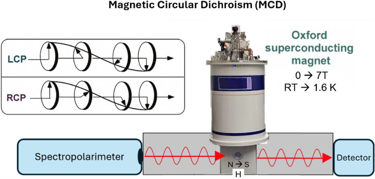
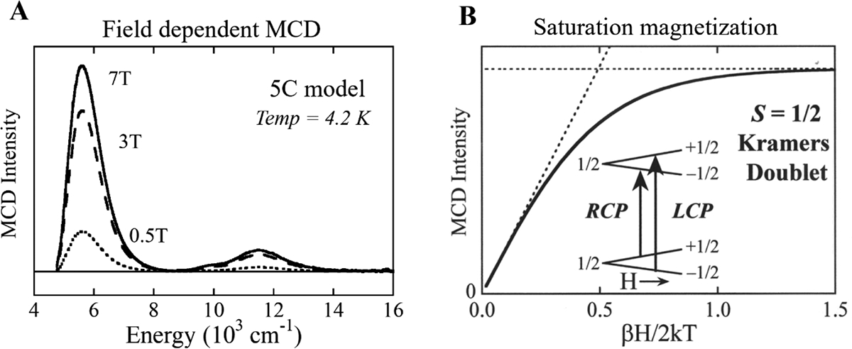
Non-heme iron enzymes play key roles in antibiotic, neurotransmitter, and natural product biosynthesis, DNA repair, hypoxia regulation, and disease states. These enzymes had been refractory to traditional bioinorganic spectroscopic methods. Thus, we developed variable-temperature variable-field magnetic circular dichroism (VTVH MCD) spectroscopy to experimentally define the excited and ground ligand field states of non-heme ferrous enzymes (Solomon et al., 1995). This method provides detailed geometric and electronic structure insight and thus enables a molecular level understanding of catalytic mechanisms. Application of this method across the five classes of non-heme ferrous enzymes has defined that a general mechanistic strategy is utilized where O2 activation is controlled to occur only in the presence of all cosubstrates.

Keywords: Ferrous enzyme mechanisms; Ligand field theory; Magnetic circular dichroism spectroscopy; Non-Kramers’ ions; Non-heme iron enzymes; Saturation magnetization.


Figures

Figure 1.
Figure 1.
Schematic structures of non-heme ferrous active sites.
Figure 2.
Figure 2.
Tanabe-Sugano Diagram quantifying the energy the states of a d6 transition metal ion in an octahedral ligand field in terms of Dq and B. Used with permission from John Wiley & Sons from (Solomon & Hanson, 2006).
Figure 3.
Figure 3.
Ligand field splitting of the high spin quintet d6 LF states with coordination number and structure. Adapted with permission from Solomon, E. I., Brunold, T. C., Davis, M. I., Kemsley, J. N., Lee, S. K., Lehnert, N., Neese, F., Skulan, A. J., Yang, Y. S., & Zhou, J. (2000). Geometric and Electronic Structure/Function Correlations in Non-Heme Iron Enzymes. Chemical Reviews, 100(1), 235–349. https://doi.org/10.1021/cr9900275 (Solomon et al., 2000). Copyright 2000 American Chemical Society.
Figure 4.
Figure 4.
Experimental configuration for VTVH MCD.
Figure 5.
Figure 5.
LT MCD spectra of representative structurally defined FeII model complexes. (A) 6C, octahedral [Fe(H2O)6](SiF6). (B) 5C, square-pyramidal [Fe(HB(3,5-iPr2pz)3)(OAc)]. (C) 5C, trigonal-bipyramidal [Fe(tris(2-(dimethylamino)ethyl)amine)Br]+. (D) 4C, tetrahedral [Fe(HB(3,5-iPr2pz)3)(Cl)]. Adapted with permission from Pavel, E. G., Kitajima, N., & Solomon, E. I. (1998). Magnetic Circular Dichroism Spectroscopic Studies of Mononuclear Non-Heme Ferrous Model Complexes. Correlation of Excited- and Ground-State Electronic Structure with Geometry. Journal of the American Chemical Society, 120(16), 3949–3962. https://doi.org/10.1021/JA973735L (Pavel et al., 1998). Copyright 1998 American Chemical Society.
Figure 6.
Figure 6.
(A) Field dependence of the MCD signal from the 5C complex in Figure 5. (B) Saturation magnetization MCD for an S = 1/2 Kramers doublet; signal as a function of field and temperature. Inset shows Kramers doublet in a magnetic field and MCD allowed transitions. Adapted with permission from Solomon, E. I., Brunold, T. C., Davis, M. I., Kemsley, J. N., Lee, S. K., Lehnert, N., Neese, F., Skulan, A. J., Yang, Y. S., & Zhou, J. (2000). Geometric and Electronic Structure/Function Correlations in Non-Heme Iron Enzymes. Chemical Reviews, 100(1), 235–349. https://doi.org/10.1021/cr9900275 (Solomon et al., 2000). Copyright 2000 American Chemical Society.
Figure 7.
Figure 7.
Saturation magnetization data and fits using equation 2 for the ferrous 5C complex in Figure 5 and 6A recorded at 6,000 cm−1. (A) Nesting behavior shown in plot of intensity versus βH/2kT; (B) and intensity versus temperature for fixed field increments. Adapted from Solomon, E. I., Pavel, E. G., Loeb, K. E., & Campochiaro, C. (1995). Magnetic circular dichroism spectroscopy as a probe of the geometric and electronic structure of non-heme ferrous enzymes. Coordination Chemistry Reviews, 144, 369–460. https://doi.org/10.1016/0010-8545(95)01150-N (Solomon et al., 1995) with permission from Elsevier.
Figure 8.
Figure 8.
Energy splittings of the S = 2 sublevels for an axial −ZFS and a rhombic distortion and then magnetic field splitting and mixing of an MS = ±2 non-Kramers doublet. Adapted with permission from Campochiaro, C., Pavel, E. G., & Solomon, E. I. (1995). Saturation Magnetization Magnetic Circular Dichroism Spectroscopy of Systems with Positive Zero-Field Splittings: Application to FeSiF6·6H2O. Inorganic Chemistry, 34(18), 4669–4675. https://doi.org/10.1021/IC00122A025 (Campochiaro et al., 1995). Copyright 1995 American Chemical Society.
Figure 9.
Figure 9.
LF splitting of the t2 dπ orbitals (A), including SOC (B) and the Zeeman effect (inset). Adapted from (A) Solomon, E. I., & Hanson, M. A. (2006). Bioinorganic Spectroscopy. In E. I. Solomon & A. B. P. Lever (Eds.), Inorganic Electronic Structure and Spectroscopy (Vol. 2, pp. 1–129). John Wiley & Sons, Inc. https://www.wiley.com/en-us/Inorganic+Electronic+Structure+and+Spectroscopy%2C+Volume+II-p-9780471971146 (Solomon & Hanson, 2006) with permission from John Wiley & Sons and (B) Solomon, E. I., Pavel, E. G., Loeb, K. E., & Campochiaro, C. (1995). Magnetic circular dichroism spectroscopy as a probe of the geometric and electronic structure of non-heme ferrous enzymes. Coordination Chemistry Reviews, 144, 369–460. https://doi.org/10.1016/0010-8545(95)01150-N (Solomon et al., 1995) with permission from Elsevier.
Figure 10.
Figure 10.
Correlations of the g (top) and δ (bottom) with the axial (Δ) and rhombic (V) LF splitting of the t2 dπ orbitals. Adapted from Solomon, E. I., Pavel, E. G., Loeb, K. E., & Campochiaro, C. (1995). Magnetic circular dichroism spectroscopy as a probe of the geometric and electronic structure of non-heme ferrous enzymes. Coordination Chemistry Reviews, 144, 369–460. https://doi.org/10.1016/0010-8545(95)01150-N (Solomon et al., 1995) with permission from Elsevier.
Figure 11.
Figure 11.
Experimental Ligand-Field splittings of the five d orbitals for the complexes in Figure 5. Adapted with permission from Pavel, E. G., Kitajima, N., & Solomon, E. I. (1998). Magnetic Circular Dichroism Spectroscopic Studies of Mononuclear Non-Heme Ferrous Model Complexes. Correlation of Excited- and Ground-State Electronic Structure with Geometry. Journal of the American Chemical Society, 120(16), 3949–3962. https://doi.org/10.1021/JA973735L (Pavel et al., 1998). Copyright 1998 American Chemical Society.
Figure 12.
Figure 12.
MCD elucidation of the cosubstrate activation mechanism of the Fe(II) site in PAH. (A) LT MCD, (B) VTVH MCD, and (C) experimentally determined LF splitting of the d orbitals. Adapted with permission from Kemsley, J. N., Mitić, N., Zaleski, K. L., Caradonna, J. P., & Solomon, E. I. (1999). Circular Dichroism and Magnetic Circular Dichroism Spectroscopy of the Catalytically Competent Ferrous Active Site of Phenylalanine Hydroxylase and Its Interaction with Pterin Cofactor. Journal of the American Chemical Society, 121(7), 1528–1536. https://doi.org/10.1021/JA9833063 (Kemsley et al., 1999) and Loeb, K. E., Westre, T. E., Kappock, T. J., Mitić, N., Glasfeld, E., Caradonna, J. P., Hedman, B., Hodgson, K. O., & Solomon, E. I. (1997). Spectroscopic characterization of the catalytically competent ferrous site of the resting, activated, and substrate-bound forms of phenylalanine hydroxylase. Journal of the American Chemical Society, 119(8), 1901–1915. https://doi.org/10.1021/JA962269H (Loeb et al., 1997). Copyright 1999 and 1997 American Chemical Society.
Figure 13.
Figure 13.
General Mechanistic Strategy for Cofactor-Dependent NHFe Enzymes. Adapted with permission from Solomon, E. I., Deweese, D. E., & Babicz, J. T. (2021). Mechanisms of O2 Activation by Mononuclear Non-Heme Iron Enzymes. Biochemistry, 60(46), 3497–3506. https://doi.org/10.1021/acs.biochem.1c00370 (Solomon et al., 2021). Copyright 2021 American Chemical Society.
Figure 14.
Figure 14.
(A) Absorption (top) and VT MCD spectra (bottom) of SyrB2 Br–FeIV═O. Transition I is the dπ* →dσ* LF transition, and II and III are oxo to iron charge transfer transitions. The * indicates a minor heme contaminant in the sample. Adapted with permission from Srnec, M., Wong, S. D., Matthews, M. L., Krebs, C., Bollinger, J. M., & Solomon, E. I. (2016). Electronic Structure of the Ferryl Intermediate in the α-Ketoglutarate Dependent Non-Heme Iron Halogenase SyrB2: Contributions to H Atom Abstraction Reactivity. Journal of the American Chemical Society, 138, 5110–5122. https://doi.org/10.1021/jacs.6b01151 (Srnec et al., 2016). Copyright 2016 American Chemical Society. (B) Schematic showing the relevant FMOs and the orbitals participating in the dπ* →dσ* LF transition.

Similar articles

References

    1. Babicz JT, Rogers MS, DeWeese DE, Sutherlin KD, Banerjee R, Böttger LH, Yoda Y, Nagasawa N, Saito M, Kitao S, Kurokuzu M, Kobayashi Y, Tamasaku K, Seto M, Lipscomb JD, & Solomon EI (2023). Nuclear Resonance Vibrational Spectroscopy Definition of Peroxy Intermediates in Catechol Dioxygenases: Factors that Determine Extra-versus Intradiol Cleavage. Journal of the American Chemical Society. 10.1021/JACS.3C02242 - DOI - PMC - PubMed
    1. Baldwin JE, & Abraham E (1988). The biosynthesis of penicillins and cephalosporins. Natural Product Reports, 5(2), 129–145. 10.1039/NP9880500129 - DOI - PubMed
    1. Barry SM, & Challis GL (2013). Mechanism and Catalytic Diversity of Rieske Non-Heme Iron-Dependent Oxygenases. ACS Catalysis, 3(10), 2362–2370. 10.1021/cs400087p - DOI - PMC - PubMed
    1. Blasiak LC, Vaillancourt FH, Walsh CT, & Drennan CL (2006). Crystal structure of the non-haem iron halogenase SyrB2 in syringomycin biosynthesis. Nature, 440(7082), 368–371. 10.1038/nature04544 - DOI - PubMed
    1. Buckingham AD, & Stephens PJ (1966). Magnetic Optical Activity. Annual Review of Physical Chemistry, 17(1), 399–432. 10.1146/annurev.pc.17.100166.002151 - DOI

LinkOut - more resources