. 2024 Sep 11;81(1):396.
doi: 10.1007/s00018-024-05438-2.

High sugar diet promotes tumor progression paradoxically through aberrant upregulation of pepck1

Affiliations

High sugar diet promotes tumor progression paradoxically through aberrant upregulation of pepck1

Che-Wei Chang et al. Cell Mol Life Sci. .

Abstract

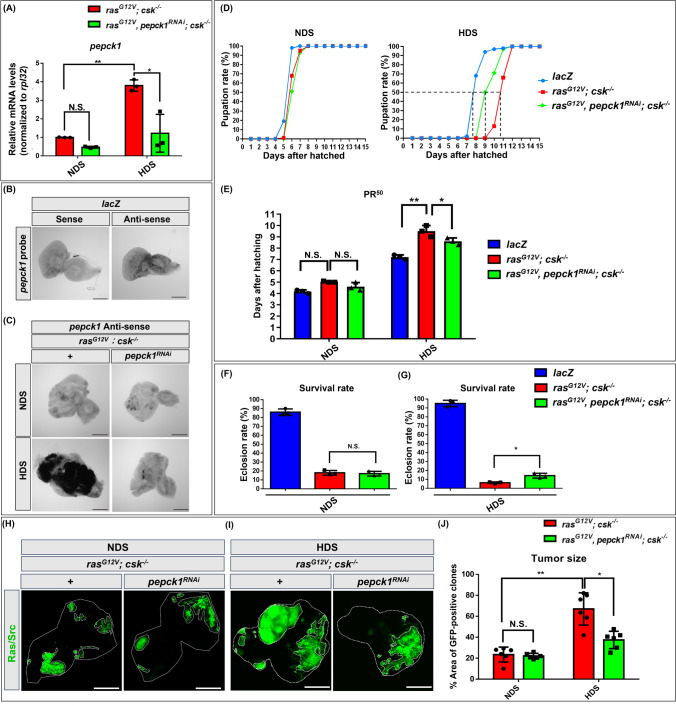
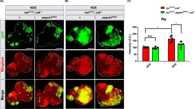
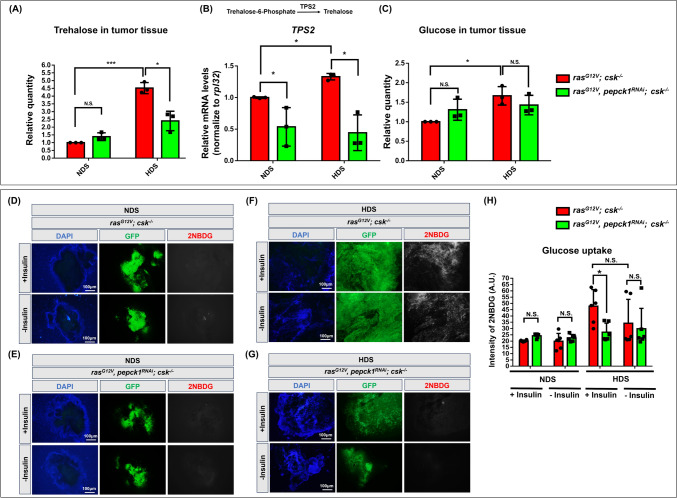
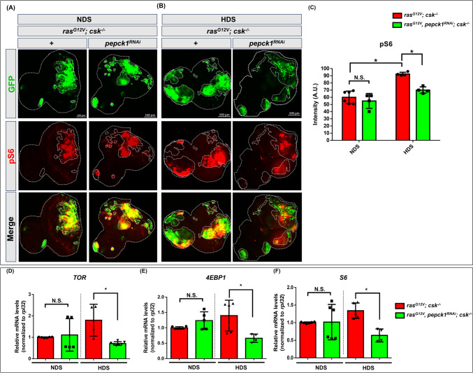
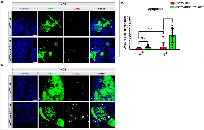
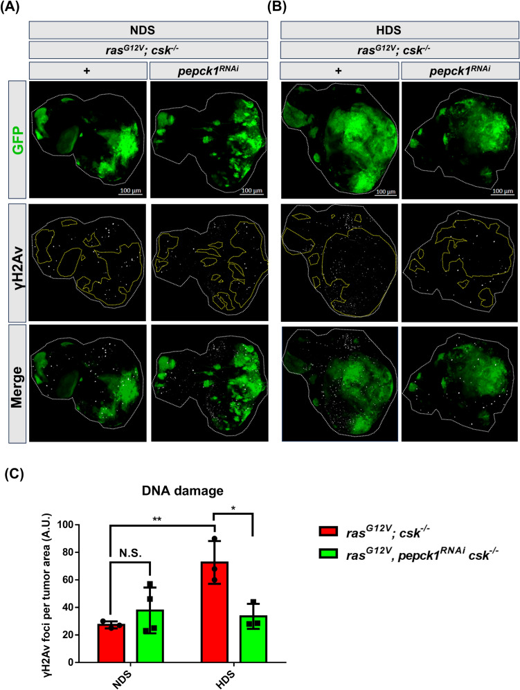
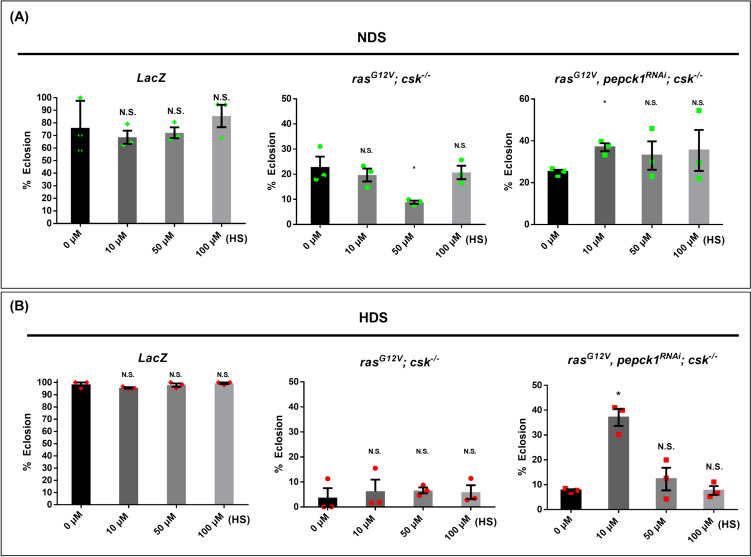
High dietary sugar (HDS), a contemporary dietary concern due to excessive intake of added sugars and carbohydrates, escalates the risk of metabolic disorders and concomitant cancers. However, the molecular mechanisms underlying HDS-induced cancer progression are not completely understood. We found that phosphoenolpyruvate carboxykinase 1 (PEPCK1), a pivotal enzyme in gluconeogenesis, is paradoxically upregulated in tumors by HDS, but not by normal dietary sugar (NDS), during tumor progression. Targeted knockdown of pepck1, but not pepck2, specifically in tumor tissue in Drosophila in vivo, not only attenuates HDS-induced tumor growth but also significantly improves the survival of Ras/Src tumor-bearing animals fed HDS. Interestingly, HP1a-mediated heterochromatin interacts directly with the pepck1 gene and downregulates pepck1 gene expression in wild-type Drosophila. Mechanistically, we demonstrated that, under HDS conditions, pepck1 knockdown reduces both wingless and TOR signaling, decreases evasion of apoptosis, reduces genome instability, and suppresses glucose uptake and trehalose levels in tumor cells in vivo. Moreover, rational pharmacological inhibition of PEPCK1, using hydrazinium sulfate, greatly improves the survival of tumor-bearing animals with pepck1 knockdown under HDS. This study is the first to show that elevated levels of dietary sugar induce aberrant upregulation of PEPCK1, which promotes tumor progression through altered cell signaling, evasion of apoptosis, genome instability, and reprogramming of carbohydrate metabolism. These findings contribute to our understanding of the complex relationship between diet and cancer at the molecular, cellular, and organismal levels and reveal PEPCK1 as a potential target for the prevention and treatment of cancers associated with metabolic disorders.

Keywords: Cancer metabolism; Epigenetics; Gene regulation; Hallmarks of cancer; Model organisms; Signal transduction.


Conflict of interest statement

The authors declare no competing interests.

Figures

Fig. 1
Fig. 1
Reducing HDS-induced upregulation of pepck1 alleviates developmental delay and reduces lethality and tumor size among Ras/Src tumor-bearing animals. Drosophila with the following genotypes were used in these experiments: lacZ (control), rasG12V; csk−/− (tumor-bearing), and rasG12V, pepck1RNAi; csk−/− (tumor-bearing with pepck1 knockdown). A Relative pepck1 mRNA levels were determined based on RNA extracted from 30 eye discs of combined 3rd instar male and female larvae (n = 15) fed HDS. Results were normalized to rpl32. B pepck1 mRNA expression patterns were examined using DIG-labeled pepck1 sense (negative control) or antisense RNA probes in WT male eye discs. C pepck1 mRNA expression patterns were analyzed using DIG-labeled pepck1 antisense RNA probes in the eye discs of male rasG12V; csk−/− and rasG12V, pepck1RNAi; csk−/− Drosophila under NDS or HDS. D Pupation rates of combined male and female animals fed a 0.15 M sucrose diet (NDS) and a 0.75 M sucrose diet (HDS). E Number of days (PR50) until pupation rate reached 50% among control and tumor-bearing animals. F, G Eclosion rates of combined male and female animals fed NDS or HDS. (H) Eye discs from 3rd instar rasG12V; csk−/− and rasG12V, pepck1RNAi; csk−/− female Drosophila larvae fed NDS, with GFP-labeled tumor cells. I Eye discs from 3rd instar rasG12V; csk−/− and rasG12V, pepck1RNAi; csk−/− female Drosophila larvae fed HDS, with GFP-labeled tumor cells. J Percentage of GFP-positive tumor cells normalized to total eye disc area from female Drosophila fed NDS or HDS. Results are shown as mean ± SD. Asterisks indicate statistically significant differences via two-way ANOVA with paired controls (*P < 0.05, **P < 0.01). GFP green fluorescent protein, HDS high dietary sugar, NDS normal dietary sugar, N.S. not significant; SD standard deviation
Fig. 2
Fig. 2
Knockdown of pepck1 inhibits the expression of HDS-induced wingless in tumor cells. Eye discs from 3rd instar rasG12V; csk−/− and rasG12V, pepck1RNAi; csk−/− Drosophila larvae, fed NDS or HDS, with GFP-labeled tumor cells and wingless immunostaining (red); Scale bar: 100 µm; n = 9 eye discs per group. A rasG12V; csk−/− and rasG12V, pepck1RNAi; csk−/− female Drosophila larvae fed NDS. B rasG12V; csk−/− and rasG12V, pepck1RNAi; csk−/− female Drosophila larvae fed HDS. C Quantification of wingless expression in tumor cells from female Drosophila fed NDS or HDS. n = 9 eye discs per group. Fluorescence intensity was quantified using the ZEISS ZEN Blue software. Results are shown as mean ± SD. Asterisk indicates a statistically significant difference via two-way ANOVA with paired control (*P < 0.05). GFP green fluorescent protein, HDS high dietary sugar, NDS normal dietary sugar, SD standard deviation
Fig. 3
Fig. 3
Knockdown of pepck1 reduces HDS-induced elevated trehalose levels and glucose uptake in tumor cells. A Trehalose levels were measured using 60 eye discs from combined 3rd instar rasG12V; csk−/− and rasG12V, pepck1RNAi; csk−/− male and female Drosophila larvae fed NDS or HDS. B Relative levels of TPS2 (trehalose-6-phosphate phosphatase) mRNA were determined based on RNA extracted from 30 eye discs of combined 3rd instar rasG12V; csk−/− and rasG12V, pepck1RNAi; csk−/− male and female Drosophila larvae fed NDS or HDS. C Glucose levels were assessed using 60 eye discs from combined 3rd instar rasG12V; csk−/− and rasG12V, pepck1RNAi; csk−/− male and female Drosophila larvae fed NDS or HDS. Results are shown as mean ± SD. Asterisk indicates a statistically significant difference via two-way ANOVA with paired control (*P < 0.05; ***P < 0.001). D, E 2NBDG expression (red) in eye discs from 3rd instar rasG12V; csk−/− and rasG12V, pepck1RNAi; csk−/− female Drosophila larvae fed NDS, with GFP-labeled tumor cells (green). F, G Expression of 2NBDG (red) in eye discs from 3rd instar rasG12V; csk−/− and rasG12V, pepck1RNAi; csk−/− female Drosophila larvae fed HDS, with GFP-labeled tumor cells (green). H Quantification of 2NBDG levels (red) via fluorescence intensity in tumor cells (green) in eye discs from 3rd instar rasG12V; csk−/− and rasG12V, pepck1RNAi; csk−/− female Drosophila larvae fed NDS or HDS. Fluorescence intensity was quantified using the ZEISS ZEN Blue software. Results are shown as mean ± SD. Asterisks indicate statistically significant differences via two-way ANOVA with paired control (*P < 0.05). GFP green fluorescent protein, HDS high dietary sugar, NDS normal dietary sugar, N.S. not significant, SD standard deviation
Fig. 4
Fig. 4
Knockdown of pepck1 decreases TOR signaling in tumor cells under NDS and HDS. Eye discs from rasG12V; csk−/− and rasG12V, pepck1RNAi; csk−/− 3rd instar larvae, fed NDS or HDS, with GFP-labeled tumor cells (green) and pS6 immunostaining (red). Scale bar: 100 µm. A rasG12V; csk−/− and rasG12V, pepck1RNAi; csk−/− female larvae fed NDS. B rasG12V; csk−/− and rasG12V, pepck1RNAi; csk−/− female larvae fed HDS on day 7 AEL. C Quantification of pS6 fluorescence intensity in tumor cells from female larvae fed HDS; n = 9 eye discs per group. Fluorescence intensity was quantified using the ZEISS ZEN Blue software. Results are shown as mean ± SD of individual eye discs. Differences between groups were assessed via two-way ANOVA; *P < 0.05. Relative levels of TOR (D), 4EBP1 (E) and S6 (F) mRNA were determined based on RNA extracted from 50 eye discs from combined 3rd instar rasG12V; csk−/− and rasG12V, pepck1RNAi; csk−/− male and female Drosophila larvae fed NDS or HDS. Results are shown as mean ± SEM. Asterisks indicate statistically significant differences via Student’s t-test (*P < 0.05). AEL after egg laying, GFP green fluorescent protein, HDS high dietary sugar, NDS normal dietary sugar, SD standard deviation
Fig. 5
Fig. 5
Knockdown of pepck1 induces apoptosis in tumor cells. A, B Eye discs from rasG12V; csk−/− and rasG12V, pepck1RNAi; csk−/− female Drosophila 3rd instar larvae, fed NDS or HDS, with GFP-labeled tumor cells (green) and TUNEL staining (white). Scale bar: 20 μm. C Quantification of TUNEL foci in tumor cells per tumor clone; n = 6 female eye discs per group. Results are shown as mean ± SD. Asterisk indicates a statistically significant difference via two-way ANOVA with paired control (*P < 0.05). GFP green fluorescent protein, Blue Hoechst, HDS high dietary sugar, NDS normal dietary sugar, N.S. not significant, SD standard deviation
Fig. 6
Fig. 6
Knockdown of pepck1 reduces HDS-induced DNA damage in tumor cells. A, B Eye discs from rasG12V; csk−/− and rasG12V, pepck1RNAi; csk−/− 3rd instar female Drosophila larvae, fed NDS or HDS, with GFP-labeled tumor cells (green) and γH2AX immunostaining (white). Scale bar: 100 μm. C Quantification of γH2AX foci in tumor cells; n = 6 female eye discs per group. Results are shown as mean ± SD. Asterisks indicate statistically significant differences via two-way ANOVA with paired control (*P < 0.05; **P < 0.01). GFP green fluorescent protein, HDS high dietary sugar, NDS normal dietary sugar, N.S. not significant, SD standard deviation
Fig. 7
Fig. 7
Hydrazinium sulfate (HS), an inhibitor of PEPCK, enhances the survival of tumor-bearing animals with pepck1 knockdown. Drosophila larvae with the following genotypes were fed either NDS or HDS and used in the experiments: lacZ, and rasG12V; csk−/− and rasG12V; pepck1RNAi; csk−/−. A total of 200 combined male and female larvae per genotype were used for each experiment. A Eclosion rates of animals fed NDS with treatments of 0 µM, 10 µM, 50 µM, or 100 µM HS. B Eclosion rates of animals fed HDS with treatments of 0 µM, 10 µM, 50 µM, or 100 µM HS. Results are shown as mean ± SD. Asterisks indicate statistically significant differences via two-way ANOVA with paired controls (*P < 0.05). HDS high dietary sugar, HS hydrazinium sulfate, NDS normal dietary sugar, N.S. not significant, SD standard deviation
Fig. 8
Fig. 8
High sugar diet induces upregulation of pepck1, which promotes cancer progression by acivating wingless/dWnt and TOR signaling, decreasing apoptosis, inducing genome instability, and reprogramming carbohydrate metabolism. In the presence of HDS, tumor cells upregulate PEPCK1 to increase tumor burden and decrease the survival rate of tumor-bearing animals. Additionally, the heterochromatin formed via an HP1a-mediated process directly suppresses pepck1 expression in tumor cells. Mechanistically, PEPCK1 facilitates HDS-induced tumor progression by elevating wingless expression and TOR signaling, suppressing apoptosis, inducing genome instability, and reprogramming carbohydrate metabolism through increased glucose uptake and elevated trehalose levels

References

    1. Ling S et al (2020) Association of Type 2 Diabetes with cancer: a meta-analysis with bias analysis for unmeasured confounding in 151 Cohorts Comprising 32 Million People. Diabetes Care 43(9):2313–2322 10.2337/dc20-0204 - DOI - PubMed
    1. Swanton C et al (2024) Embracing cancer complexity: Hallmarks of systemic disease. Cell 187(7):1589–1616 10.1016/j.cell.2024.02.009 - DOI - PubMed
    1. Goncalves MD et al (2019) High-fructose corn syrup enhances intestinal tumor growth in mice. Science 363(6433):1345–1349 10.1126/science.aat8515 - DOI - PMC - PubMed
    1. Jiang Y et al (2016) A sucrose-enriched diet promotes tumorigenesis in mammary gland in part through the 12-lipoxygenase pathway. Cancer Res 76(1):24–29 10.1158/0008-5472.CAN-14-3432 - DOI - PMC - PubMed
    1. Newton H et al (2020) Systemic muscle wasting and coordinated tumour response drive tumourigenesis. Nat Commun 11(1):4653 10.1038/s41467-020-18502-9 - DOI - PMC - PubMed

MeSH terms

LinkOut - more resources