. 2024 Sep 11;14(1):21195.
doi: 10.1038/s41598-024-70659-1.

Comprehensive analysis of scRNA-Seq and bulk RNA-Seq reveals ubiquitin promotes pulmonary fibrosis in chronic pulmonary diseases

Affiliations

Comprehensive analysis of scRNA-Seq and bulk RNA-Seq reveals ubiquitin promotes pulmonary fibrosis in chronic pulmonary diseases

Zhuman Wen et al. Sci Rep. .

Abstract

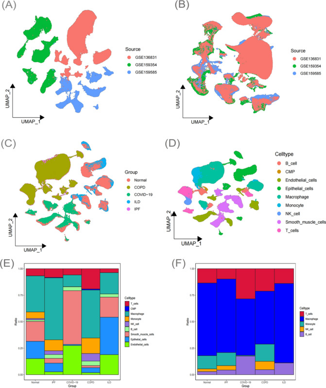
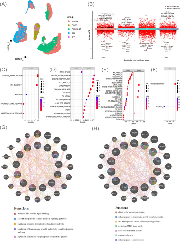
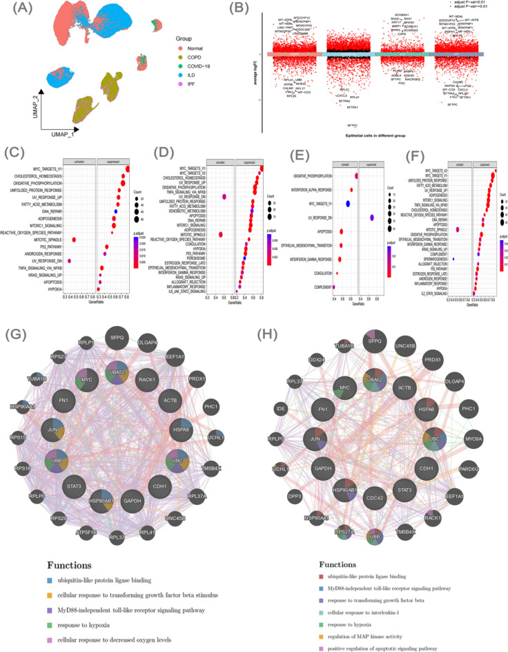
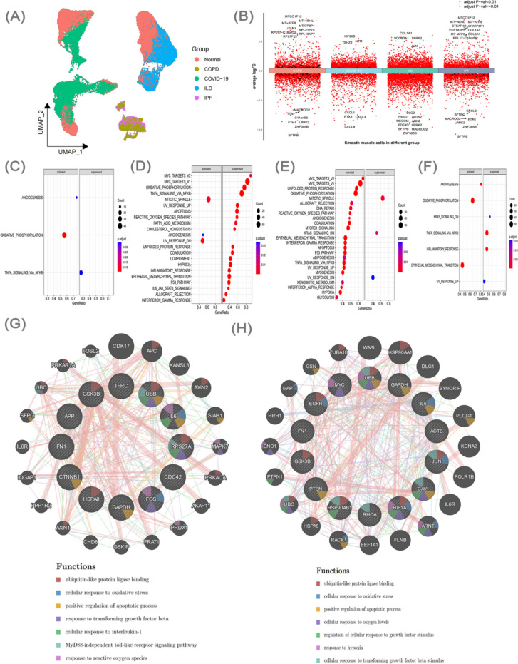
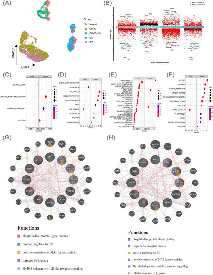
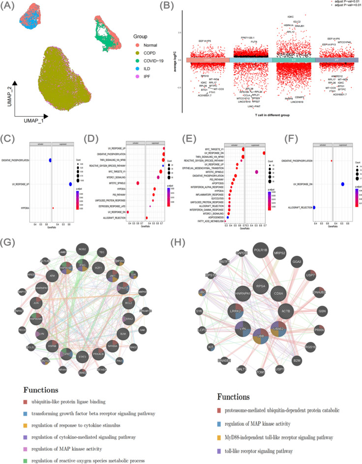
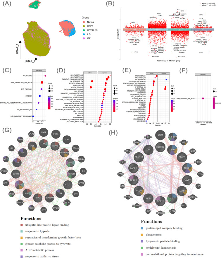
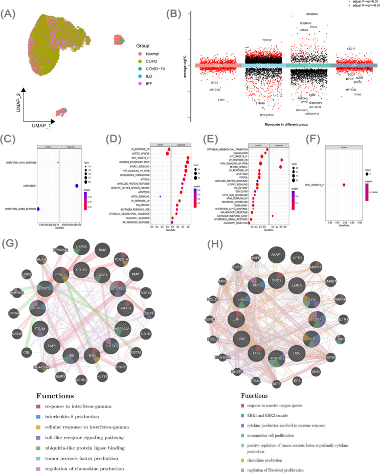
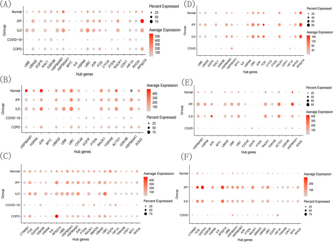
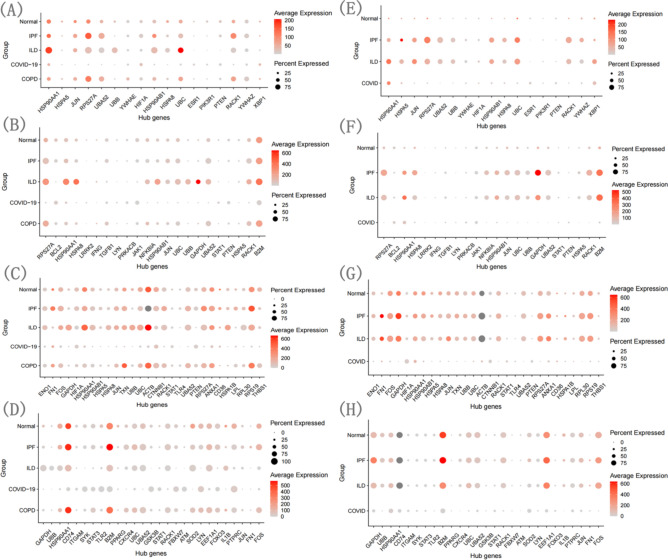
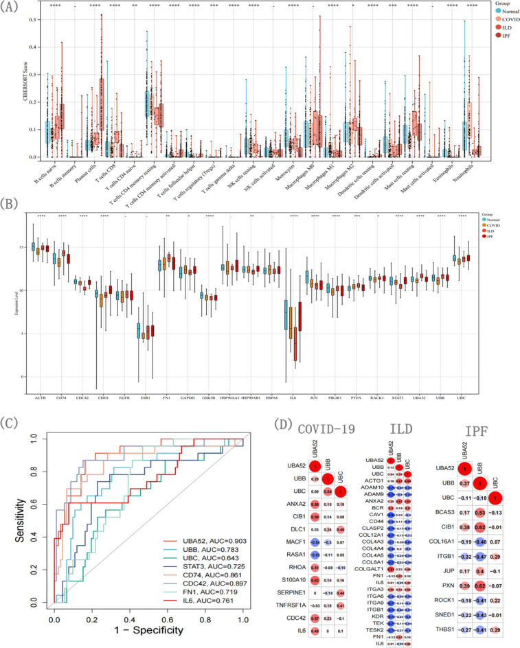
It is estimated that there are 544.9 million people suffering from chronic respiratory diseases in the world, which is the third largest chronic disease. Although there are various clinical treatment methods, there is no specific drug for chronic pulmonary diseases, including chronic obstructive pulmonary disease (COPD), interstitial lung disease (ILD) and idiopathic pulmonary fibrosis (IPF). Therefore, it is urgent to clarify the pathological mechanism and medication development. Single-cell transcriptome data of human and mouse from GEO database were integrated by "Harmony" algorithm. The data was standardized and normalized by using "Seurat" package, and "SingleR" algorithm was used for cell grouping annotation. The "Findmarker" function is used to find differentially expressed genes (DEGs), which were enriched and analyzed by using "clusterProfiler", and a protein interaction network was constructed for DEGs, and four algorithms are used to find the hub genes. The expression of hub genes were analyzed in independent human and mouse single-cell transcriptome data. Bulk RNA data were used to integrate by the "SVA" function, verify the expression levels of hub genes and build a diagnostic model. The L1000FWD platform was used to screen potential drugs. Through exploring the similarities and differences by integrated single-cell atlas, we found that the lung parenchymal cells showed abnormal oxidative stress, cell matrix adhesion and ubiquitination in COPD, corona virus disease 2019 (COVID-19), ILD and IPF. Meanwhile, the lung resident immune cells showed abnormal Toll-like receptor signals, interferon signals and ubiquitination. However, unlike acute pneumonia (COVID-19), chronic pulmonary disease shows enhanced ubiquitination. This phenomenon was confirmed in independent external human single-cell atlas, but unfortunately, it was not confirmed in mouse single-cell atlas of bleomycin-induced pulmonary fibrosis model and influenza virus-infected mouse model, which means that the model needs to be optimized. In addition, the bulk RNA-Seq data of COVID-19, ILD and IPF was integrated, and we found that the immune infiltration of lung tissue was enhanced, consistent with the single-cell level, UBA52, UBB and UBC were low expressed in COVID-19 and high expressed in ILD, and had a strong correlation with the expression of cell matrix adhesion genes. UBA52 and UBB have good diagnostic efficacy, and salermide and SSR-69071 can be used as their candidate drugs. Our study found that the disorder of protein ubiquitination in chronic pulmonary diseases is an important cause of pathological phenotype of pulmonary fibrosis by integrating scRNA-Seq and bulk RNA-Seq, which provides a new horizons for clinicopathology, diagnosis and treatment.

Keywords: Bulk RNA sequencing; Chronic pulmonary diseases; Immune infiltration; Single-cell RNA sequencing; Ubiquitination.


Conflict of interest statement

The authors declare no competing interests.

Figures

Fig. 1
Fig. 1
Integrated single cell atlas, cell type and cell ratio of four diseases. UMAP diagram before (A) and after (B) batch removal by integrating the single cell atlas; UMAP diagram of cell grouping (C) and annotation (D); The proportion of all cell types (E) and the proportion of immune cells (F).
Fig. 2
Fig. 2
DEGs in endothelial cells and functional analysis. UMAP diagram of endothelial cell grouping (A). Compared with the normal group, the volcano map of DEGs in different disease groups (B). GSEA analysis (CF) of DEGs in COPD, COVID-19, ILD and IPF, co-expression network and enrichment analysis of hub genes of COPD (G) and IPF (H).
Fig. 3
Fig. 3
DEGs in epithelial cells and functional analysis. UMAP diagram of epithelial cell grouping (A). Compared with the normal group, the volcano map of DEGs in different disease groups (B). GSEA analysis (CF) of DEGs in COPD, COVID-19, ILD and IPF, co-expression network and enrichment analysis of hub genes of COPD (G) and IPF (H).
Fig. 4
Fig. 4
DEGs and functional analysis in smooth muscle cells. UMAP diagram of smooth muscle cell grouping (A). Compared with the normal group, the volcano map of DEGs in different disease groups (B). GSEA analysis (CF) of DEGs in COPD, COVID-19, ILD and IPF, co-expression network and enrichment analysis of hub genes of COPD (G) and IPF (H).
Fig. 5
Fig. 5
DEGs in B cells and functional analysis. UMAP diagram of B cell grouping (A). Compared with the normal group, the volcano map of DEGs in different disease groups (B). GSEA analysis (CF) of DEGs in COPD, COVID-19, ILD and IPF, co-expression network and enrichment analysis of hub genes of COPD (G) and IPF (H).
Fig. 6
Fig. 6
DEGs in T cells and functional analysis. UMAP diagram of T cell grouping (A). Compared with the normal group, the volcano map of DEGs in different disease groups (B). GSEA analysis (CF) of DEGs in COPD, COVID-19, ILD and IPF, co-expression network and enrichment analysis of hub genes of COPD (G) and IPF (H).
Fig. 7
Fig. 7
DEGs and functional analysis of macrophages. UMAP diagram of macrophage grouping (A). Compared with the normal group, the volcano map of DEGs in different disease groups (B). GSEA analysis (CF) of DEGs in COPD, COVID-19, ILD and IPF, co-expression network and enrichment analysis of hub genes of COPD (G) and IPF (H).
Fig. 8
Fig. 8
DEGs and functional analysis of monocytes. UMAP diagram of monocyte grouping (A). Compared with the normal group, the volcano map of DEGs in different disease groups (B). GSEA analysis (C–F) of DEGs in COPD, COVID-19, ILD and IPF, co-expression network and enrichment analysis of hub genes of COPD (G) and IPF (H).
Fig. 9
Fig. 9
Expression proportion and abundance of hub genes in two atlases. The expression of hub genes in endothelial cells, epithelial cells and smooth muscle cells in first atlas (AC) and second atlas (DF). The circle represents the proportion of genes in cell type, and the color represents the average expression of genes.
Fig. 10
Fig. 10
Expression proportion and abundance of hub genes in two atlases. The expression of hub genes of B cells, T cells, macrophages and monocytes in first atlas (AD) and second atlas (EH). The circle represents the proportion of genes in cell type, and the color represents the average expression of genes.
Fig. 11
Fig. 11
Bulk RNA-seq data integration analysis of COVID-19, ILD and IPF. Immune infiltration analysis (A), hub gene expression analysis (B), gene diagnosis model (C) and ECM-related genes correlation analysis (D) of three diseases.

Similar articles

Cited by

References

    1. GBD Chronic Respiratory Disease Collaborators. Prevalence and attributable health burden of chronic respiratory diseases, 1990–2017: A systematic analysis for the Global Burden of Disease Study 2017. Lancet Respir. Med.8(6), 585–596. 10.1016/S2213-2600(20)30105-3 (2020). 10.1016/S2213-2600(20)30105-3 - DOI - PMC - PubMed
    1. Naiel, S. et al. Protein misfolding and endoplasmic reticulum stress in chronic lung disease: Will cell-specific targeting be the key to the cure?. Chest157(5), 1207–1220. 10.1016/j.chest.2019.11.009 (2020). 10.1016/j.chest.2019.11.009 - DOI - PubMed
    1. Rabe, K. F. & Watz, H. Chronic obstructive pulmonary disease. Lancet389(10082), 1931–1940. 10.1016/S0140-6736(17)31222-9 (2017). 10.1016/S0140-6736(17)31222-9 - DOI - PubMed
    1. Richeldi, L. et al. Pharmacological management of progressive-fibrosing interstitial lung diseases: A review of the current evidence. Eur. Respir. Rev.27(150), 180074. 10.1183/16000617.0074-2018 (2018). 10.1183/16000617.0074-2018 - DOI - PMC - PubMed
    1. Cunha, B. A. Pneumonia in the elderly. Clin. Microbiol. Infectig.7(11), 581–588. 10.1046/j.1198-743x.2001.00328.x (2001).10.1046/j.1198-743x.2001.00328.x - DOI - PubMed

MeSH terms

LinkOut - more resources