. 2024 Sep 11;14(1):21257.
doi: 10.1038/s41598-024-71706-7.

Alpha-1 antitrypsin inhibits Clostridium botulinum C2 toxin, Corynebacterium diphtheriae diphtheria toxin and B. anthracis fusion toxin

Affiliations

Alpha-1 antitrypsin inhibits Clostridium botulinum C2 toxin, Corynebacterium diphtheriae diphtheria toxin and B. anthracis fusion toxin

Stefanie Lietz et al. Sci Rep. .

Abstract

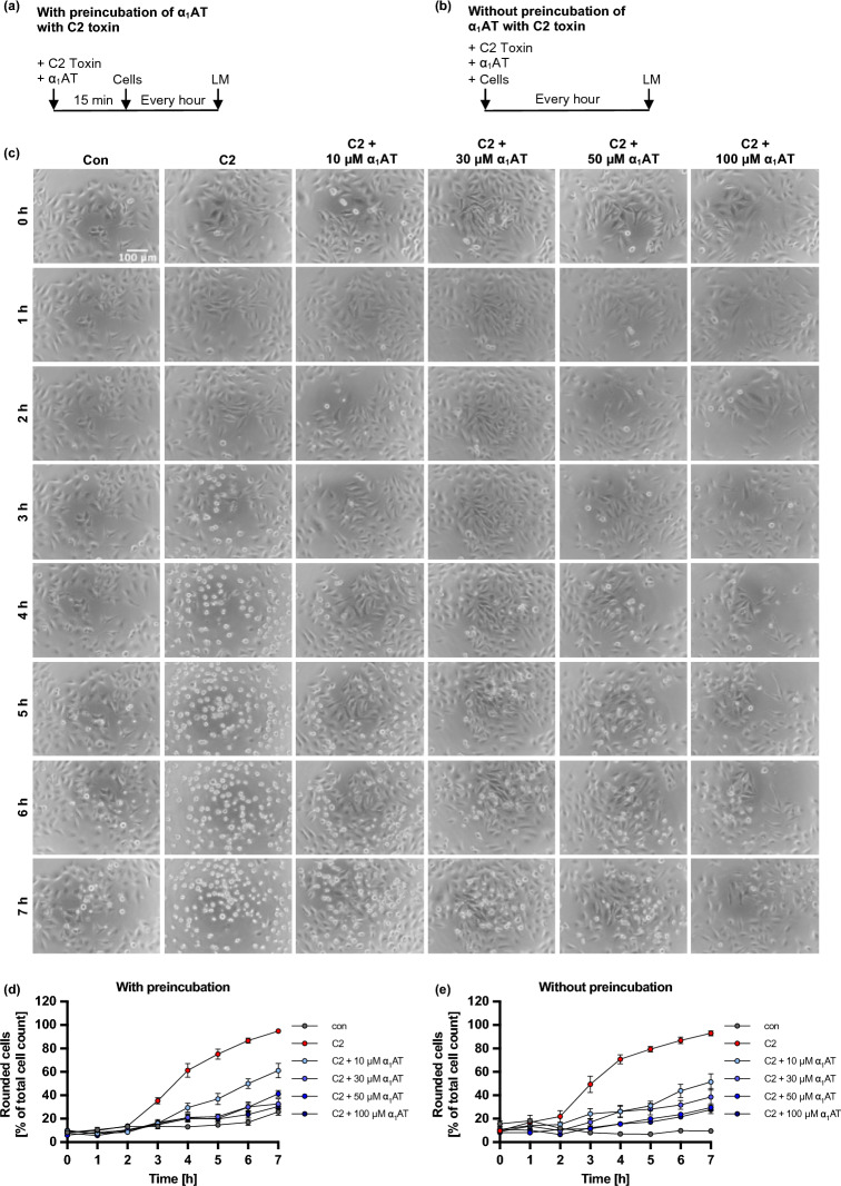
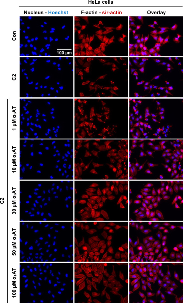
The bacterium Clostridium botulinum, well-known for producing botulinum neurotoxins, which cause the severe paralytic illness known as botulism, produces C2 toxin, a binary AB-toxin with ADP-ribosyltranferase activity. C2 toxin possesses two separate protein components, an enzymatically active A-component C2I and the binding and translocation B-component C2II. After proteolytic activation of C2II to C2IIa, the heptameric structure binds C2I and is taken up via receptor-mediated endocytosis into the target cells. Due to acidification of endosomes, the C2IIa/C2I complex undergoes conformational changes and consequently C2IIa forms a pore into the endosomal membrane and C2I can translocate into the cytoplasm, where it ADP-ribosylates G-actin, a key component of the cytoskeleton. This modification disrupts the actin cytoskeleton, resulting in the collapse of cytoskeleton and ultimately cell death. Here, we show that the serine-protease inhibitor α1-antitrypsin (α1AT) which we identified previously from a hemofiltrate library screen for PT from Bordetella pertussis is a multitoxin inhibitor. α1AT inhibits intoxication of cells with C2 toxin via inhibition of binding to cells and inhibition of enzyme activity of C2I. Moreover, diphtheria toxin and an anthrax fusion toxin are inhibited by α1AT. Since α1AT is commercially available as a drug for treatment of the α1AT deficiency, it could be repurposed for treatment of toxin-mediated diseases.

Keywords: Clostridium botulinum C2 toxin; Anthrax toxin; Diphtheria toxin; Drug repurposing; Toxin inhibitor; α1-Antitrypsin.


Conflict of interest statement

The authors declare no competing interests.

Figures

Fig. 1
Fig. 1
Effect of α1AT on C2-toxin mediated cell rounding of HeLa cells. (a, b) Schematic representation of experimental setup for the cell morphology assay. C2 toxin and α1AT were either preincubated for 15 min before addition to cells (a) or added simultaneously (b). Then, cells were incubated for 7 h at 37 °C, and pictures were taken every hour using the light microscope (LM). (ce) Different concentrations of α1AT or the respective amount of its solvent (H2O) were preincubated for 15 min at RT with C2 toxin (C2 toxin = C2I/C2IIa: 100/200 ng/ml) in FCS-free medium before addition to HeLa cells (c, d) or added directly (e). The cells were incubated for 7 h at 37 °C, and pictures were taken every hour. Rounded cells are given as percent of the total cell count, mean + /− SEM (at least n = 6 and at most n = 9 values from three independent experiments). (c) Representative pictures are shown for a representative experiment where C2 toxin was preincubated for 15 min with different concentrations of α1AT before addition to HeLa cells.
Fig. 2
Fig. 2
Effect of α1AT on F-actin during intoxication of HeLa cells with C2 toxin. C2 toxin (C2I/C2IIa: 100/200 ng/ml) and different concentrations α1AT or the respective amount of solvent (H2O) were added directly to HeLa cells and incubated for 4 h at 37 °C. Cells were left untreated as control (Con). Subsequently, the cells were washed, fixed, permeabilized, and quenching was performed. Blocking was performed and F-actin was stained using sir-actin (red), and nuclei were stained using Hoechst (blue). Representative images are shown from three independent experiments (n = 3).
Fig. 3
Fig. 3
Effect of α1AT on enzyme activity of C2I in vitro. (a) Schematic representation of experimental setup for the enzyme activity assay. C2I and α1AT were directly added to HeLa cell lysate and biotin-NAD+, and incubated for 30 min at 37 °C. (bd) C2I (20 fmol = 0.001 µM) and different concentrations α1AT or the respective amount of solvent (H2O) (Con) were added directly to HeLa cell lysate and biotin-NAD+ and incubated for 30 min at 37 °C. Cell lysate was left untreated with biotin-NAD+ as further control (Lysate). G-Actin which was ADP-ribosylated and biotin-labeled via the incubation with C2I and biotin-labeled NAD+ was detected via Western Blot (WB), whereas Hsp90 or Ponceau-S staining served as control for equal protein loading. The bar graph (b) shows the quantifications of Western Blot signals from nine independent experiments, while (c, d) show blots of representative experiments. The intensity values of the bar graph are given as x-fold of the control (con), normalized to Hsp90 or Ponceau-S staining, mean + /− SEM (at least n = 4 at most n = 16 values from nine independent experiments). (b) Significance was tested using one-way ANOVA followed by Dunnett’s multiple comparison test and refers to untreated controls (con) (* p < 0.1, ** p < 0.01, *** p < 0.001, **** p < 0.0001, ns not significant).
Fig. 4
Fig. 4
Effect of α1AT on binding of labeled C2 toxin to HeLa cells. (ad) 488-labeled C2IIa (800 ng/ml) and 405-labeled C2I (800 ng/ml) and 100 µM of α1AT or the respective amount of solvent (H2O) were added directly to HeLa cells and incubated for 15 min at 4 °C to enable binding of C2 toxin to cells but no internalization. Cells were left untreated (PBS controls from labeling process) as control. After that, cells were washed by centrifugation and 488-labeled C2IIa (a) and 405-labeled C2I (c) bound to cell surfaces was measured using flow cytometry. Values of median are given as x-fold of the untreated control (Con), mean + /− SEM (n = 9 from three independent experiments). (b, d) Histograms show fluorescence intensity of cells for one representative experiment. (ad) Significance was tested using one-way ANOVA followed by Dunnett’s multiple comparison test and refers to toxin-treated controls (* p < 0.1, ** p < 0.01, *** p < 0.001, **** p < 0.0001, ns not significant).
Fig. 5
Fig. 5
Effect of α1AT on binding of C2 toxin to HeLa cells. (a) Schematic representation of experimental setup for the cell morphology binding assay. The toxin and α1AT were added directly to the cells and were incubated for 40 min at 4 °C. Then, medium exchange to FCS-free medium was performed. The cells were incubated for 7 h at 37 °C, and pictures were taken every hour using the light microscope. (b) C2 toxin (C2I/C2IIa: 100/200 ng/ml) and different concentrations of α1AT or the respective amount of solvent (H2O) were added in FCS-free medium to HeLa cells and incubated for 40 min at 4 °C to enable C2 binding but not internalization. After that medium exchange to FCS-free medium was performed. The cells were incubated for 7 h at 37 °C, and pictures were taken every hour. Rounded cells are given as percent of the total cell count, mean + /− SEM (for each time point n = 9 values from three independent experiments). (c) C2 toxin (C2IIa-488/C2I: 33.2/20 nM) and different concentrations α1AT or the respective amount of solvent (H2O) were mixed, centrifuged, and supernatants were added directly to HeLa cells and incubated for 40 min at 4 °C to enable C2 binding but not internalization. Cells were left untreated as control (Con) or treated with respective amount of PBS-488 (control from toxin labeling) and solvent (H2O) (Con + PBS-488). Subsequently, the cells were washed, fixed, permeabilized (as indicated), and quenching was performed. Blocking was performed, and the cells were incubated with a primary antibody for α1AT (purple). Primary antibody was detected via a fluorescently labeled secondary antibody, F-actin was stained using sir-actin (red), and nuclei were stained using Hoechst (blue). Representative images are shown from three independent experiments (n = 3).
Fig. 6
Fig. 6
Effect of α1AT on endocytosed C2IIa-488 signal in HeLa cells. C2 toxin (C2IIa-488/C2I: 33.2/20 nM) and different concentrations α1AT or the respective amount of solvent (H2O) were mixed, centrifuged, and supernatants were added directly to HeLa cells and incubated for 30 min at 37 °C. Cells were left untreated as control (Con) or treated with respective amounts of PBS-488 (control from toxin labeling) and solvent (H2O) (Con + PBS-488). Subsequently, the cells were washed, fixed, permeabilized (as indicated), and quenching was performed. Blocking was performed, and the cells were incubated with a primary antibody for α1AT (purple). Primary antibody was detected via a fluorescently labeled secondary antibody, F-actin was stained using sir-actin (red), and nuclei were stained using Hoechst (blue). Labelled C2IIa is shown in green. Representative images are shown from three independent experiments (n = 3).
Fig. 7
Fig. 7
Effect of α1AT on other bacterial AB-toxins. (a) Schematic representation of experimental setup for the cell morphology assay. The toxins and α1AT were preincubated for 15 min before addition to cells. The cells were incubated for 7 h at 37 °C, and pictures were taken every hour using the light microscope (LM). (bi) Different concentrations of α1AT or the respective amount of solvent (H2O) were preincubated for 15 min at RT with the respective toxin in FCS-free medium before addition to HeLa (b, c, e, f, h, i) or Vero (d, f) cells. Toxin concentrations: nDT: 800 ng/ml, DT 5 nM, TcdA: 180 pM, TcdB: 10 pM, CDT = His_CDTa/CDTb: 5/2 nM, C2IIa + His_TcdB-GTD: 30 nM + 150 nM, PA63 + His_TcdB-GTD: 10 nM + 50 nM, PA63 + LFN-DTA: 0.5 nM + 0.25 nM. The cells were incubated for 7 h at 37 °C, and pictures were taken every hour. Rounded cells are given as percent of the total cell count, mean + /− SEM (at least n = 7 and at most n = 12 from three to four independent experiments).

Similar articles

Cited by

  • Alpha-1 antitrypsin inhibits pertussis toxin.
    Lietz S, Sommer A, Sokolowski LM, Kling C, Rodríguez Alfonso AA, Preising N, Alpízar-Pedraza D, King J, Streit L, Schröppel B, van Erp R, Barth E, Schneider M, Münch J, Michaelis J, Ständker L, Wiese S, Barth H, Pulliainen AT, Scanlon K, Ernst K. Lietz S, et al. J Biol Chem. 2024 Dec;300(12):107950. doi: 10.1016/j.jbc.2024.107950. Epub 2024 Oct 30. J Biol Chem. 2024. PMID: 39481600 Free PMC article.
  • The antimicrobial peptide Angie 5 inhibits TcdA and TcdB from Clostridioides difficile.
    Lietz S, Sokolowski LM, Lindner K, Rodríguez AA, Ständker L, Vogel V, Spellerberg B, Stenger S, Alpízar-Pedraza D, Ernst K, Papatheodorou P, Barth H. Lietz S, et al. Cell Mol Life Sci. 2025 Jun 30;82(1):265. doi: 10.1007/s00018-025-05799-2. Cell Mol Life Sci. 2025. PMID: 40586877 Free PMC article.

References

    1. Stiles, B. G., Wigelsworth, D. J., Popoff, M. R. & Barth, H. Clostridial binary toxins: Iota and C2 family portraits. Front. Cell Infect. Microbiol.10.3389/fcimb.2011.00011 (2011). 10.3389/fcimb.2011.00011 - DOI - PMC - PubMed
    1. Ohishi, I. Lethal and vascular permeability activities of botulinum C2 toxin induced by separate injections of the two toxin components. Infect. Immun.40, 336–339 (1983). 10.1128/iai.40.1.336-339.1983 - DOI - PMC - PubMed
    1. Ohishi, I. Response of mouse intestinal loop to botulinum C2 toxin: Enterotoxic activity induced by cooperation of nonlinked protein components. Infect. Immun.40, 691–695 (1983). 10.1128/iai.40.2.691-695.1983 - DOI - PMC - PubMed
    1. Ohishi, I., Iwasaki, M. & Sakaguchi, G. Purification and characterization of two components of botulinum C2 toxin. Infect. Immun.30, 668–673 (1980). 10.1128/iai.30.3.668-673.1980 - DOI - PMC - PubMed
    1. Aktories, K. et al. Botulinum C2 toxin ADP-ribosylates actin. Nature322, 390–392 (1986). 10.1038/322390a0 - DOI - PubMed

MeSH terms

LinkOut - more resources