. 2024 Nov;38(11):2429-2442.
doi: 10.1038/s41375-024-02379-4. Epub 2024 Sep 11.

The non-canonical BAF chromatin remodeling complex is a novel target of spliceosome dysregulation in SF3B1-mutated chronic lymphocytic leukemia

Affiliations

The non-canonical BAF chromatin remodeling complex is a novel target of spliceosome dysregulation in SF3B1-mutated chronic lymphocytic leukemia

Daniel Hägerstrand et al. Leukemia. 2024 Nov.

Abstract

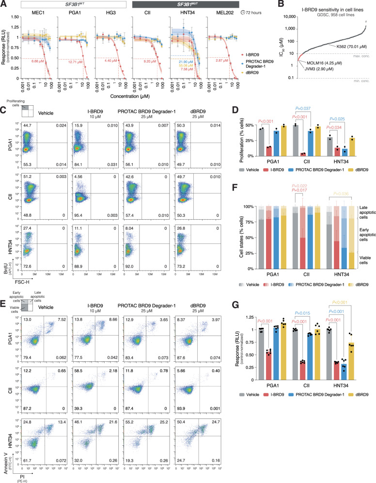
SF3B1 mutations are recurrent in chronic lymphocytic leukemia (CLL), particularly enriched in clinically aggressive stereotyped subset #2. To investigate their impact, we conducted RNA-sequencing of 18 SF3B1MUT and 17 SF3B1WT subset #2 cases and identified 80 significant alternative splicing events (ASEs). Notable ASEs concerned exon inclusion in the non-canonical BAF (ncBAF) chromatin remodeling complex subunit, BRD9, and splice variants in eight additional ncBAF complex interactors. Long-read RNA-sequencing confirmed the presence of splice variants, and extended analysis of 139 CLL cases corroborated their association with SF3B1 mutations. Overexpression of SF3B1K700E induced exon inclusion in BRD9, resulting in a novel splice isoform with an alternative C-terminus. Protein interactome analysis of the BRD9 splice isoform revealed augmented ncBAF complex interaction, while exhibiting decreased binding of auxiliary proteins, including SPEN, BRCA2, and CHD9. Additionally, integrative multi-omics analysis identified a ncBAF complex-bound gene quartet on chromosome 1 with higher expression levels and more accessible chromatin in SF3B1MUT CLL. Finally, Cancer Dependency Map analysis and BRD9 inhibition displayed BRD9 dependency and sensitivity in cell lines and primary CLL cells. In conclusion, spliceosome dysregulation caused by SF3B1 mutations leads to multiple ASEs and an altered ncBAF complex interactome, highlighting a novel pathobiological mechanism in SF3B1MUT CLL.


Conflict of interest statement

NEK advisory board: AbbVie, AstraZeneca, BeiGene, Behring, Cytomx Therapy, Dava Oncology, Janssen, Juno Therapeutics/Celgene, ONCOtracker, Pharmacyclics, and Targeted Oncology; research funding: AbbVie, Acerta Pharma/AstraZeneca, Bristol Meyer Squib, Celgene, Genentech, MEI Pharma, Pharmacyclics, Sunesis, TG Therapeutics, and Tolero Pharmaceuticals. AWL honoraria: Janssen and Roche. PG honoraria/advisory board: AbbVie, Acerta Pharma/AstraZeneca, Adaptive, ArQule/MSD, BeiGene, Juno Therapeutics/Celgene, Gilead, Janssen, Loxo Oncology/Lilly, and Sunesis; research funding: AbbVie, Gilead, Janssen, Novartis, and Sunesis. KS honoraria: Janssen, Abbvie, and AstraZeneca; research funding: Janssen, Gilead Sciences, and AbbVie. LAS honoraria: AbbVie and Janssen. RR honoraria: AbbVie, AstraZeneca, Janssen, Illumina, and Roche. The other authors declare no competing interests.

Figures

Fig. 1
Fig. 1. Alternative splicing analysis of SF3B1-mutated subset #2 CLL.
A ASE categories, including skipped exon (SE), alternative 3′ splice site usage (A3SS), alternative 5′ splice site usage (A5SS), mutually exclusive exon usage (MXE), and intron retention (RI). B Bar plot displaying numbers of all identified ASEs in the comparison of 18 SF3B1MUT and 17 SF3B1WT subset #2 cases across different ASE categories. 163,097 ASEs were detected and involved transcripts of 10,437 genes. C Volcano plot depicting all identified ASEs in the comparison of SF3B1MUT and SF3B1WT subset #2 cases. Red dots indicate ASEs that are considered significant (|ΔPSI | ≥ 20% and FDR ≤ 0.01; 80 ASEs). D Bar plot displaying numbers of significant ASEs in the comparison of SF3B1MUT and SF3B1WT subset #2 cases across different ASE categories. 80 ASEs were considered significant and involved transcripts of 62 genes. E Heatmap illustrating the individual PSI values for the 80 significant ASEs detected in the comparison of SF3B1MUT and SF3B1WT subset #2 cases. 33 ASEs occurred within the same gene at least twice. ASEs are displayed based on the ASE category. The bar plot to the right of the heatmap shows ΔPSI values. The gene affected by each ASE and the corresponding unique ASE ID are listed. Genes that encode ncBAF complex-interacting proteins are depicted in color. For multiple ASEs per gene, only the top ASE is colored, while the others are marked with black asterisks. Additional details of these ASEs are provided in Supplementary Table 3A, B. F Model of the ncBAF chromatin remodeling complex and previously reported ncBAF complex interactors. Alternatively spliced transcripts identified in SF3B1MUT subset #2 CLL that encode ncBAF complex-related proteins are depicted in color. G Scattered box plot showing the PSI value distribution, ΔPSI values, and P values (Wilcoxon rank-sum test) for significant ncBAF complex-related ASEs identified in the comparison of SF3B1MUT and SF3B1WT subset #2 cases. The specific ASEs for ZEB1, BRD9, PLSCR1, TENT4B, CXXC1, DCAF16, UBP1, DLST, and SERBP1 have the unique ASE IDs ZEB1_SE_27141, BRD9_SE_53886, PLSCR1_SE_92684, PAPD5_SE_94061, CXXC1_SE_102397, DCAF16_SE_4627, UBP1_SE_85897, DLST_A3SS_2409, and SERBP1_A3SS_1321, respectively, in Supplementary Table 3A, B. The boxes represent the median and the interquartile range (IQR), while the whiskers extend to 1.5 times the IQR from the first and third quartiles. ASE: alternative splicing event; WT: wildtype; MUT: mutated; FDR: false discovery rate; PSI: percent spliced in; VAF: variant allele frequency.
Fig. 2
Fig. 2. Long-read RNA-sequencing validates the predicted alternative transcripts related to the ncBAF complex in SF3B1-mutated CLL.
Sashimi plots illustrating the identified ASEs and alternative splicing patterns in four genes that encode ncBAF complex-related proteins in SF3B1WT versus SF3B1MUT CLL; ZEB1 and BRD9 exhibit the top two ASEs in the SE category, while DLST and SERBP1 showcase the top two ASEs in the A3SS category. Sashimi plots for other ncBAF complex-related genes, PLSCR1, TENT4B, CXXC1, DCAF16, and UBP1, are presented in Supplementary Fig. 3. For each gene, the top two sashimi plots within the gray box illustrate the predicted splice variants in SF3B1WT versus SF3B1MUT CLL. The colored arc highlights the primary ASE, while lighter arcs represent additional ASEs if present. The gene map indicates the relative location and order of the detected exons in relation to the sequencing results. For each corresponding gene, the lower two sashimi plots show the coverage and splice junction count data from the aligned long-read RNA-seq data from an SF3B1WT case (RS24) and an SF3B1MUT case (RS55), both belonging to subset #2 CLL. The direction of the genes is arranged from left to right. WT: wildtype; MUT: mutated; PSI: percent spliced in.
Fig. 3
Fig. 3. SF3B1 mutations induce alternative splicing of BRD9 resulting in a splice isoform with an alternative C-terminus.
A Scheme illustrating lentiviral constructs for SF3B1WT and SF3B1K700E overexpression under the control of a DOX-inducible promoter. B Bar plots depicting expression levels of endogenous and exogenous SF3B1 upon overexpression of SF3B1WT and SF3B1K700E in MEC1 and PCL12 cell lines. The bar plots display the mean values of triplicates with error bars representing the standard deviation. C Scheme illustrating the identified ASE in BRD9 with the regular and alternative splicing pattern. The blue and red boxes illustrate constant and alternative exons, respectively. D Increased expression of the alternative BRD9 transcript upon overexpression of SF3B1K700E as compared to SF3B1WT in MEC1 and PCL12 cell lines. The checkerboard scheme at the top of the panel illustrates the experimental conditions. The bar plots display the expression levels of BRD9 transcripts in general (transcripts containing exon 14), the regular BRD9 transcript (transcripts containing exon 14 followed by exon 16), and the alternative BRD9 transcript (transcripts containing exon 14 followed by exon 15) as determined by qPCR. Results are normalized on GAPDH levels and expressed as fold difference between DOX+ and DOX− conditions with corresponding P values (Student’s t-test). The bar plots display the mean values of experimental triplicates, with error bars representing the standard deviation. E Agarose gel electrophoresis images of PCR products spanning from exon 14 to 16 and allowing for exon 15 inclusion show a higher abundance of alternative BRD9 transcript in SF3B1K700E DOX+. For a complete gel image see Supplementary Fig. 13A. F Assessment of BRD9 splice variants in SF3B1WT and SF3B1MUT CLL, AML, and UVM cell lines. MEC1, PGA1, and HG3 are SF3B1WT CLL cell lines, CII an SF3B1K666E CLL cell line, HNT34 an SF3B1K700E AML cell line, and MEL202 an SF3B1R625G UVM cell line. The bar plots display the expression levels of BRD9 transcripts in general, the regular BRD9 transcript, and the alternative BRD9 transcript as determined by qPCR. Results for BRD9 transcripts in general are normalized on GAPDH levels, whereas regular and alternative BRD9 transcript levels are normalized on levels of BRD9 transcripts in general and expressed as fold difference between MEC1 and other cell lines with corresponding P values (Student’s t-test). The bar plots display the mean values of experimental triplicates, with error bars representing the standard deviation. G Agarose gel electrophoresis image of PCR products spanning from exon 14 to 16 and allowing for exon 15 inclusion shows a higher abundance of alternative BRD9 transcript in SF3B1MUT cell lines. For a complete gel image see Supplementary Fig. 13A. H Sashimi plots from the SF3B1WT CLL cell line HG3 and the SF3B1MUT AML and UVM cell lines, HNT34 and MEL202, respectively, illustrating the alternatively spliced exon 15 in BRD9. The plots for HG3 are based on direct long-read RNA-seq, and HNT34 (SRR8616208) and MEL202 (SRR12354765) on short-read RNA-seq. I BRD9 splice isoform expression in SF3B1WT and SF3B1MUT CLL cell lines determined by isoform-specific antibodies. An antibody specific to the C-terminus of the regular BRD9 isoform detected a single band in both SF3B1WT and SF3B1MUT cell lines. Conversely, an antibody targeting a constant epitope within the central part of BRD9, detected two bands in the SF3B1MUT cell line CII and one band in the SF3B1WT cell lines MEC1 and PGA1, corresponding to the regular higher molecular weight BRD9 isoform and the alternative lower molecular weight BRD9 isoform. For complete blot images see Supplementary Fig. 13A. WT: wildtype; MUT: mutated; DOX: doxycycline.
Fig. 4
Fig. 4. The regular and alternative BRD9 isoforms display different interactomes.
A Scheme illustrating BRD9 splice variants and predicted protein isoforms according to NCBI RefSeq (Annotation release GCF_000001405.40-RS_2023_03). The zoom-in view highlights the predicted protein sequence stemming from exon 15 inclusion in BRD9. Upon translation of exon 15, a stop codon emerges near the end of the exon, resulting in a shorter splice isoform with an alternative C-terminus. B Scheme depicting lentiviral constructs for overexpression of the regular and alternative BRD9 splice variants. The corresponding DNA and protein sequences are available in Supplementary Information Appendix 1. C Heatmap illustrating the detected ncBAF complex subunits upon co-immunoprecipitation and subsequent mass spectrometry analysis with the regular or alternative BRD9 isoforms stably overexpressed in the HEK293T cell line. The relative protein levels are based on the total number of identified peptide-spectrum matches (PSMs) for the corresponding protein from the mass spectrometry analysis. The ncBAF complex model to the right of the heatmap illustrates that the alternative BRD9 isoform precipitated a majority of the ncBAF complex subunits. ACTB was not detected among the co-immunoprecipitates. The control cells expressing FLAG-V5-tagged YFP were used. D Volcano plot displaying differential interaction analysis of proteins that selectively interacted more with the regular or alternative BRD9 isoforms (multiple Student’s t-tests with Benjamini–Hochberg multiple testing correction). The top 5 candidates are highlighted in red. E Venn diagram showing a comparison of the significant differentially interacting proteins with the regular or alternative BRD9 isoforms and BRD9-interacting proteins reported by Gaudio et al. [46]. The intersecting proteins are SPEN, BRCA2, and CHD9. F Western blot analysis of V5-tag and BICRA co-immunoprecipitates in the HEK293T cell line with stable overexpression of the regular and alternative BRD9 isoforms. In the V5-tag-immunoprecipitations, both overexpressed BRD9 isoforms precipitated BICRA together with SMARCA4 and SMARCC1. Reciprocal BICRA co-immunoprecipitations precipitated both overexpressed BRD9 isoforms, SMARCA4 and SMARCC1. The control cells were transduced with an empty lentiviral vector. For complete blot images see Supplementary Fig. 13A. G Bar plots showing V5-tag and BICRA co-immunoprecipitation efficiency with BRD9, BICRA, SMARCA4, and SMARCC1. Results are expressed as fold differences between the regular and alternative BRD9 isoforms with corresponding P values (Student’s t-test). The bar plots display the mean values from three repeated experiments, with error bars representing the standard deviation. For detailed quantification and calculation see Supplementary Fig. 13B. PSM: peptide-spectrum matches; FDR: false discovery rate; IP: immunoprecipitation.
Fig. 5
Fig. 5. BRD9 binds a gene quartet on chromosome 1 and displays selective dependency in DepMap.
A Volcano plot depicting differentially expressed genes between 18 SF3B1MUT and 17 SF3B1WT subset #2 cases with 22 downregulated and 55 upregulated genes ( | log2FC | ≥ 0.58 and FDR < 0.01). B Unsupervised clustermap illustrating the distinct clustering of SF3B1MUT and SF3B1WT subset #2 cases based on differential gene expression. The four neighboring genes, NOL9, TAS1R1, ZBTB48, and KLHL21, located on chromosome 1 are depicted in color. The Ward method and the Euclidean metric were employed. C Karyoplot of chromosome 1 displaying differentially expressed genes between SF3B1MUT and SF3B1WT subset #2 cases. Examination of chromosomal positions revealed four neighboring genes, NOL9, TAS1R1, ZBTB48, and KLHL21. D Venn diagram showing the overlap of genes with differential expression between SF3B1MUT and SF3B1WT cases in three independent datasets; subset #2 CLL, ICGC CLLE-ES [30, 31], and TCGA CLL [29, 33, 34]. For the differential gene expression analyses of the latter two datasets see Supplementary Fig. 10A–E. ZBTB48 appeared upregulated in all three datasets, while TAS1R1 in subset #2 CLL and ICGC CLLE-ES. E Venn diagram showing the overlap of genes with higher expression levels in SF3B1MUT subset #2 cases and increased chromatin accessibility in SF3B1MUT CLL cases from Beekman et al. [47]. The overlap comprises NOL9, TAS1R1, ZBTB48, and KLHL21. F Coverage tracks from ChIP-seq data sourced from the ENCODE database [35, 36], based on the CML cell line K562, illustrating the binding of BRD9 and the ncBAF complex subunits SMARCA4 and SMARCC2 to the gene quartet region on chromosome 1. The promoters for these genes appear active based on the signal for H3K27ac, H3K4me3, H3K9ac, and H3K79me2, and transcriptionally active based on the H3K36me3 signal, whereas transcriptional repression is low based on the low signal for H3K27me3 and H3K9me3. G Gene essentiality map showing the relationship between efficacy and selectivity of genes in shinyDepMap [37, 38]. SF3B1 appears as a non-selective dependency with high efficacy, while BRD9 as a selective dependency with high efficacy. BRD9 is in the dependency region of PIK3R1, EZH2, and FBXW7. H Functional similarity clustering showing a chromatin remodeling dependency cluster connected to the BRD9 dependency signature in shinyDepMap. BRD9 clusters with SMARCD1, a BAF complex subunit, subunits of the Mediator complex, p300/CBP subunits, and subunits of the SAGA complex. I BRD9 dependency ranking of the 1086 DepMap (22Q2) cell lines. For CLL cell lines, gene effect scores are given within the parenthesis. WT: wildtype; MUT: mutated; FDR: false discovery rate; FC: fold change.
Fig. 6
Fig. 6. BRD9 inhibition exhibits anti-proliferative and pro-apoptotic effects in SF3B1-mutated cell lines.
A Dose-response analysis of I-BRD9, PROTAC BRD9 Degrader-1, and dBRD9 treatments in 3 SF3B1WT cell lines, MEC1, PGA1, and HG3 (all CLL), and 3 SF3B1MUT cell lines, CII (CLL), HNT34 (AML), and MEL202 (UVM). The cell lines were treated with drug concentrations ranging from 0.001 to 50 µM for 3 days, and cell viability was determined by CellTiter-Glo 2.0. Complete cell killing was exclusively observed with I-BRD9 treatment, allowing for the determination of corresponding IC50 values for each cell line. The HNT34 cell line exhibited sensitivity to all three drugs, thereby allowing for the determination of IC50 values for all conditions. Dose-response curves are shown with 95% confidence intervals, while individual dots display the mean values of triplicates, with error bars representing the standard deviation. B I-BRD9 sensitivity profile in 958 cancer cell lines from GDSC [39]. C Assessment of proliferation in an SF3B1WT cell line, PGA1, and SF3B1MUT cell lines, CII and HNT34, upon treatment with 10 µM I-BRD9, 25 µM PROTAC BRD9 Degrader-1, or 25 µM dBRD9. Cell lines were treated for 3 days with a 5-hour exposure to BrdU at the end, and proliferation was quantified by flow cytometry, measured as BrdU+ cells. Vehicle (DMSO) was used as the negative control for drug treatment, while cells not exposed to BrdU served as the negative control to assess the specificity of the anti-BrdU antibody. The upper left quadrants (BrdU+ cells) in the density plots represent the percentages of proliferating cells. D Bar plot displaying differences in the percentages of proliferating cells compared to negative controls with corresponding P values (one-way ANOVA). The bar plot displays the mean values from two repeated experiments, with error bars representing the standard deviation. E Assessment of apoptosis in the same cell lines and the identical samples as in (C). Treatment with 5 µM Camptothecin and vehicle (DMSO) were used as the positive and negative controls, respectively. Apoptosis was evaluated by Annexin V/PI staining and subsequent flow cytometry analysis. The lower left (Annexin V-/PI- cells), upper left (Annexin V+/PI- cells), and upper right (Annexin V+/PI+ cells) quadrants in the density plots represent the percentages of viable, early apoptotic, and late apoptotic cells, respectively. F Stacked bar plot displaying differences in the percentages of viable, early apoptotic, and late apoptotic cells compared to negative controls with corresponding P values (one-way ANOVA). The bar plot displays the mean values from two repeated experiments, with error bars representing the standard deviation. G Bar plot showing cell viability differences as determined by CellTiter-Glo 2.0 in the same cell lines under the same experimental conditions as in panels (C, E) with corresponding P values (one-way ANOVA). The bar plot displays the mean values from two repeated experiments in triplicates, with error bars representing the standard deviation. WT: wildtype; MUT: mutated; IC50: half-maximal inhibitory concentration; RLU: relative luminescence unit.
Fig. 7
Fig. 7. BRD9 inhibition induces potent pro-apoptotic effects in primary CLL cells.
A Dose-response analysis of I-BRD9 treatment in 3 SF3B1WT (SKL48, SKL152, SKL53) and 3 SF3B1MUT (SKL147, SKL47, SKL157) primary CLL cell samples. The cell lines were treated with drug concentrations ranging from 0.001 to 50 µM for 2 days, and cell viability was determined by CellTiter-Glo 2.0. Dose-response curves are shown with 95% confidence intervals, while individual dots display the mean values of triplicates, with error bars representing the standard deviation. B Stacked density plots showing apoptosis assessment in the same primary CLL cell samples as in (A). Treatment with 5 µM Camptothecin and vehicle (DMSO) were used as the positive and negative controls, respectively. Apoptosis was evaluated by Annexin V/PI staining and subsequent flow cytometry analysis. The lower left (Annexin V-/PI- cells), upper left (Annexin V+/PI- cells), and upper right (Annexin V+/PI+ cells) quadrants in the density plots represent the percentages of viable, early apoptotic, and late apoptotic cells, respectively. C Stacked bar plot displaying differences in the percentages of viable, early apoptotic, and late apoptotic cells compared to negative controls with corresponding P values (Student’s t-test). The bar plot displays the mean values, with error bars representing the standard deviation. WT: wildtype; MUT: mutated; IC50: half-maximal inhibitory concentration; RLU: relative luminescence unit.

Similar articles

Cited by

References

    1. Wahl MC, Will CL, Lührmann R. The Spliceosome: design principles of a dynamic RNP machine. Cell. 2009;136:701–18. - PubMed
    1. DeBoever C, Ghia EM, Shepard PJ, Rassenti L, Barrett CL, Jepsen K, et al. Transcriptome sequencing reveals potential mechanism of cryptic 3’ splice site selection in SF3B1-mutated cancers. PLoS Comput Biol. 2015;11:e1004105. - PMC - PubMed
    1. Wang L, Brooks AN, Fan J, Wan Y, Gambe R, Li S, et al. Transcriptomic characterization of SF3B1 mutation reveals its pleiotropic effects in chronic lymphocytic leukemia. Cancer Cell. 2016;30:750–63. - PMC - PubMed
    1. Mansouri L, Thorvaldsdottir B, Sutton L-A, Karakatsoulis G, Meggendorfer M, Parker H, et al. Different prognostic impact of recurrent gene mutations in chronic lymphocytic leukemia depending on IGHV gene somatic hypermutation status: a study by ERIC in HARMONY. Leukemia. 2023;37:339–47. - PMC - PubMed
    1. Quesada V, Conde L, Villamor N, Ordóñez GR, Jares P, Bassaganyas L, et al. Exome sequencing identifies recurrent mutations of the splicing factor SF3B1 gene in chronic lymphocytic leukemia. Nat Genet. 2012;44:47–52. - PubMed

MeSH terms

LinkOut - more resources