. 2024 Sep 11;14(1):21252.
doi: 10.1038/s41598-024-72272-8.

FOS mapping reveals two complementary circuits for spatial navigation in mouse

Affiliations

FOS mapping reveals two complementary circuits for spatial navigation in mouse

Edyta Balcerek et al. Sci Rep. .

Abstract

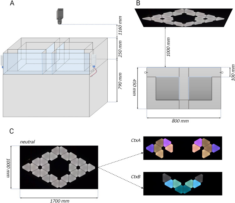
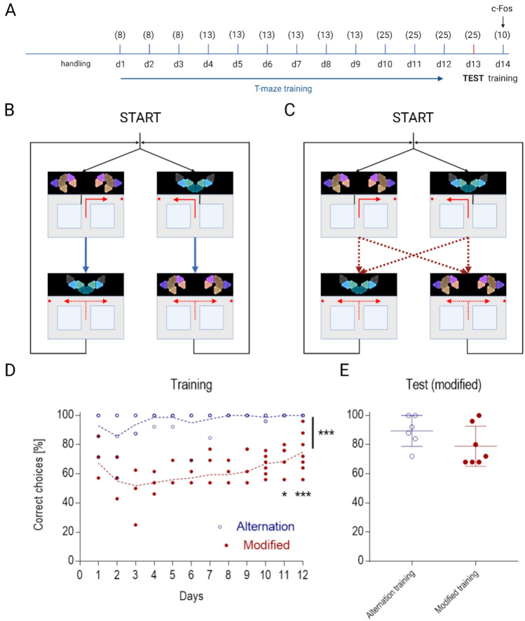
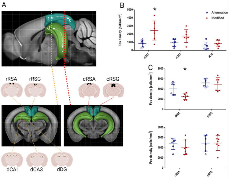
Here, we show that during continuous navigation in a dynamic external environment, mice are capable of developing a foraging strategy based exclusively on changing distal (allothetic) information and that this process may involve two alternative components of the spatial memory circuit: the hippocampus and retrosplenial cortex. To this end, we designed a novel custom apparatus and implemented a behavioral protocol based on the figure-8-maze paradigm with two goal locations associated with distinct contexts. We assessed whether mice are able to learn to retrieve a sequence of rewards guided exclusively by the changing context. We found out that training mice in the apparatus leads to change in strategy from the internal tendency to alternate into navigation based exclusively on visual information. This effect could be achieved using two different training protocols: prolonged alternation training, or a flexible protocol with unpredictable turn succession. Based on the c-FOS mapping we also provide evidence of opposing levels of engagement of hippocampus and retrosplenial cortex after training of mice in these two different regimens. This supports the hypothesis of the existence of parallel circuits guiding spatial navigation, one based on the well-described hippocampal representation, and another, RSC-dependent.


Conflict of interest statement

The authors declare no competing interests.

Figures

Fig. 1
Fig. 1
Figure-8-maze apparatus (see text for details) (A) Schematic front view with camera and reward dispenser locations. (B) Schematic top view with LED panels placed 1000 mm ahead of the transparent, front wall showing neutral context used for habituation protocol as well as for starting and ending of each daily session of behavioral procedure. Transparent walls are light blue colored. All inner doors are pneumatically movable in order to enable continuous movement of the animals and to force a desired turn direction when necessary. (C) Realistic view of displayed visual cues. Neutral context (left) showing the full array of triangular LED panels, CtxA (right, top) associated with right turn, CtxB (right, down), associated with left turn.
Fig. 2
Fig. 2
Behavioral experiment. (A) Timeline representation of the experimental design (see Methods for details). Numbers in brackets indicate subsequent numbers of forced-choice trials on each day of spaced training. The final number of right and left turns (corresponding with visual cue) was equal on each day. (B) Schematic representation of “Alternation” training strategy. A rigid order of turns was imposed. (C) Schematic representation of “Modified” training strategy. Any sequence of turn directions was possible between forced and choice phase while maintaining an equal proportion of every possible sequence. (D) Learning curve for “Alternation” and “Modified” groups. Data points show the percentage of rewarded decisions (congruent with the displayed context) by individual animals in each session. Significant effects labeled: (***) training effect, F(1,11) = 1.499, P < 0.0001, two-way repeated measures (RM) ANOVA; training day for modified group: (*) day 11 (P = 0.023), (***) day 12 (P = 0.0002), post hoc Tukey test. (E) Test session in “Modified” layout. Data points show the percentage of decisions congruent with the displayed context.
Fig. 3
Fig. 3
Comparison of c-FOS-positive cell density between training protocols across brain regions. (A) Visualization of the position of central slices for each region of interest in the mouse brain using Allen Brain Atlas. In the upper panel, the hippocampus and retrosplenial cortex are marked with colors, and the rostro-caudal position of the slices is indicated. The lower panel shows the position of ROIs for c-FOS positive cell density collection on the coronal slices. (B) Density of c-FOS immunopositive cells in the ROIs of the dorsal hippocampus, compared between both experimental groups. A statistically significant difference between training protocols has been revealed for CA1, where c-FOS protein level proved to be higher in mice trained in the “Modified” protocol (P = 0.027, Holm-Sidak multiple t test). A similar trend (however, with no statistical significance, P = 0.088) was observed for CA3. (C) Group differences of activation of the rostral part of granular (rRSG) and agranular (rRSA) subdivisions of RSC. A significant suppression of the c-FOS protein level in the “Modified” protocol was observed for rRSA (P = 0.023, Holm-Sidak multiple t test). A similar analysis for the caudal part or RSC shows no visible difference. This result proves the existence of an activation gradient along the rostro-caudal axis for the agranular retrosplenial cortex.

Similar articles

References

    1. Bures, J., Fenton, A. A., Kaminsky, Y. & Zinyuk, L. Place cells and place navigation. Proc. Natl. Acad. Sci. U S A.94, 343–350. 10.1073/pnas.94.1.343 (1997). 10.1073/pnas.94.1.343 - DOI - PMC - PubMed
    1. O’Keefe, J. & Dostrovsky, J. The hippocampus as a spatial map. Preliminary evidence from unit activity in the freely-moving rat. Brain Res.34, 171–175. 10.1016/0006-8993(71)90358-1 (1971). 10.1016/0006-8993(71)90358-1 - DOI - PubMed
    1. Spiers, H. J. & Maguire, E. A. Thoughts, behaviour, and brain dynamics during navigation in the real world. NeuroImage.31, 1826–1840. 10.1016/j.neuroimage.2006.01.037 (2006). 10.1016/j.neuroimage.2006.01.037 - DOI - PubMed
    1. O’Keefe, J. Place units in the hippocampus of the freely moving rat. Exp. Neurol.51, 78–109. 10.1016/0014-4886(76)90055-8 (1976). 10.1016/0014-4886(76)90055-8 - DOI - PubMed
    1. O’Keefe, J. & Nadel, L. Nadel RP of PL (Clarendon Press, 1978).

Substances

LinkOut - more resources