

doi: 10.1038/s41592-024-02413-4.
Navigate: an open-source platform for smart light-sheet microscopy
Affiliations
- PMID: 39261640
- PMCID: PMC11540721
- DOI: 10.1038/s41592-024-02413-4
Item in Clipboard
Navigate: an open-source platform for smart light-sheet microscopy
Nat Methods.
2024 Nov.
No abstract available
Figures
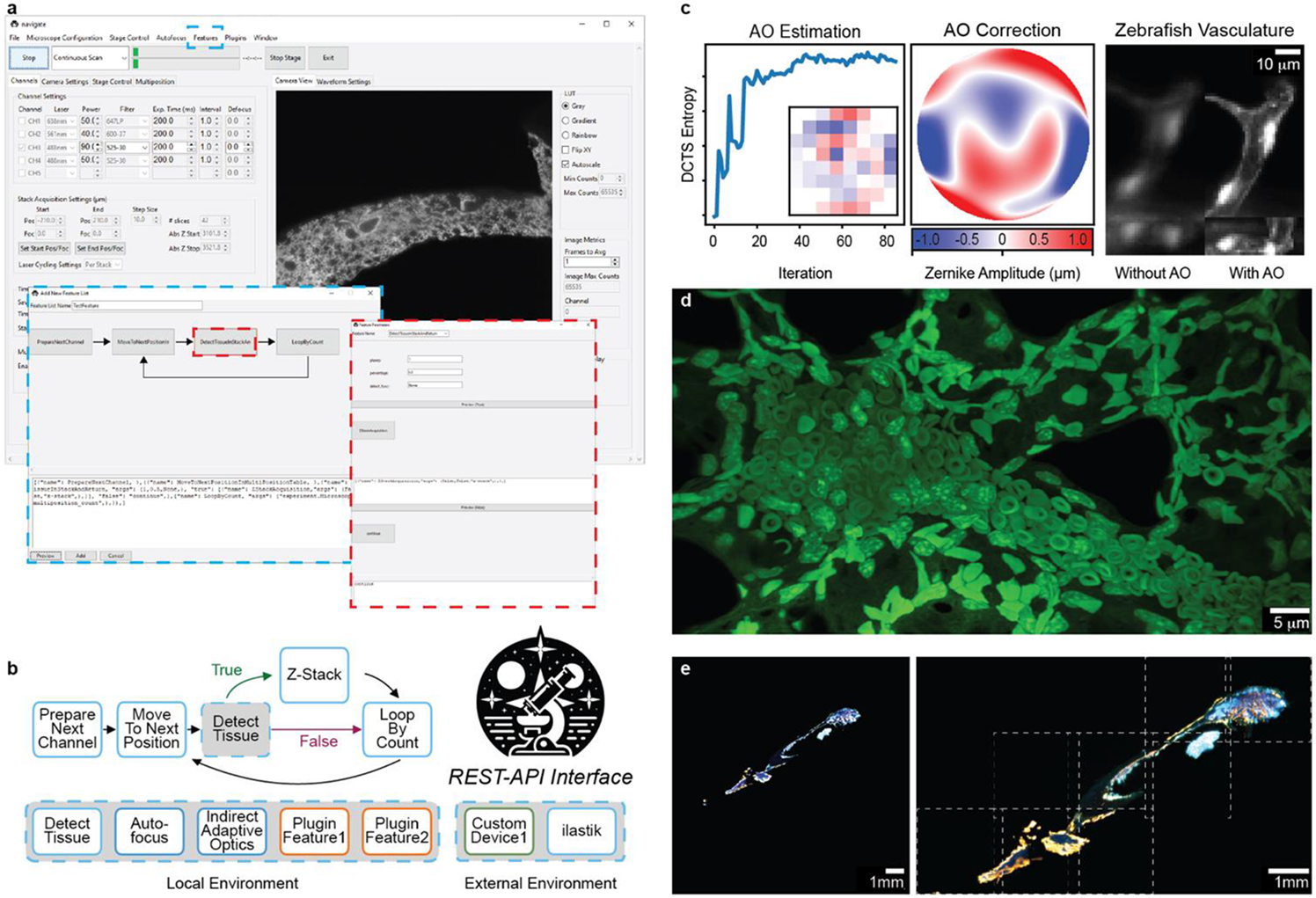
navigate provides a GUI with access to reconfigurable smart acquisition pipelines and runs on multiple types of light-sheet configurations. a, Screenshot of the navigate GUI running a mesoSPIM with GUI controls for the smart acquisition routine diagrammed in b. The blue dashed boxes show the Feature menu, where the controls are accessed, and the corresponding editor for the smart routine. The red dashed boxes show a decision node and the corresponding editor for this decision node. b, An example of a reconfigurable, decision-based pipeline that acquires a Z-Stack if tissue is present at a particular stage position. The user guide in the Supplementary Documentation features a tutorial on how to build this pipeline. Individual features can be swapped for other features that ship with the software or with custom-built plugin features. c, navigate can integrate advanced optical techniques, such as adaptive optics. (Left) An iterative sensorless adaptive optics scheme uses the Discrete Cosine Transform Shannon (DCTS) Entropy to estimate aberrations. Inset shows the final actuator positions of the deformable mirror. (Middle) Final estimated adaptive optics wavefront. (Right) 3D stack of a ventricle within the vasculature of a zebrafish embryo (labelled with kdrl:eGFP), before and after adaptive optics. d, Example image of an expanded mouse lung, captured with an upright ctASLM using navigate. e, Results of smart imaging workflow from a and b, captured on a mesoSPIM. Bone tissue imaged at 1x magnification (left) was automatically segmented and tiled at 6x magnification (right). Imaged regions, indicated by dashed white boxes, were saved to BDV and displayed in a tiled format.
Update of
-
navigate: an open-source platform for smart light-sheet microscopy.bioRxiv [Preprint]. 2024 Feb 11:2024.02.09.579083. doi: 10.1101/2024.02.09.579083. bioRxiv. 2024. Update in: Nat Methods. 2024 Nov;21(11):1967-1969. doi: 10.1038/s41592-024-02413-4. PMID: 38370811 Free PMC article. Updated. Preprint.
References
Publication types
Grants and funding
- RM1 GM145399/GM/NIGMS NIH HHS/United States
- U54 CA268072/CA/NCI NIH HHS/United States
- R35GM133522/U.S. Department of Health & Human Services | National Institutes of Health (NIH)
- RR1900371/Cancer Prevention and Research Institute of Texas (Cancer Prevention Research Institute of Texas)
- R35 GM133522/GM/NIGMS NIH HHS/United States
- RM1GM145399/U.S. Department of Health & Human Services | National Institutes of Health (NIH)
- U54CA268072/U.S. Department of Health & Human Services | National Institutes of Health (NIH)
- R01 CA238519/CA/NCI NIH HHS/United States
- R01CA238519/U.S. Department of Health & Human Services | NIH | National Cancer Institute (NCI)
LinkOut - more resources
Full Text Sources
