. 2024 Sep;56(9):2052-2064.
doi: 10.1038/s12276-024-01304-0. Epub 2024 Sep 12.

IKKε-deficient macrophages impede cardiac repair after myocardial infarction by enhancing the macrophage-myofibroblast transition

Affiliations

IKKε-deficient macrophages impede cardiac repair after myocardial infarction by enhancing the macrophage-myofibroblast transition

Hyang Hee Cho et al. Exp Mol Med. 2024 Sep.

Abstract

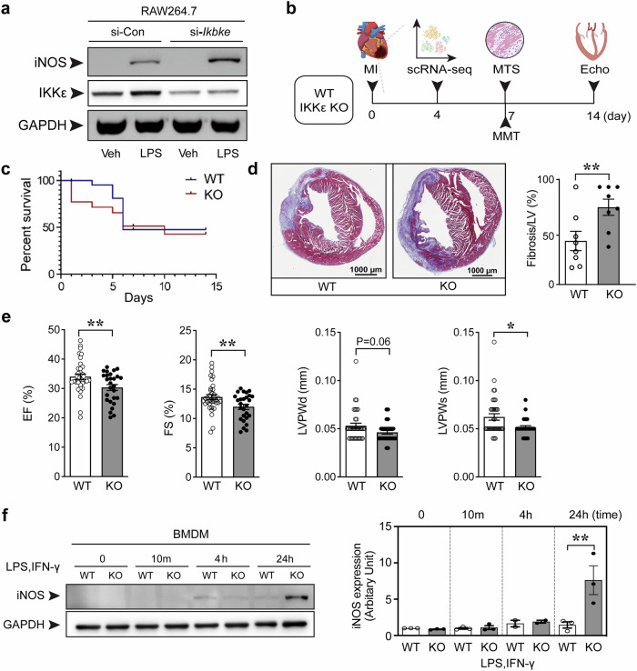
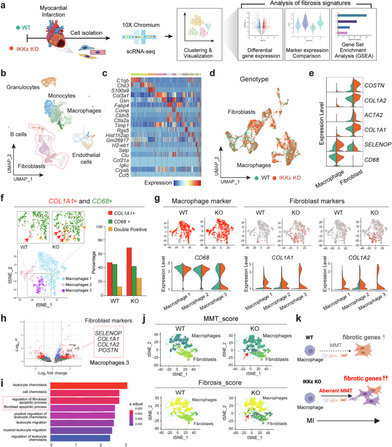
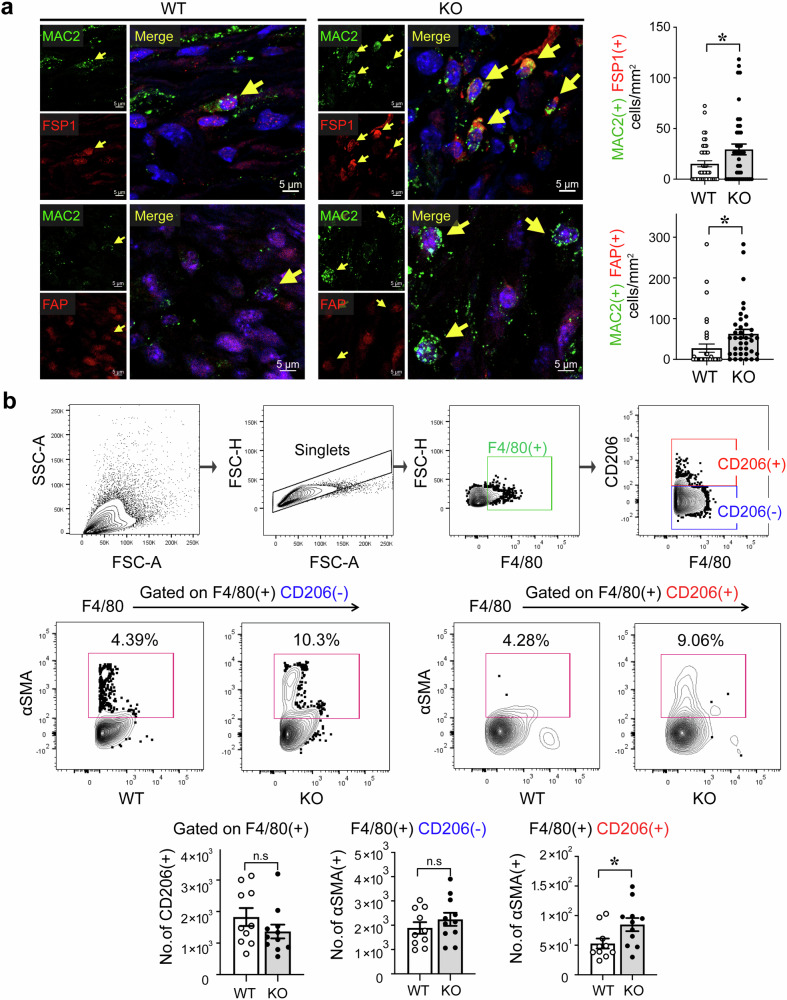
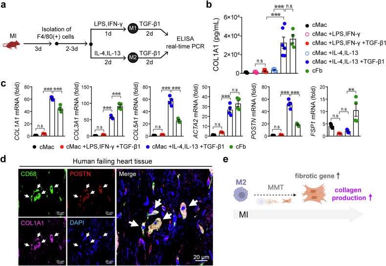
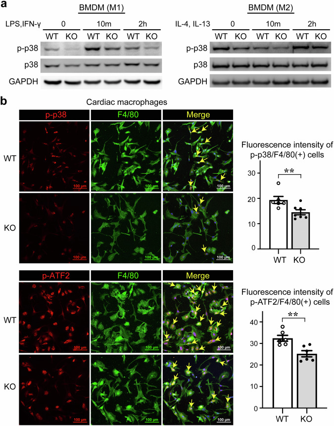
The regulatory role of the inhibitor of NF-kB kinase ε (IKKε) in postmyocardial infarction (MI) inflammation remains uncertain. Using an MI mouse model, we examined the cardiac outcomes of IKKε knockout (KO) mice and wild-type mice. We employed single-cell RNA sequencing (scRNA-seq) and phosphorylated protein array techniques to profile cardiac macrophages. IKKε KO mice exhibited compromised survival, heightened inflammation, pronounced cardiac fibrosis, and a reduced ejection fraction. A distinct cardiac macrophage subset in IKKε KO mice exhibited increased fibrotic marker expression and decreased phosphorylated p38 (p-p38) levels, indicating an enhanced macrophage-myofibroblast transition (MMT) post-MI. While cardiac inflammation is crucial for initiating compensatory pathways, the timely resolution of inflammation was impaired in the IKKε KO group, while the MMT in macrophages accelerated post-MI, leading to cardiac failure. Additionally, our study highlighted the potential of 5-azacytidine (5-Aza), known for its anti-inflammatory and cardioprotective effects, in restoring p-p38 levels in stimulated macrophages. The administration of 5-Aza significantly reduced the MMT in cardiac macrophages from the IKKε KO group. These findings underscore the regulation of the inflammatory response and macrophage transition by the IKKε-p38 axis, indicating that the MMT is a promising therapeutic target for ischemic heart disease.


Conflict of interest statement

The authors declare no competing interests.

Figures

Fig. 1
Fig. 1. Cardiac injury and inflammation postmyocardial infarction in IKKε knockout mice.
a RAW264.7 cells were transfected with the indicated siRNAs and stimulated with 100 ng/mL LPS for 4 h. b Schedule of the animal experiments. Adult (8 weeks) WT or IKKε KO mice were subjected to myocardial infarction (MI) or sham surgery, and hearts were harvested at the indicated times. Single-cell RNA sequencing (scRNA- seq), Masson’s trichrome staining (MTS), macrophage–myofibroblast transition (MMT) assays, and echocardiography (Echo) were performed. c The survival rates of the WT and IKKε KO mice after MI surgery. d Representative images and quantification of MTS to assess the scar size in mice 7 days after MI. Scale bars: 1000 μm. e Cardiac function was measured using echocardiography 14 days after MI. The ejection fraction (EF), fractional shortening (FS), left ventricular posterior wall thickness in diastole (LVPWd), and left ventricular posterior wall thickness in systole (LVPWs) were measured. f Bone marrow-derived macrophages (BMDMs) were isolated from WT and IKKε KO mice and cultured. Then, BMDMs were stimulated with 100 ng/mL LPS and 30 ng/mL IFN-γ for the indicated times. The induction of iNOS expression in BMDMs was assessed in WT and IKKε KO mice using Western blotting. The data are presented as the means ± SEMs. *P < 0.05 and **P < 0.01 (by Student’s t-test).
Fig. 2
Fig. 2. Single-cell RNA sequencing shows the proportion of macrophages expressing fibrotic markers.
Cells were isolated from the infarcted myocardium for single-cell RNA sequencing. a Diagram of the experimental workflow for WT and IKKε KO mice postmyocardial infarction, followed by cell isolation, scRNA-seq, and a fibrosis analysis. b UMAP plot of cell clusters identified via scRNA-seq, including granulocytes, monocytes, macrophages, B cells, fibroblasts, and endothelial cells. c Heatmap displaying gene expression patterns across different cell types. d Genotype distinction in the UMAP plot showing WT and IKKε KO fibroblast and macrophage populations. e Violin plots showing fibrosis-associated gene expression levels in macrophages and fibroblasts. f t-SNE plots and quantitative analysis of COL1A1 and CD68 expression in WT and IKKε KO cells. g Comparison of the expression of macrophage and fibroblast markers between WT and IKKε KO samples via feature and violin plots. h Volcano plot showing the differential expression of fibroblast markers between WT and IKKε KO mice. i Gene Ontology enrichment analysis comparing the WT and IKKε KO transcriptomes. j t-SNE plots representing the differences in MMT scores between WT and IKKε KO cells. k A diagram showing the increased expression of fibrotic genes and aberrant MMT in IKKε KO mice postinfarction compared to those in WT mice.
Fig. 3
Fig. 3. Fibrotic marker expression in macrophages in the infarcted myocardium.
a Immunofluorescence staining showing the expression of fibrotic markers, including FSP1 and FAP, in MAC2(+) macrophages in the hearts of WT mice and IKKε KO mice at 7 days post myocardial infarction (MI). The number of double-positive cells was measured and is shown in the graph. Scale bars: 5 μm. b Cells were isolated from heart tissues at 7 days post-MI, and αSMA expression in F4/80(+)CD206(−) and F4/80(+)CD206(+) macrophages was analyzed via flow cytometry. The data are presented as the means ± SEMs. *P < 0.05 (Student’s t-test).
Fig. 4
Fig. 4. Macrophage–myofibroblast transition (MMT)-induced macrophages acquire fibrotic features after myocardial infarction.
a Experimental protocol for inducing the MMT in cardiac macrophages (cMacs) isolated from WT mice with myocardial infarction (MI). cMacs were differentiated into inflammatory M1 macrophages with 100 ng/mL LPS and 30 ng/mL IFN-γ or into anti-inflammatory M2 macrophages with 30 ng/mL IL-4 and 30 ng/mL IL-13. Then, the cells were stimulated with the fibrosis inducer TGF-β1 (5 ng/mL) in culture media supplemented with 1% FBS. Isolated cardiac fibroblasts (cFbs) were used as a control group. b The amount of COL1A1 protein released in the culture media was measured. c Fibrotic genes were analyzed in cMacs and cFbs, as indicated. d Human heart tissues were isolated from consenting recipients with ischemic cardiomyopathy. Immunofluorescence staining revealed that periostin (POSTN)-expressing CD68(+) macrophages produced the collagen protein. White arrows, POSTN(+)CD68(+)COL1A1(+) cells. Scale bars: 20 μm. e Schematic representation showing that macrophages, particularly M2 macrophages, lead to the MMT and increased collagen production. The data are presented as the means ± SEMs. **P < 0.01 and ***P < 0.001 (one-way ANOVA with Tukey’s multiple comparisons test).
Fig. 5
Fig. 5. IKKε knockout mice have lower levels of phosphorylated p38 in macrophages.
a Bone marrow-derived macrophages (BMDMs) were stimulated with 100 ng/mL LPS, 30 ng/mL IFN-γ, 30 ng/mL IL-4, and 30 ng/mL IL-13 for the indicated times. The protein levels of p-p38 and p38 in BMDMs were assessed in WT and IKKε KO mice using Western blotting. b Cardiac macrophages were isolated from heart tissues 3 days after myocardial infarction and immunofluorescence staining revealed the phosphorylation of p38 and its substrate ATF2. The fluorescence intensity indicating the phosphorylation of p38 and ATF2 was quantified in both the WT and IKKε KO groups. Scale bars: 100 μm. The data are presented as the means ± SEMs. **P < 0.01 (Student’s t-test).
Fig. 6
Fig. 6. Active p38 in macrophages is involved in the macrophage–myofibroblast transition (MMT).
a Experimental protocol used to induce the MMT in THP-1 cells. Monocytic THP-1 cells were differentiated into macrophages by treatment with 250 nM phorbol 12-myristate 13-acetate (PMA). Then, the cells were differentiated into an anti-inflammatory phenotype by treatment with 30 ng/mL IL-4 and 30 ng/mL IL-13. Next, 5 ng/mL TGF-β1 was added to induce the MMT, and during this process, the p38 inhibitor SB203580 (SB), was added to the culture media containing 1% FBS at a concentration of 10 μM. Representative images of cell morphology after MMT induction. The number of MMT cells was quantified by counting the number of cells with morphological changes. Scale bars: 100 μm. b The expression of fibrotic proteins, including FAP and vimentin (VIM), was assessed using Western blotting. c αSMA expression in MMT-induced THP-1 cells was assessed using flow cytometry. d The experimental protocol used to induce the MMT in human CD14(+) monocyte-derived macrophages treated with or without SB is shown. Cells were treated with 30 ng/mL M-CSF, 20 ng/mL IL-4, 20 ng/mL IL-6, and 20 ng/mL IL-13. A representative image of cell morphology is shown, and cellular changes were quantified. Scale bars: 100 μm. e αSMA expression in CD206(+) human anti-inflammatory macrophages, as shown by immunofluorescence staining. Scale bars: 100 μm. The data are presented as the means ± SEMs. **P < 0.01 and ***P < 0.001 (Student’s t-test).
Fig. 7
Fig. 7. Control of the MMT by 5-Aza through the restoration of phosphorylated p38 levels in macrophages.
a, b Stimulated bone marrow-derived macrophages (BMDMs) and RAW264.7 cells were treated with 10 μM 5-azacytidine (5-Aza) to examine the involvement of p-p38 activity in anti-inflammatory activity. The protein levels of iNOS, p-p38, and total p38 were assessed by Western blotting. c Bone marrow cells isolated from WT and IKKε KO mice were differentiated into BMDMs and then treated with 30 ng/mL IL-4 and 30 ng/mL IL-13, followed by treatment with 5 ng/mL TGF-β1 and 10 μM 5-Aza. Finally, POSTN and COL1A1 expression were analyzed using real-time PCR. d Experimental protocol used to examine the effect of 5-Aza on MMT induction. Saline or 5-Aza was administered to mice with myocardial infarction (MI), and cardiac macrophages were isolated for flow cytometry analysis to quantify the MMT. e Cardiac cells were isolated from mice, and the phenotypes of cardiac macrophages were compared among the following groups: WT+saline, WT+5-Aza, IKKε KO+saline, and IKKε KO+5-Aza. f Schematic illustrations showing that the MMT was strongly induced in IKKε-deficient macrophages with low p38 activity. An aberrant MMT was associated with enhanced dysfunction and remodeling after MI. The data are presented as the means ± SEMs. *P < 0.05, **P < 0.01, and ***P < 0.001 (one-way ANOVA with Tukey’s multiple comparisons test).

References

    1. Lawrence, T., Gilroy, D. W., Colville-Nash, P. R. & Willoughby, D. A. Possible new role for NF-kappaB in the resolution of inflammation. Nat. Med.7, 1291–1297 (2001). - PubMed
    1. Gadjeva, M. et al. A role for NF-kappa B subunits p50 and p65 in the inhibition of lipopolysaccharide-induced shock. J. Immunol.173, 5786–5793 (2004). - PubMed
    1. Pradere, J. P. et al. Negative regulation of NF-kappaB p65 activity by serine 536 phosphorylation. Sci. Signal.9, ra85 (2016). - PMC - PubMed
    1. Shimada, T. et al. IKK-i, a novel lipopolysaccharide-inducible kinase that is related to IkappaB kinases. Int. Immunol.11, 1357–1362 (1999). - PubMed
    1. Peters, R. T., Liao, S. M. & Maniatis, T. IKKepsilon is part of a novel PMA-inducible IkappaB kinase complex. Mol. Cell5, 513–522 (2000). - PubMed