. 2024 Oct;43(20):4625-4655.
doi: 10.1038/s44318-024-00193-3. Epub 2024 Sep 11.

Interaction of chikungunya virus glycoproteins with macrophage factors controls virion production

Affiliations

Interaction of chikungunya virus glycoproteins with macrophage factors controls virion production

Zhenlan Yao et al. EMBO J. 2024 Oct.

Abstract

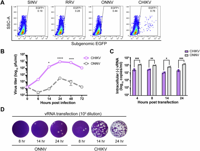
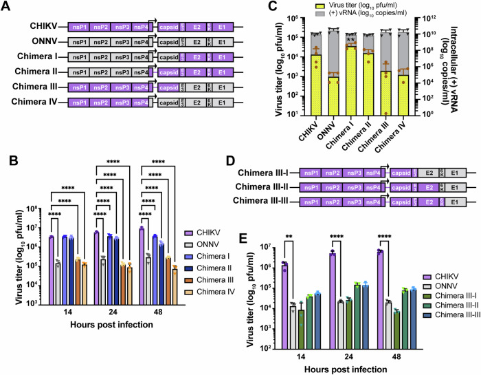
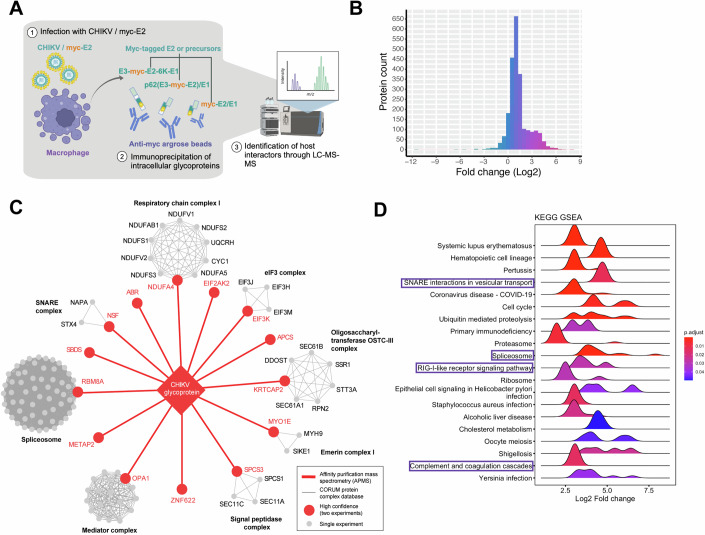
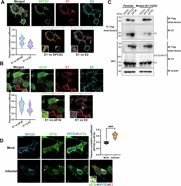
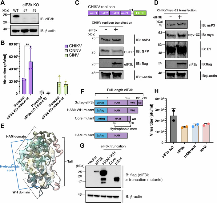
Despite their role as innate sentinels, macrophages can serve as cellular reservoirs of chikungunya virus (CHIKV), a highly-pathogenic arthropod-borne alphavirus that has caused large outbreaks among human populations. Here, with the use of viral chimeras and evolutionary selection analysis, we define CHIKV glycoproteins E1 and E2 as critical for virion production in THP-1 derived human macrophages. Through proteomic analysis and functional validation, we further identify signal peptidase complex subunit 3 (SPCS3) and eukaryotic translation initiation factor 3 subunit K (eIF3k) as E1-binding host proteins with anti-CHIKV activities. We find that E1 residue V220, which has undergone positive selection, is indispensable for CHIKV production in macrophages, as its mutation attenuates E1 interaction with the host restriction factors SPCS3 and eIF3k. Finally, we show that the antiviral activity of eIF3k is translation-independent, and that CHIKV infection promotes eIF3k translocation from the nucleus to the cytoplasm, where it associates with SPCS3. These functions of CHIKV glycoproteins late in the viral life cycle provide a new example of an intracellular evolutionary arms race with host restriction factors, as well as potential targets for therapeutic intervention.

Keywords: Alphavirus E1 Glycoprotein; Chikungunya Virus; Evolutionary Selection; Macrophage; eIF3k.


Conflict of interest statement

The authors declare no competing interests.

Figures

Figure 1
Figure 1. Efficient CHIKV infection in human macrophages depends on a high level of virion production.
(A) Human peripheral monocyte-derived macrophages were infected with EGFP-labeled alphaviruses (SINV TE/5’2 J, RRV strain T48, ONNV strain SG650, and CHIKV vaccine strain 181/clone 25) at MOI of 5 for 24 h. Levels of infection with different alphaviruses were determined by percent EGFP-positive cells evaluated by flow cytometry. Data are representative of 2 independent experiments performed in biological duplicates. (B) THP-1-derived macrophages were infected with CHIKV 181/clone 25 or ONNV SG650 at MOI 5. Titration of supernatant virus samples was performed at 0, 6, 14, 24, 48, and 72 h.p.i by plaque assay on BHK-21 cells. Data were representative of two independent experiments. Mean values of biological duplicates were plotted with SD. Asterisks indicate statistically significant differences as compared to ONNV (two-way ANOVA and Šidák’s multiple comparisons test: 14 h *p = 0.0128; 24 h and 48 h ****p < 0.0001). (C) Levels of intracellular (−) vRNAs, the viral replicative intermediate, at 4, 8, 14, and 24 h post transfection of THP-1 derived macrophages with CHIKV 181/clone 25 or ONNV SG650 viral RNAs (vRNAs) were quantified through RT-qPCR with specific TaqMan probes. Data were representative of two independent experiments. Mean values of biological duplicates measured in technical duplicates were plotted with SD (Two-way ANOVA and Šidák’s multiple comparisons test: 4 h ***p = 0.0006; 8 h **p = 0.004; 14 h *p = 0.0299; 24 h ***p = 0.0001). (D) CHIKV and ONNV titers of supernatant samples collected from transfected THP-1 derived macrophages in (C) were determined by plaque assay. The incubation period for plaque assay took 40 h. Representative plaques of CHIKV and ONNV from two independent experiments (1:100 dilution) are shown. Source data are available online for this figure.
Figure 2
Figure 2. Viral glycoproteins are critical determinants for macrophage tropism of CHIKV.
(A) Schematic representation of CHIKV, ONNV, Chimera I, II, III, and IV. These chimeras consist of genomes from CHIKV vaccine strain 181/clone 25 and ONNV SG650 in different ratios: Chimera I contains the ONNV genome from nsP1 to capsid and the CHIKV genome from E3 to E1. Chimera II contains the ONNV genome from nsP1 to the region prior to the subgenomic promoter in nsP4 and the CHIKV genome from the subgenomic promoter to E1. Chimera III contains the CHIKV genome from nsP1 to capsid and the ONNV genome from E3 to E1. Chimera IV contains the CHIKV genome from nsP1 to the region prior to the subgenomic promoter in nsP4 and the ONNV genome from the subgenomic promoter to E1. (B) Titration of supernatant samples from THP-1 derived macrophages infected with CHIKV 181/clone 25, ONNV SG650, and 4 chimeras (I, II, III, IV). The macrophages were inoculated with the virus at MOI 5, and the supernatant samples were collected at 14, 24, and 48 h.p.i for plaque assay analysis. The incubation period for plaque assay took 28 h. Data were representative of three independent experiments. Mean values of biological triplicates were plotted with SD. Asterisks indicate statistically significant differences as compared to CHIKV (Two-way ANOVA and Dunnett’s multiple comparisons test: 14, 24, and 48 h ****p < 0.0001). (C) THP-1-derived macrophages were transfected with 0.5 μg RNA of CHIKV 181/clone 25, ONNV SG650, or chimeras (I, II, III, IV). Virion productions were determined by intracellular (+) vRNA transcript levels and supernatant infectious particle titers through RT-qPCR and plaque assay, respectively. The incubation period for plaque assay took 40 h. Data were plotted with the mean value of four biological replicates from two independent experiments. The error bar represents SD. Asterisks indicate statistically significant differences as compared to CHIKV (two-way ANOVA and Dunnett’s multiple comparisons test: viral titer of CHIKV vs Chimera I **p = 0.0036). (D) Schematic representation of Chimera III-I, III-II, and III-III. Chimera III-I contains the CHIKV genome from nsP1 to E3 and the ONNV genome from E2 to E1. Chimera III-II contains the CHIKV genome from nsP1 to E2 and the ONNV genome from 6K to E1. Chimera III-III contains the CHIKV genome from nsP1 to 6 K and ONNV E1. (E) Titration of supernatant samples from THP-1 derived macrophages infected with CHIKV 181/clone 25, ONNV SG650, or chimeras (III-I, III-II, III-III) for 14, 24, and 48 h. The infection conditions and virus titer assessments were performed as previously described in (B). Data were representative of two independent experiments. Mean values of biological triplicates measured were plotted with SD. Asterisks indicate statistically significant differences as compared to ONNV (two-way ANOVA and Dunnett’s multiple comparisons test: 14 h CHIKV vs ONNV **p = 0.0092; 24 and 48 h CHIKV vs ONNV ****p < 0.0001). Source data are available online for this figure.
Figure 3
Figure 3. CHIKV E2 and E1 dramatically increase the specific infectivity of viral particles secreted from human macrophages without affecting viral RNA replication.
(A) Schematic representation of chimera ONNV/CHIKV E2, ONNV/CHIKV E1 and ONNV/CHIKV E2 + E1. These three chimeric viruses were built on ONNV backbone with the replacement of CHIKV E2 (ONNV/CHIKV E2), E1 (ONNV/CHIKV E1), or both E2 and E1 (ONNV/CHIKV E2 + E1). (B) Titration of supernatant samples from THP-1 derived macrophages infected with CHIKV vaccine strain 181/clone 25, ONNV SG650, ONNV/CHIKV E2, ONNV/CHIKV E1, and ONNV/CHIKV E2 + E1. Macrophages were inoculated with the viruses at MOI 5, and the supernatant samples were collected at 24 h.p.i for plaque assay analysis. The incubation period for plaque assay took 28 h. Data were representative of two independent experiments. Mean values of biological duplicates were plotted with SD. Asterisks indicate statistically significant differences as compared to ONNV (two-way ANOVA and Dunnett’s multiple comparisons test: ONNV vs CHIKV **p = 0.004; ONNV vs ONNV/CHIKV E2 + E1****p < 0.0001). (C) THP-1 derived macrophages were transfected with 0.5 μg RNA of CHIKV 181/clone 25, ONNV SG650, ONNV/CHIKV E2, ONNV/CHIKV E1, or ONNV/CHIKV E2 + E1. Virion production was determined by intracellular (+) vRNA transcript levels and supernatant infectious particle titers through RT-qPCR and plaque assay, respectively. The incubation period for plaque assay took 40 h. Data were representative of three independent experiments. Mean values of biological duplicates were plotted with SD. Asterisks indicate statistically significant differences as compared to ONNV (one-way ANOVA and Dunnett’s multiple comparisons test: viral titer of ONNV vs CHIKV ****p < 0.0001; viral titer of ONNV vs ONNV/CHIKV E2 + E1**p = 0.0058; viral copies of ONNV vs CHIKV **p = 0.0031; viral copies of ONNV vs ONNV/CHIKV E2 **p = 0.0034; viral copies of ONNV vs ONNV/CHIKV E1 **p = 0.0019). (D, E) Particle-to-PFU ratios of ONNV, CHIKV, and chimeric viruses containing CHIKV glycoproteins. THP-1-derived macrophages were infected with ONNV SG650, CHIKV 181/clone 25, Chimera I (refer to Fig. 2A schematic), and ONNV/CHIKV E2 + E1 at MOI 5 for 24 h. The viral particle numbers in the supernatant were quantified by TaqMan qPCR assay with specific probes targeting nsP1 in (+) RNA. Virus titers were determined by plaque assay on BHK-21 cells. The incubation period for plaque assay took 28 h. Data were representative of two independent experiments, each of which has biological duplicate samples. The viral copy numbers and titers of each duplicate and their averaged values are shown in (D) and summarized as bar charts in (E). Asterisks indicate statistically significant differences as compared to ONNV (one-way ANOVA and Dunnett’s multiple comparisons test: ONNV vs CHIKV***p = 0.0005; ONNV vs Chimera I***p = 0.0008; ONNV vs ONNV/CHIKV E2 + E1***p = 0.0006). (F) Schematic representation of modified chimeras based on parental ONNV/CHIKV E1 + E2 that contain hybrid E2 or E1. E2 has three domains: A and B connected to A and C by two flanking β-ribbon arches, and C. E1 has three domains: I, II, and III, with a fusion loop in II. Chimera containing hybrid E2 that has arch-B-arch-C (E2-I + E1), or only domain C (E2-II + E1) from ONNV. Chimera containing hybrid E1 has domains II and III (E2 + E1-I), or only domain III (E2 + E1-II) from ONNV. (G) THP-1 derived macrophages were transfected with 0.5 μg RNA of ONNV SG650, ONNV/CHIKV E1 and chimeras (E2-I + E1, E2-II + E1), ONNV/CHIKV E2 and chimeras (E2 + E1-I, E2 + E1-II). Virion production was determined through RT-qPCR and plaque assays as described in (C). Data were representative of four independent experiments. Mean values of biological duplicates were plotted with SD. Asterisks indicate statistically significant differences as compared to ONNV (one-way ANOVA and Dunnett’s multiple comparisons test: viral titer of ONNV vs E2-II + E1***p = 0.0008; viral titer of ONNV vs E2 + E1-II****p < 0.0001). Source data are available online for this figure.
Figure 4
Figure 4. CHIKV E2 and E1 residues under positive selection are essential for virion production in human macrophages.
(A) The pipeline for analyzing natural selection in the evolution of CHIKV structural proteins in human hosts. 397 CHIKV sequences isolated from infected individuals globally were downloaded from the NCBI virus database, and structural polyprotein sequence alignment was performed by MUSCLE(Edgar, 2004). The phylogenetic tree of CHIKV was constructed based on the maximum-likelihood (ML) optimality criterion with IQ-TREE (Minh et al, ; Trifinopoulos et al, 2016). The sites under positive selection were identified using mixed effects model of evolution (MEME) (Murrell et al, 2012) and fixed effects likelihood (FEL) (Kosakovsky Pond and Frost, 2005). (B) The positively selected sites identified by FEL or MEME are annotated in each CHIKV structural protein. The sites identified by MEME are colored in dark gray. Four of these sites (E2-164, 6K-47, E1-145, and E1-211) were identified with both methods and are colored in orange. (C) The positively selected sites in CHIKV structural proteins are plotted with the y-axis of −log10 P values (determined by MEME or FEL) and the x-axis of the amino acid locations in the full-length structural polyprotein (from the beginning of capsid to the end of E1). The P values are generated by the FEL or MEME algorithm and adjusted with Benjamini–Hochberg correction. The statistically significant sites identified by MEME (p < 0.05) are in blue, the ones identified by FEL (FEL p < 0.05) are in orange, and the ones identified by both methods (MEME p < 0.05 and FEL p < 0.05) are in orange with blue circles. (D) Comparison of virion production of CHIKV positive selection site mutants in THP-1 derived macrophages. The positive selection site in E2 or E1 of CHIKV 181/clone 25 was mutated to the homologous residue in ONNV, respectively, to generate six CHIKV mutants (E2-V135L, E2-A164T, E2-A246S, E1-E211K, E1-V220I, and E1-R366K). Macrophages were transfected with 0.5 μg RNA of CHIKV, ONNV, or CHIKV positive selection site mutants, and virion productions were determined by intracellular (+) vRNA transcript levels and supernatant infectious particle titers as previously described. Data were representative of three independent experiments. Mean values of biological duplicates were plotted with SD. Asterisks indicate statistically significant differences as compared to CHIKV (One-way ANOVA and Dunnett’s multiple comparisons test: viral titer of CHIKV vs E211K *p = 0.0414; viral copies of CHIKV vs ONNV ****p < 0.0001; viral copies of CHIKV vs V135L *p = 0.0228). (E) Representative plaque images of CHIKV E1 positive selection site mutants (E1-E211K, E1-V220I, E1-R366K) in comparison with CHIKV and ONNV. Plaque assays were performed on supernatant samples from transfected THP-1-derived macrophages as mentioned in (D). The incubation period for plaque assay is 40 h. The representative plaques from the 1:100 dilution are shown here. (F) The expression levels of viral nonstructural and structural proteins of CHIKV wild-type, E2-V135L, and E1-V220I mutants in THP-1 derived macrophages. The THP-1-derived macrophages were transfected with viral RNAs of CHIKV, E2-V135L, or E1-V220I mutant for 48 h. The expression levels of viral nsP3, E2, and E1 proteins were evaluated through immunoblotting. (G) Visualization of positively selected sites in single E2/E1 heterodimer with the presence of E3 from infectious CHIKV 181/clone 25 virus particle. The heterodimer structure was downloaded from PDB (6NK7)(Basore et al, 2019) and visualized in Chimera X (Pettersen et al, 2021). The positively selected sites E2-V135, E2-A164, and E2-A246 are located in β-ribbon arches flanking domain B in E2. The positively selected sites E1-E211 and E1-V220 are located in domain II in E1. The positively selected site E1-R366 is in domain III in E1. (H) The locations of E2-V135 (yellow nodes) and E1-V220 (orange nodes) in trimerized E2/E1 heterodimers (PDB: 6NK7). The E2 (cyan), E1 (purple), and E3 (gray) were annotated to show a single heterodimer unit. Source data are available online for this figure.
Figure 5
Figure 5. Identifying host factors interacting with CHIKV glycoproteins in infected human macrophages by affinity purification-mass spectrometry (AP-MS).
(A) The workflow of AP-MS analysis to identify host factors in THP-1 derived macrophages that interact with CHIKV glycoproteins. After 48 h infection with CHIKV/myc-E2, different forms of myc-tagged glycoproteins (polyprotein E3-myc-E2-6K-E1, E3-myc-E2/E1, myc-E2/E1) were pulled down by anti-myc agarose beads from the infected macrophage lysates and submitted to LC-MS/MS analysis to identify co-immunoprecipitated host factors. The co-immunoprecipitated proteins from untagged CHIKV vaccine strain 181/clone 25 (CHIKV WT)-infected macrophages serve as negative controls for proteomic analysis (not elaborated in this diagram). (B) The histogram of fold change distribution of all the identified macrophage proteins in the second independent AP-MS experiment that interact with myc-tagged glycoproteins (poly-glycoprotein E3-myc-E2-6K-E1, E3-myc-E2/E1, myc-E2/E1). (C) The interaction network between CHIKV glycoproteins and macrophage proteins. CHIKV glycoproteins interacting partners in THP-1 derived macrophages that were significantly enriched in both independent mass spectrometry experiments are depicted in red. Candidate interactors significantly enriched in at least one mass spectrometry experiment that belongs to existing red protein complexes are colored in gray. The protein complexes were identified through the CORUM database. (D) Gene set enrichment analysis (GSEA) of top 20 KEGG pathways in identified host factors summarized in ridgeplot. All the identified host factors are ranked according to the log2 expression fold change of proteins co-immunoprecipitated from CHIKV/myc-E2 infected macrophages with respect to proteins from CHIKV 181/clone 25 infected macrophages (x-axis). The significance of the KEGG enrichment is shown in a continuous color scale based on the adjusted P values, which are generated by Fisher’s exact test and corrected by Benjamini–Hochberg. The histogram in each KEGG term is defined by the number of genes with a specific log2 fold change value. Created with BioRender.com.
Figure 6
Figure 6. CHIKV E1 interacts with macrophage host factors that block virion production.
(A) Table of identified host factors that were chosen for siRNA knockdown assays in (B, C). Statistical analysis for protein differential expression is a moderated t-test from R package ArtMS3. The P values are adjusted with Benjamini–Hochberg for the multiple hypothesis correction. The gray-highlighted genes are significantly detected in 2 independent AP-MS experiments. (B, C) Evaluation of CHIKV infection (B) and production (C) in human macrophages with OAS3, NSF, CHCHD2, RBM8A, S100A9, SBDS, SPCS3, KRTCAP2, APOBEC3F, ZNF622, METAP2, EIF3K, or PKR knocked down. THP-1-derived macrophages were transfected with pooled siRNAs targeting specific host factors or nontargeting siRNAs (NT) for 48 h. The cells were then infected with CHIKV 181/clone 25 (MOI 5) (B) or transfected with CHIKV vRNA (C) for 24 h. The supernatant virus titers from cells treated with siRNAs targeting host factors were determined by plaque assay and compared to the titers from cells treated with NT siRNA to assess the anti- or proviral effects of specific host genes on CHIKV production. G3BP1 and G3BP2 (G3BP1 + 2) known to be proviral for CHIKV replication were knocked down together as control. For (B), data were representative of two independent experiments. The mean values of biological duplicates were plotted with SD (one-way ANOVA and Dunnett’s multiple comparisons test: si-NT vs si-SPCS3 *p = 0.031; si-NT vs si-EIF3K *p = 0.0421.) For (C), data were representative of two independent experiments. The plaque assay results were plotted from biological duplicates with the mean values (one-way ANOVA and Dunnett’s multiple comparisons test: viral titer of si-NT vs si-OAS3 **p = 0.01; viral titer of si-NT vs si-SPCS3 ***p = 0.0008; viral titer of si-NT vs si-EIF3K *p = 0.0194). The qPCR results were plotted from biological triplicates with the mean values (one-way ANOVA and Brown-Forsythe test: viral copy of si-NT vs si-RBM8A ****p < 0.0001; viral copy of si-NT vs si-KRTCAP2 **p = 0.0054; viral copy of si-NT vs si-ZNF622 ****p < 0.0001). (D) 293T cells were transfected with plasmids expressing 3xflag-tagged host factors (TRIM25, OAS3, PKR, SPCS3, eIF3k, and APOBEC3F) or empty vector control for 24 h and later transfected with vRNA of CHIKV/myc-E2. The cells were lysed and immunoprecipitated by anti-flag agarose beads. Immunoblot was probed to check for E2/E1 binding to these host factors. 3xflag tagged TRIM25 (tripartite motif containing 25) was transfected into 293T cells for immunoprecipitation control. Data were representative of three independent experiments. Source data are available online for this figure.
Figure 7
Figure 7. A pool of free E1 separate from E2 associates with SPCS3 and eIF3k through the positively selected site E1-V220.
(A) Colocalization analysis of CHIKV E1 with E2 or with SPCS3 through immunofluorescence. 293T cells were transfected with plasmids expressing 3xflag-SPCS3 and infected with CHIKV 181/clone 25 for 24 h 1 day later. flag-tagged SPCS3 (green), and CHIKV E1 (red) and E2 (cyan) were labeled through indirect staining with primary antibodies against flag, E1, and E2. The representative colocalization regions are enlarged on the bottom left of the overlaid images. Colocalization between CHIKV E1 and SPCS3 (E1 vs SPCS3) and between CHIKV E1 and E2 (E1 vs E2) are compared through Pearson correlation analysis and shown as violin plots. Pearson correlation coefficient values range from 1 to −1, where 1 is a total positive correlation, −1 is a total negative correlation, and 0 is no correlation. Scale bar: 5 μm. Representative results from two independent are shown here. Two field images were taken for each sample in each independent experiment, and four cells from one independent experiment were designated as region of interests (ROIs) for colocalization analysis. (B) Colocalization analysis of CHIKV E1 with E2 or with eIF3k through immunofluorescence. 293T cells were transfected with plasmids expressing 3xflag-eIF3k and infected with CHIKV 181/clone 25 for 24 h one day later. 3xflag-tagged eIF3k (green), and CHIKV E1 (red) and E2 (cyan) were labeled as previously described. The representative colocalization regions are shown in the bottom left of the overlaid images. Colocalization between CHIKV E1 and eIF3k (E1 vs eIF3k) and between CHIKV E1 and E2 (E1 vs E2) is compared through Pearson correlation analysis (refer to 7A) and shown as violin plots. Scale bar: 5 μm. Representative results from two independent are shown here. Two field images were taken for each sample in each independent experiment, and four cells from one independent experiment were designated as region of interests (ROIs) for colocalization analysis. (C) 293T cells were transfected with plasmids expressing 3xflag-tagged host factors (SPCS3, eIF3k) or empty vector control for 24 h followed by transfection with a plasmid expressing parental or E1-V220I-containing CHIKV poly-glycoprotein (E3-myc-E2-6K-E1). The cells were lysed for immunoprecipitation with anti-flag agarose beads. Immunoblot was probed for parental or mutant E1 binding to host factors. Data are representative of 2 independent experiments. (D) Colocalization analysis of SPCS3 and eIF3k in uninfected and CHIKV-infected cells through immunofluorescence. 293T cells were co-transfected with plasmids expressing 3xflag-eIF3k and V5-SPCS3 followed by mock or CHIKV infection for 24 h 1 day later. V5-SPCS3 (cyan), 3xflag-eIF3k (green), and CHIKV E1 (red) were labeled through indirect staining with antibodies against V5, flag, and E1. The representative colocalization regions are shown on the bottom left of the overlaid images. Colocalization between SPCS3 and eIF3k (SPCS3 vs eIF3k) is compared in mock and CHIKV-infected 293T cells through Pearson correlation analysis (refer to 7A) and shown as violin plots (unpaired t-test: Mock vs Infected ***p = 0.0004). Scale bar: 5 μm. Representative results from two independent are shown here. Two field images were taken for each sample in each independent experiment, and four cells from one independent experiment were designated as region of interests (ROIs) for colocalization analysis. Source data are available online for this figure.
Figure 8
Figure 8. The specific anti-CHIKV activity of eIF3k is translation-independent and mediated by its HAM domain.
(A) Immunoblot validation of EIF3K CRISPR KO in 293T clones 7 and 9. (B) eIF3k KO 293T cells (clone 9) were infected with CHIKV 181/clone 25, ONNV SG650, or SINV Toto1101 at MOI 1 for 24 h. Virion production was evaluated by titering the supernatant infectious particles through plaque assay. Data were representative results of three independent experiments. The mean values of biological triplicates were plotted with SD (two-way ANOVA and Šidák’s multiple comparisons test: Parental vs eIF3k KO for CHIKV infection **p = 0.0066). (C) Translation of CHIKV replicon in eIF3k KO 293T cells (clone 7) with or without eIF3k reconstitution. The schematic for the CHIKV replicon is shown on top. Viral structural polyprotein downstream of the subgenomic promoter is replaced with the EGFP reporter. The eIF3k KO 293T cells were first transfected with an empty vector or plasmid expressing 3xflag-eIF3k followed by transfection with the CHIKV replicon RNA one day later. Twenty-four hours after the second transfection, protein expression of nsP3, EGFP, and 3xflag-eIF3k was detected by immunoblotting. As a GFP variant, EGFP was detected by a GFP antibody. Data were representative of three independent experiments. (D) The translation of CHIKV/myc-E2 in eIF3k KO 293T cells (clone 7) with or without eIF3k reconstitution. As mentioned in Fig. 5A, the myc tag is inserted at the N-terminal end of E2. The eIF3k KO 293T cells were first transfected with an empty vector or plasmid expressing 3xflag-eIF3k followed by transfection with CHIKV/myc-E2 RNA one day later. Protein expression of nsP3, myc (E2), E1, and (flag) eIF3k was detected by immunoblotting 24 h after the second transfection. Data were representative of three independent experiments. (E) The structure of human eIF3k with the protein domains labeled. The eIF3k crystal structure is downloaded from PDB (1RZ4)(Wei et al, 2004) and visualized in Chimera X. eIF3k consists of a HAM domain (khaki), WH domain (blue), and a long C-terminal tail region with α-helix at both ends (pink). The HAM domain contains a leading α-helix and 3 HEAT analogous repeats followed by a short helix. The WH domain contains three α-helices and three β-strands. (F) The diagram of eIF3k truncation mutants. The HAM + WH mutant that lacks the C-terminal tail terminates after residue S191 of full-length eIF3k. The Core mutant with a truncated WH domain terminates after residue Y150 of full-length eIF3k and includes the hydrophobic core formed by the highly conserved hydrophobic residues from HAM and the first helix of WH. The HAM-only mutant terminates after residue T132 of full-length eIF3k. All the eIF3k truncation mutants are tagged with an N-terminal 3xflag. (G) Validation of expression of eIF3k truncation mutants in eIF3k KO 293 T cells through immunoblotting. Since the Core mutant cannot be expressed, it is not followed up in (8H). (H) The anti-CHIKV activities of eIF3k truncation mutants. The eIF3k KO 293T cells (clone 7) were transfected with plasmids expressing full-length eIF3k or different truncation mutants. The cells were then infected with CHIKV at MOI 1 for 24 h 1 day following transfection. Levels of infectious particle production in supernatant samples were determined by plaque assay on BHK-21 cells. The incubation period for plaque assay is 28 h. Data were representative of five independent experiments. The mean values of biological duplicates were plotted with SD. Source data are available online for this figure.
Figure EV1
Figure EV1. The advantage of virus production in macrophages is also recapitulated by pathogenic CHIKV and depends more on the host secretory pathway.
(A) THP-1-derived macrophages were infected with ONNV SG650, CHIKV La Réunion strain (LR2006 OPY1), and CHIKV Asian strain (AF15561) at MOI 5. Titration of supernatant infectious particles was performed at 24 h.p.i by plaque assay on BHK-21 cells. The incubation period for plaque assay takes 28 h. Data were representative of three independent experiments. Mean values of biological duplicates were plotted with SD. Asterisks indicate statistically significant differences as compared to ONNV (One-way ANOVA and Dunnett’s multiple comparisons test: ONNV vs LR2006 OPY1 **p = 0.0024; ONNV vs AF15561 **p = 0.0082). (B) The influence of secretary pathway inhibition on the infections of ONNV, CHIKV, Chimera I, and ONNV/CHIKV E2 + E1. The THP-1-derived macrophages were pretreated with 10 μM FLI-06 or GCA for 30 min prior to 1-h inoculation with ONNV, CHIKV, Chimera I, or ONNV/ CHIKV E2 + E1. The cells were then cultured with the inhibitors at the same concentration (10 μM) for 24 h. The virus titers from supernatants were analyzed by plaque assay as previously described. Data were representative of two independent experiments. Mean values of biological duplicates were plotted with SD. Asterisks indicate statistically significant differences as compared to ONNV (one-way ANOVA and Dunnett’s multiple comparisons test: DMSO vs FLI-06/GCA with the infection of CHIKV, Chimera I, or ONNV/CHIKV E2 + E1 ****p < 0.0001).
Figure EV2
Figure EV2. Evolutionary selection analysis on CHIKV structural proteins.
(A) Phylogenetic tree constructed by IQ-tree (Minh et al, 2020) using an alignment of the CHIKV structural polyprotein. The tree was visualized by ggtree (Yu et al, 2017). Tree branches were colored according to the latest CHIKV lineage classification (de Bernardi Schneider et al, 2019) used in CHIKVnext v3 (nextstrain.org/groups/ViennaRNA/CHIKVnext/v3.0). AUL-Am Asian urban + American lineage, AUL Asian urban lineage, EAL Eastern African lineage, IOL Indian Ocean lineage, MAL Middle African lineage, SAL South American lineage, WA Western African lineage. (B) Comparison of CHIKV positively selected sites with homologous sites in ONNV. MEME and FEL were used to analyze the positively selected sites in CHIKV structural proteins and generate P values. The P values are corrected with Benjamini–Hochberg. The positively selected CHIKV amino acids that are different from the homologous residues in ONNV were colored in red and highlighted in gray. (C) The heterogeneity of residues at E2-135 and E1-220 in 397 CHIKV patient isolates from NCBI Virus database. (D) The E2 alignment of different ONNV and CHIKV strains to compare the amino acid residues at E2-135, E2-164, and E2-246. CHIKV 37997 belongs to the West African lineage. CHIKV LR2006 OPY1 and CHIKV SL15649 belong to the East/Central/South African (ECSA) lineage. CHIKV Caribbean and CHIKV AF15561 belong to the Asian lineage. CHIKV AF15561 is the parental strain of CHIKV vaccine strain 181/clone 25. The alignment is visualized through ggmsa (Zhou et al, 2022). (E) The E1 alignment of different ONNV and CHIKV strains to compare the amino acid residues at E1-211, E1-220, and E1-366.
Figure EV3
Figure EV3. The superior virus production conferred by CHIKV structural proteins is macrophage-specific.
(A) CHIKV, ONNV, Chimera I, Chimera III, and ONNV/CHIKV E2 + E1 infection in 293T cells. Virion production in the supernatant of infected 293T cells was titrated through plaque assay on BHK-21 cells as previously described. Mean values of biological duplicates were plotted with SD. Data were representative of two independent experiments. Asterisks indicate statistically significant differences as compared to CHIKV (one-way ANOVA and Dunnett’s multiple comparisons test: CHIKV vs ONNV/CHIKV E2 + E1 ****p < 0.0001). (B, C) Infection of 293T (B) and BHK-21 (C) cells with CHIKV vaccine strain 181/clone 25 positive selection site mutants. Viral replication and production of positive selection site mutants (E2-V135L, E2-A164T, E2-A246S, E1-E211K, E1-V220I, and E1-R366K) were determined by levels of intracellular (+) vRNAs and secreted infectious particles as previously described. For EV3B, data were representative of two independent experiments. The plaque assay results were plotted from biological duplicates with the mean values. Error bars represent SD (one-way ANOVA and Dunnett’s multiple comparisons test: viral titer of CHIKV vs ONNV ***p = 0.0004; viral titer of CHIKV vs E2-V135L ***p = 0.0004; viral titer of CHIKV vs E1-E211K *p = 0.017; viral titer of CHIKV vs E1-V220I ***p = 0.0004). The qPCR results were plotted from biological duplicates with the mean values. Error bars represent SD (one-way ANOVA and Brown-Forsythe test: viral copies of CHIKV vs E2-V135L *p = 0.0116; viral copies of CHIKV vs E2-A164T **p = 0.0036; viral copies of CHIKV vs E2-A246S *p = 0.0156; viral copies of CHIKV vs E1-E211K ****p < 0.0001; viral copies of CHIKV vs E1-V220I *p = 0.011; viral copies of CHIKV vs E1-R366K *p = 0.0274). For EV3C, data were representative of two independent experiments. The plaque assay results were plotted from biological duplicates with the mean values. Error bars represent SD (one-way ANOVA and Dunnett’s multiple comparisons test: viral titer of CHIKV vs ONNV **p = 0.006; viral titer of CHIKV vs E2-V135L **p = 0.0058; viral titer of CHIKV vs E1-V220I **p = 0.0057). The qPCR results were plotted from biological duplicates with the mean values. Error bars represent SD (one-way ANOVA and Brown-Forsythe test: viral copies of CHIKV vs E2-V135L **p = 0.0027; viral copies of CHIKV vs E1-V220I **p = 0.0019).
Figure EV4
Figure EV4. The macrophage host factors identified by AP-MS and representative biological processes of significantly enriched host factors.
(A) Volcano plot depicting cellular interactors of CHIKV glycoproteins identified by mass spectrometry. A moderated t-test from R package ArtMS3 was used to generate the P values which were adjusted with Benjamini–Hochberg for the multiple hypothesis correction. The volcano plot is scattered by −log10 P value (y-axis) and log2 expression fold change (FC) of proteins co-immunoprecipitated from CHIKV/myc-E2 infected cells with respect to the proteins from CHIKV WT infected cells (x-axis). The dashed cut-offs of the adjusted P value and expression fold change are 0.05 (−log10 P value = 1.30103) and 2 (log2FC = 1), respectively. CHIKV glycoproteins (E3, E2, E1) and host factors for further investigation in Fig. 6A are annotated here. (B) Enrichment map that summarizes over-represented biological processes of identified host factors in groups. The enriched proteins identified by mass spectrometry were clustered by biological processes and organized into a network with edges connecting overlapping gene sets to reveal functional modules.
Figure EV5
Figure EV5. New antiviral host factors, SPCS3 and eIF3k, specifically interact with CHIKV E1.
(A) The macrophages were transfected with 25 nM nontargeting siRNAs (NT) or pooled siRNAs targeting host factors (G3BP1, G3BP2, OAS3, NSF, CHCHD2, RBM8A, S100A9, SBDS, SPCS3, KRTCAP2, APOBEC3F, ZNF622, METAP2, EIF3K, and PKR). mRNAs of cells treated with siRNAs were extracted 48 h post transfection for RT-qPCR to evaluate the host factor knockdown efficiencies. Data were representative of two independent experiments. The mean values of biological duplicates were plotted with SD (two-way ANOVA and Šidák’s multiple comparisons test: si-NT vs si-OAS3 *p = 0.0118; si-NT vs si-ZNF622 **p = 0.0026; si-NT vs si-NSF ***p = 0.0006; si-NT vs si-S100A9 ***p = 0.0009; si-NT vs si-G3BP1/G3BP2/CHCHD2/RBM8A/SBDS/SPCS3/KRTCAP2/APOBEC3F/METAP2/EIF3K/PKR ****p < 0.0001). (B) 293T cells were transfected with plasmids expressing 3xflag-tagged host factors (TRIM25, OAS3, SPCS3, APOBEC3F, eIF3k, and PKR) or empty vector control for 24 h, and later transfected with plasmid expressing CHIKV glycoproteins (E3-myc-E2-6K-E1). The cells were lysed and immunoprecipitated by anti-flag agarose beads. Immunoblot was probed to check for E2/E1 binding to these host factors. TRIM25-3xflag was transfected into 293T cells for immunoprecipitation control. Data were representative of three independent experiments. (C) 293T cells were transfected with plasmids expressing 3xflag-tagged host factors (SPCS3, eIF3k) or empty vector control for 24 h, followed by transfection with the plasmid expressing CHIKV E3-myc-E2-6K-E1. The cells were lysed for immunoprecipitation with Dynabeads Protein G conjugated with E2 antibody (CHK-48) (Fox et al, 2015). Immunoblot was probed for host factor (SPCS3, eIF3k) binding to E2. Data were representative of two independent experiments. (D) 293T cells were transfected with plasmids expressing 3xflag-tagged host factors (SPCS3, eIF3k) or empty vector control for 24 h, followed by transfection with the plasmid expressing CHIKV E3-myc-E2-6K-E1-3xflag. The cells were lysed for immunoprecipitation with Dynabeads Protein G conjugated with E1 antibody. Immunoblot was probed for host factor (SPCS3, eIF3k) binding to E1. Data were representative of two independent experiments.

References

    1. Aitken CE, Beznosková P, Vlčkova V, Chiu W-L, Zhou F, Valášek LS, Hinnebusch AG, Lorsch JR (2016) Eukaryotic translation initiation factor 3 plays distinct roles at the mRNA entry and exit channels of the ribosomal preinitiation complex. eLife 5:e20934 - PMC - PubMed
    1. Basore K, Kim AS, Nelson CA, Zhang R, Smith BK, Uranga C, Vang L, Cheng M, Gross ML, Smith J et al (2019) Cryo-EM structure of chikungunya virus in complex with the Mxra8 receptor. Cell 177:1725–1737.e16 - PMC - PubMed
    1. Böhni PC, Deshaies RJ, Schekman RW (1988) SEC11 is required for signal peptide processing and yeast cell growth. J Cell Biol 106:1035–1042 - PMC - PubMed
    1. Brault AC, Foy BD, Myles KM, Kelly CLH, Higgs S, Weaver SC, Olson KE, Miller BR, Powers AM (2004) Infection patterns of o’nyong nyong virus in the malaria-transmitting mosquito, Anopheles gambiae. Insect Mol Biol 13:625–635 - PubMed
    1. Bréhin A-C, Casadémont I, Frenkiel M-P, Julier C, Sakuntabhai A, Desprès P (2009) The large form of human 2′,5′-oligoadenylate synthetase (OAS3) exerts antiviral effect against chikungunya virus. Virology 384:216–222 - PubMed

Substances