. 2024 Nov;43(21):5018-5036.
doi: 10.1038/s44318-024-00220-3. Epub 2024 Sep 11.

TNFSF14+ natural killer cells prevent spontaneous abortion by restricting leucine-mediated decidual stromal cell senescence

Affiliations

TNFSF14+ natural killer cells prevent spontaneous abortion by restricting leucine-mediated decidual stromal cell senescence

Jia-Wei Shi et al. EMBO J. 2024 Nov.

Abstract

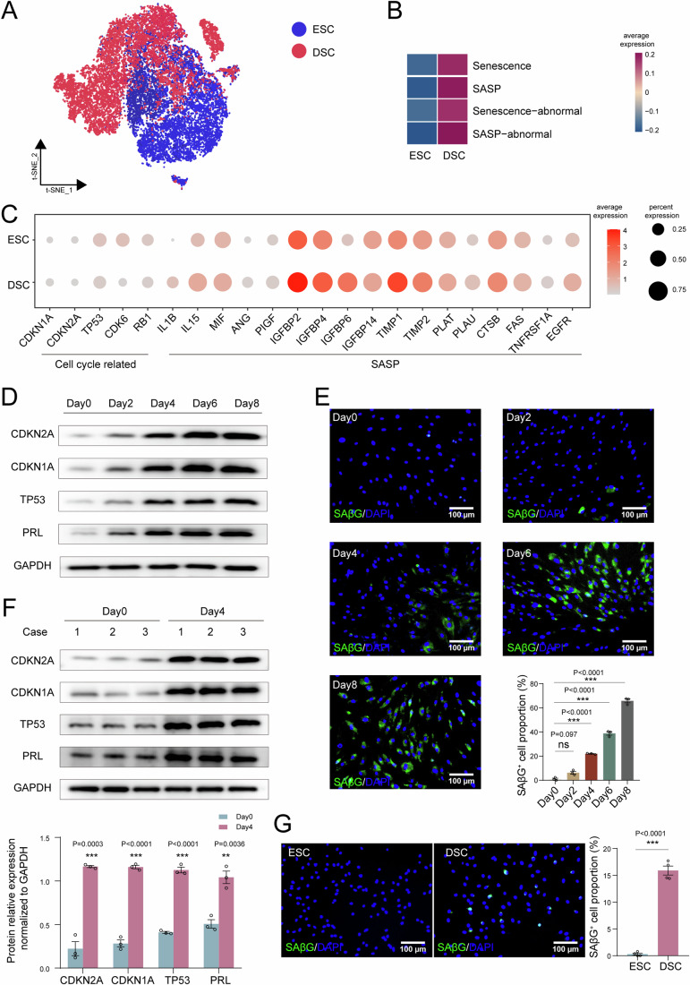
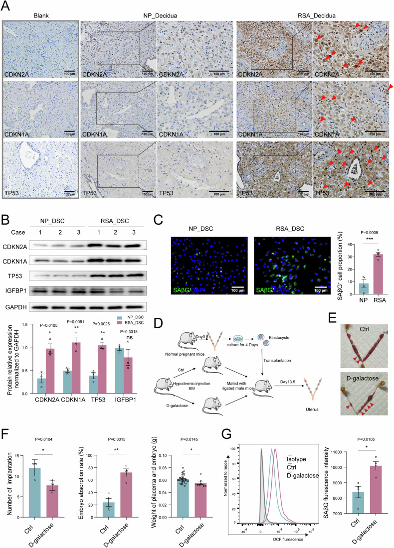
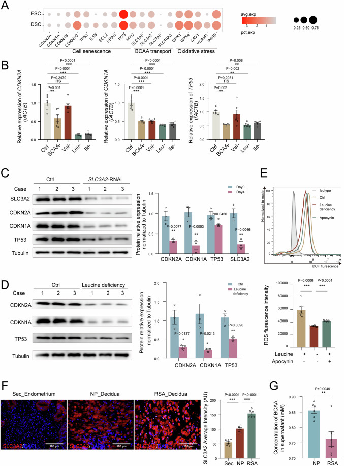
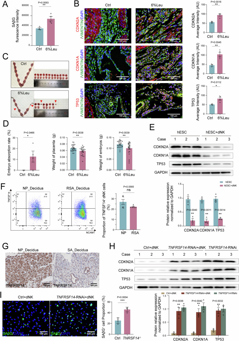
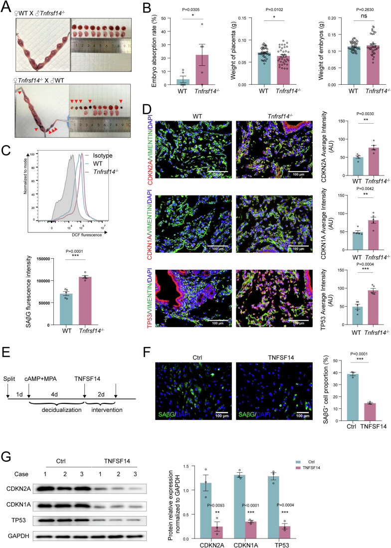
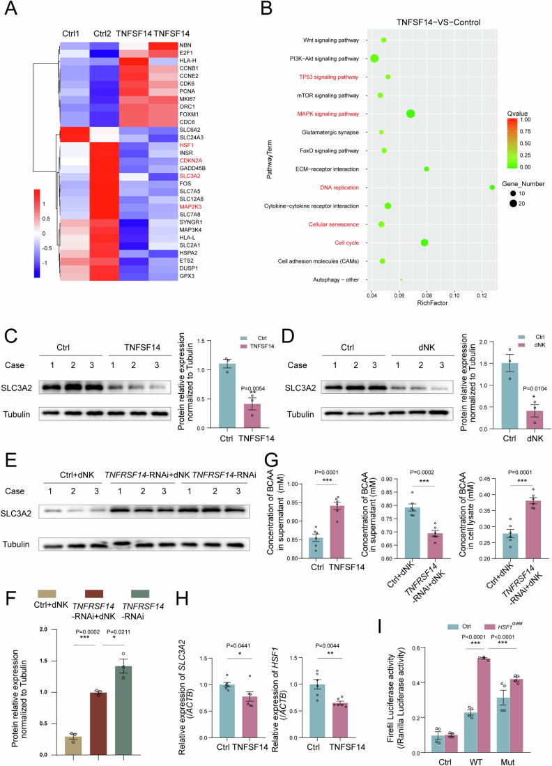
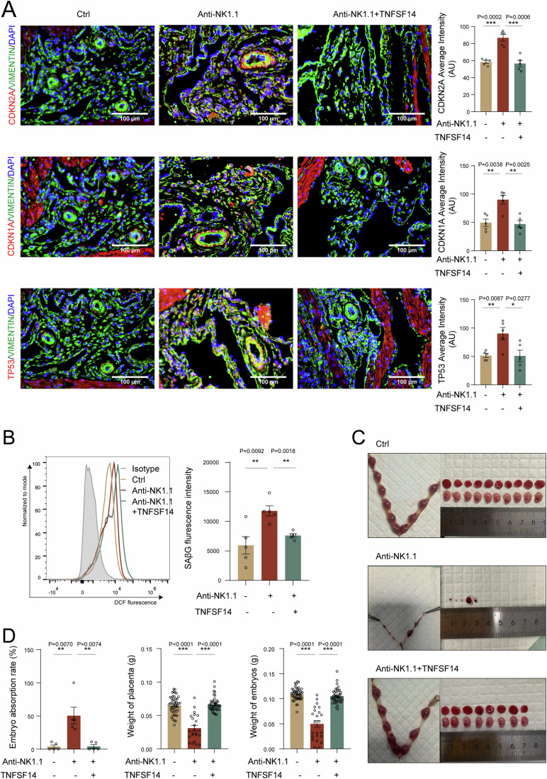
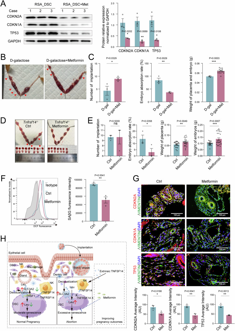
In preparation for a potential pregnancy, the endometrium of the uterus changes into a temporary structure called the decidua. Senescent decidual stromal cells (DSCs) are enriched in the decidua during decidualization, but the underlying mechanisms of this process remain unclear. Here, we performed single-cell RNA transcriptomics on ESCs and DSCs and found that cell senescence during decidualization is accompanied by increased levels of the branched-chain amino acid (BCAA) transporter SLC3A2. Depletion of leucine, one of the branched-chain amino acids, from cultured media decreased senescence, while high leucine diet resulted in increased senescence and high rates of embryo loss in mice. BCAAs induced senescence in DSCs via the p38 MAPK pathway. In contrast, TNFSF14+ decidual natural killer (dNK) cells were found to inhibit DSC senescence by interacting with its ligand TNFRSF14. As in mice fed high-leucine diets, both mice with NK cell depletion and Tnfrsf14-deficient mice with excessive uterine senescence experienced adverse pregnancy outcomes. Further, we found excessive uterine senescence, SLC3A2-mediated BCAA intake, and insufficient TNFRSF14 expression in the decidua of patients with recurrent spontaneous abortion. In summary, this study suggests that dNK cells maintain senescence homeostasis of DSCs via TNFSF14/TNFRSF14, providing a potential therapeutic strategy to prevent DSC senescence-associated spontaneous abortion.

Keywords: Abortion; Aging; Decidualization; Leucine; TNFRSF14.


Conflict of interest statement

The authors declare no competing interests.

Figures

Figure 1
Figure 1. The decidualization process is accompanied with senescent DSCs.
(A) Subpopulation of stromal cells of secretory endometrium and decidua in t-SNE plots through single-cell sequencing. (B) QuSAGE analysis of ESCs and DSCs through single-cell sequencing. (C) Bubble diagram presented the average expression of cell cycle and senescence-associated secretory phenotype (SASP) related genes based on the results of single-cell sequencing. (D) Human ESC cell line (hESCs) was treated with 8-bromo-cAMP (0.5 mM) plus MPA (1 μM) for different times, and western blotting assay was used to detect the expression of CDKN2A, CDKN1A, TP53 (indicator of cell senescence) and PRL. (E) SAβG staining (indicator of cell senescence) and statistical data of decidualized hESCs (n = 3 biological replicates per group). (F) Primary ESCs were treated with 8-bromo-cAMP (0.5 mM) plus MPA (1 μM) for 4 days, and the protein of CDKN2A, CDKN1A, TP53, and PRL were measured by western blotting (n = 3 biological replicates per group), relative expression levels of proteins were standardized using internal reference GAPDH. (G) SAβG staining in primary ESCs (n = 4 biological replicates) and primary DSCs (n = 4 biological replicates). Statistical data were presented as mean ± SEM. **P < 0.01, ***P < 0.001, ns, no significance, using one-way ANOVA with Bonferroni multiple comparisons test (E), using a two-tailed, unpaired Student’s t test (F, G). Source data are available online for this figure.
Figure 2
Figure 2. Excessive senescence of DSCs leads to pregnancy loss.
(A) Expression of CDKN2A, CDKN1A, and TP53 in the decidual from normal pregnancy (NP) and females with unexplained recurrent spontaneous abortion (RSA) were analyzed by immunohistochemistry (n = 6 biological replicates for each group). Scale bar, 100 μm. (B) The expression of CDKN2A, CDKN1A, TP53, and IGFBP1 in DSCs of NP (n = 3 biological replicates) and RSA (n = 3 biological replicates) was detected by western blotting, relative expression levels of proteins were standardized using internal reference GAPDH. (C) The Immunofluorescence staining of SAβG in DSCs of NP (n = 4 biological replicates) and RSA (n = 4 biological replicates). Scale bar, 100 μm. (D) Schematic diagram of aging mouse model construction. (E, F) The pregnancy outcome at the gestation of day 13.5 in the control (n = 4 biological replicates) and D-galactose injected group (n = 4 biological replicates). Number of blastocyst implantation, embryo resorption rate, and weight of the embryo and placenta in the control (n = 4 biological replicates) and D-galactose (n = 4 biological replicates) group. (G) SAβG activity of DSCs were measured by flow cytometry in the control (n = 4 biological replicates) and D-galactose (n = 4 biological replicates) group. Data were presented as mean ± SEM. *P < 0.05, **P < 0.01, ***P < 0.001, ns, no significance, using a two-tailed, unpaired Student’s t test. NP normal pregnancy, RSA recurrent spontaneous abortion. Source data are available online for this figure.
Figure 3
Figure 3. Leucine accumulation induces excessive senescence of DSC and spontaneous abortion.
(A) Bubble diagram showing the average expression of BCAA transport and oxidative stress-related genes of stromal cells from control endometrium (ESC) and decidua of normal pregnancy (DSC) through single-cell sequencing. (B) During the process of decidualization (cAMP plus MPA), hESCs were cultured with medium lacking BCAA, leucine, valine, or isoleucine, respectively, for 96 h. The mRNA expression of CDKN2A, CDKN1A, and TP53 were detected by qRT-PCR (n = 6 biological replicates per group). (C) During decidualization, the expression of SLC3A2, CDKN2A, CDKN1A, and TP53 in control and siSLC3A2 hESCs were measured by western blotting (n = 3 biological replicates per group), relative expression levels of proteins were standardized using internal reference Tubulin. (D) hESCs were cultured with media of leucine deficiency during decidualization, and CDKN2A, CDKN1A, and TP53 expression were detected by western blotting (n = 3 biological replicates per group), relative expression levels of proteins were standardized using internal reference Tubulin. (E) hESCs were cultured with normal media, leucine deficiency media, or normal media plus apocynin (10 μM) during decidualization, and ROS were measured by flow cytometry (n = 6 biological replicates per group). (F) The expression of SLC3A2 in ESCs of secretory phase endometrium, DSCs of NP and RSA were detected by immunofluorescence (n = 6 biological replicates per group). Scale bar, 100 μm. (G) Concentration of BCAA in supernatant of DSCs form NP (n = 6 biological replicates) and RSA (n = 6 biological replicates) were measured by BCAA detection kit. NP normal pregnancy, RSA recurrent spontaneous abortion. Data were presented as mean ± SEM. *P < 0.05, **P < 0.01, ***P < 0.001, ns, no significance, using one-way ANOVA with Bonferroni multiple comparisons test (B), using a two-tailed, unpaired Student’s t test (CG). Source data are available online for this figure.
Figure 4
Figure 4. dNK cell inhibits DSC senescence in a TNFSF14/TNFRSF14-dependent manner.
(AC) C57BL/6 pregnant mice were fed with control (1.2% leucine) (n = 3 biological replicates) or high leucine (6%) (n = 4 biological replicates) fodder, the SAβG activity of DSCs were measured by flow cytometry, and expression of CDKN2A, CDKN1A, and TP53 of DSCs were detected by immunofluorescence. (D) The embryo resorption rate, the weight of placenta and embryo of pregnant mice were quantified at the gestation of day 13.5 (control fodder: n = 3 biological replicates, high leucine fodder: n = 4 biological replicates). (E) Decidualized hESCs were co-cultured with dNK cells for another 48 h, and CDKN2A, CDKN1A, and TP53 expression were detected by western blotting (n = 3 biological replicates per group), relative expression levels of proteins were standardized using internal reference GAPDH. (F) Compared the expression of TNFSF14 on dNK cells of NP (n = 6 biological replicates) and SA (n = 6 biological replicates) by flow cytometry. (G) Compared the expression of TNFRSF14 on DSCs of NP (n = 6 biological replicates) and SA (n = 6 biological replicates) by Immunohistochemistry. Scale bar, 100 μm. (H, I) Decidualized siTNFRSF14-hESCs were co-cultured with dNK cells for another 48 h, western blotting indicated CDKN2A, CDKN1A, and TP53 expression (n = 3 biological replicates per group), relative expression levels of proteins were standardized using internal reference GAPDH, and immunofluorescence showed SAβG+ cells (n = 4 biological replicates per group). NP: normal pregnancy; RSA: recurrent spontaneous abortion. Data were presented as mean ± SEM. *P < 0.05, **P < 0.01, ***P < 0.001, ns, no significance, using a one-tailed (A, B, D), two-tailed, unpaired Student’s t test (E, F, H, I). Source data are available online for this figure.
Figure 5
Figure 5. TNFSF14/TNFRSF14 signal maintains DSC senescence homeostasis and normal pregnancy.
(A, B) At the gestation of day 13.5, embryo resorption, weight of placenta and embryo in Tnfrsf14 knockout pregnant mice (Tnfrsf14−/−♀×WT♂, n = 5 biological replicates) and WT pregnant mice (WT♀×Tnfrsf14−/−♂, n = 5 biological replicates) were recorded. (C, D) The SAβG activity of DSCs were measured by flow cytometry, and expression of CDKN2A, CDKN1A and TP53 of DSCs were detected by immunofluorescence (n = 5 biological replicates per group). (E) Schematic diagram of decidualized hESCs treated with recombinant TNFSF14 protein. (F, G) After decidualized hESCs were treated with TNFSF14 (250 ng/mL, 48 h), SAβG+ cells were detected by immunofluorescence, and CDKN2A, CDKN1A, and TP53 expression were measured by western blotting (n = 3 biological replicates per group), relative expression levels of proteins were standardized using internal reference GAPDH. Scale bar, 100 μm. Data were presented as mean ± SEM. *P < 0.05, **P < 0.01, ***P < 0.001, ns, no significance, using a one-tailed (BD), two-tailed, unpaired Student’s t test (F, G). Source data are available online for this figure.
Figure 6
Figure 6. TNFSF14+ dNK cell control DSC senescence by the SLC3A2/BCAA signaling pathway.
(A) Bubble diagram showed the enrichment of pathway for DSCs treated with TNFSF14. (B) Heat map showed the relative expression of differential gene for DSCs treated with TNFSF14. (C, D) Decidualized hESCs were treated with TNFSF14 (250 ng/mL), or co-cultured with dNK cells (5 × 10⁵) for 48 h, SLC3A2 expression levels were detected with western blotting (n = 3 biological replicates per group), relative expression levels of proteins were standardized using internal reference Tubulin. (E, F) Decidualized siTNFRSF14 hESCs were co-cultured with dNK cells for 48 h, SLC3A2 expression were detected by western blotting (n = 3 biological replicates per group), relative expression levels of proteins were standardized using internal reference Tubulin. (G) Decidualized hESCs were treated with TNFSF14 (250 ng/mL), or decidualized siTNFRSF14 hESCs were co-cultured with dNK cells for 48 h, and then the concentration of BCAA was measured (n = 6 biological replicates per group). (H) After decidualized hESCs were treated with TNFSF14 (250 ng/mL, 48 h), the transcriptional levels of SLC3A2 and HSF1 were detected by RT-PCR (n = 6 biological replicates per group). (I) Dual luciferase reporter assays were conducted in hESCs to verify the combination of HSF1 and WT or mutated SLC3A2 promoter region (n = 4 biological replicates per group). Data were presented as mean ± SEM. *P < 0.05, **P < 0.01, ***P < 0.001, using a two-tailed, unpaired Student’s t test. Source data are available online for this figure.
Figure 7
Figure 7. TNFSF14 reduces risk of NK cell depletion-associated pregnancy loss with excessive DSC senescence.
After C57BL/6 pregnant mice were treated with NK1.1 neutralizing antibody (n = 5 biological replicates) or isotype control IgG antibody (n = 5 biological replicates) by intraperitoneal injection, and at the same time, mice were injected with recombinant TNFSF14 protein intraperitoneally (n = 5 biological replicates). (A, B) SAβG activity of DSCs were measured by flow cytometry, and CDKN2A, CDKN1A, and TP53 expression of DSCs were verified by immunofluorescence. (C, D) The pregnancy outcome, embryo resorption, weight of placenta, and embryo were counted at the gestation of day 13.5. Data were presented as mean ± SEM. *P < 0.05, **P < 0.01, ***P < 0.01, using a two-tailed, unpaired Student’s t test. Source data are available online for this figure.
Figure 8
Figure 8. Metformin reduces the risk of excessive DSC senescence-associated pregnancy loss.
(A) DSCs of spontaneous abortion were treated with metformin (2.5 mM) for 48 h, CDKN2A, CDKN1A, and TP53 expression were detected by western blotting (n = 3 biological replicates per group), relative expression levels of proteins were standardized using internal reference GAPDH. (B, C) After treatment with metformin (200 mg/kg, every other day) in aging pregnant female mice, number of blastocyst implantation, embryo absorption, weight of placenta and embryo were counted at the gestation of day 13.5 (D-galactose group: n = 3 biological replicates, metformin group: n = 3 biological replicates). (D, E) Tnfrsf14-/- pregnant mice were treated with metformin by gavage (n = 3, biological replicates per group, 200 mg/kg, every other day), embryo resorption, weight of placenta and embryo were calculated at the gestation of day 13.5. (F, G) SAβG activity of DSCs were measured by flow cytometry, and CDKN2A, CDKN1A, and TP53 expression of DSCs were verified by immunofluorescence (n = 3 biological replicates per group). Scale bar, 100 μm. (H) Schematic roles of decidual NK cells in preventing spontaneous abortion by maintaining homeostasis of DSC senescence during early pregnancy. During decidualization, increased BCAA transport mediated by SLC3A2 induces senescent DSCs, whereas TNFSF14+ dNK cells control DSC senescence by the TNFSF14/TNFRSF14-SLC3A2/leucine regulatory axis. Excessive senescence of DSC induced by the imbalance of TNFSF14/TNFRSF14-SLC3A2/leucine regulatory axis increases the risk of adverse pregnancy outcomes. The image was drew by the Figdraw. Data were presented as mean ± SEM. *P < 0.05, **P < 0.01, ***P < 0.001 using a one-tailed (C, EG), two-tailed, unpaired Student’s t test (A). Source data are available online for this figure.

References

    1. Brand KMG, Saarelainen L, Sonajalg J, Boutmy E, Foch C, Vaarasmaki M, Morin-Papunen L, Schlachter J, Group CS, Hakkarainen KM et al (2022) Metformin in pregnancy and risk of adverse long-term outcomes: a register-based cohort study. BMJ Open Diabetes Res Care 10(1):e002363 - PMC - PubMed
    1. Brighton PJ, Maruyama Y, Fishwick K, Vrljicak P, Tewary S, Fujihara R, Muter J, Lucas ES, Yamada T, Woods L et al (2017) Clearance of senescent decidual cells by uterine natural killer cells in cycling human endometrium. Elife 6:e31274 - PMC - PubMed
    1. Brosens I, Puttemans P, Benagiano G (2019) Placental bed research: I. The placental bed: from spiral arteries remodeling to the great obstetrical syndromes. Am J Obstet Gynecol 221:437–456 - PubMed
    1. Chen S, Gan D, Lin S, Zhong Y, Chen M, Zou X, Shao Z, Xiao G (2022) Metformin in aging and aging-related diseases: clinical applications and relevant mechanisms. Theranostics 12:2722–2740 - PMC - PubMed
    1. Crespo AC, Mulik S, Dotiwala F, Ansara JA, Sen Santara S, Ingersoll K, Ovies C, Junqueira C, Tilburgs T, Strominger JL et al (2020) Decidual NK cells transfer granulysin to selectively kill bacteria in trophoblasts. Cell 182:1125–1139.e1118 - PMC - PubMed

LinkOut - more resources