. 2024 Oct;634(8033):482-491.
doi: 10.1038/s41586-024-07910-2. Epub 2024 Sep 11.

Promotion of DNA end resection by BRCA1-BARD1 in homologous recombination

Affiliations

Promotion of DNA end resection by BRCA1-BARD1 in homologous recombination

Sameer Salunkhe et al. Nature. 2024 Oct.

Abstract

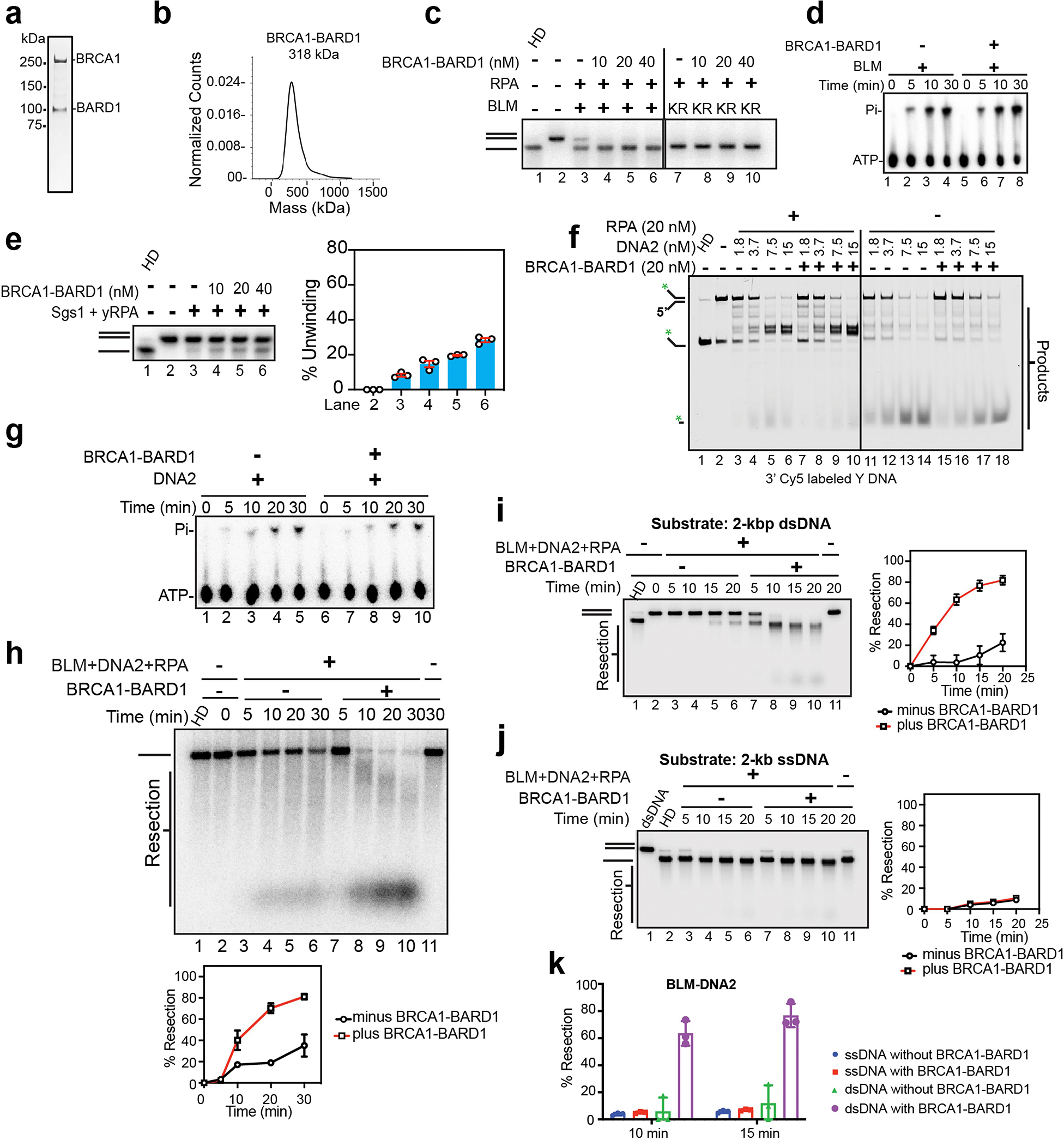
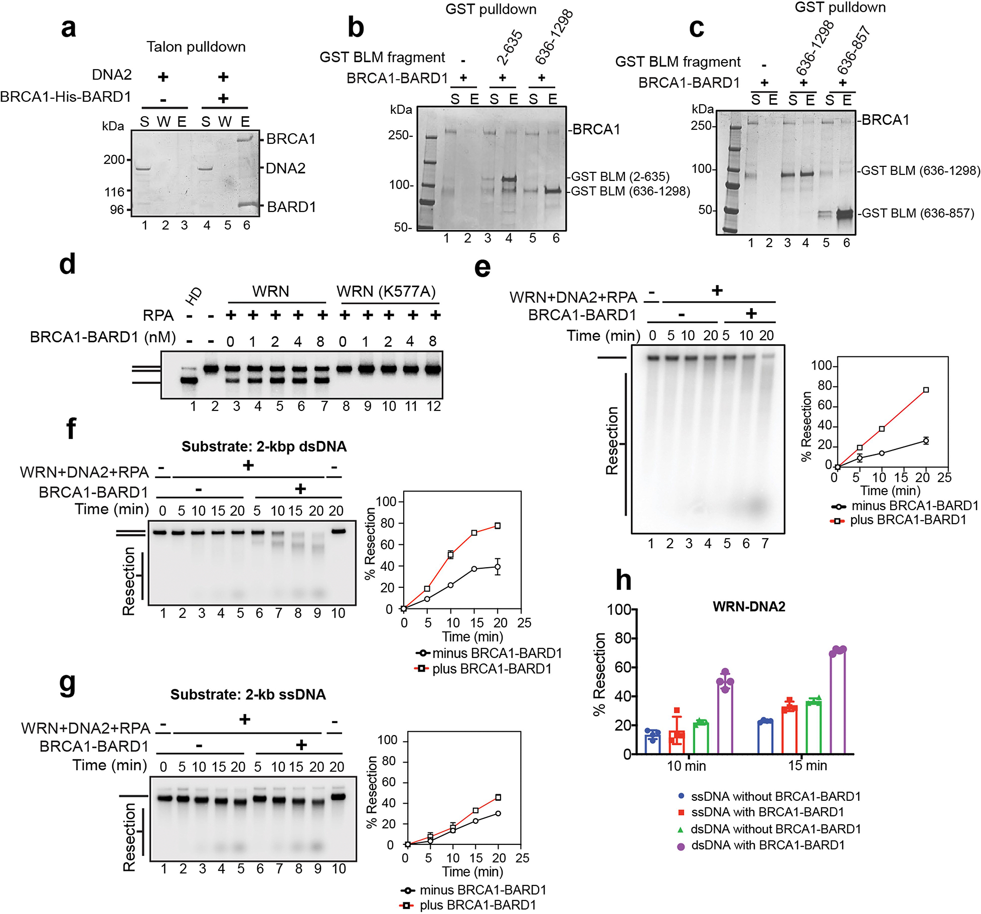
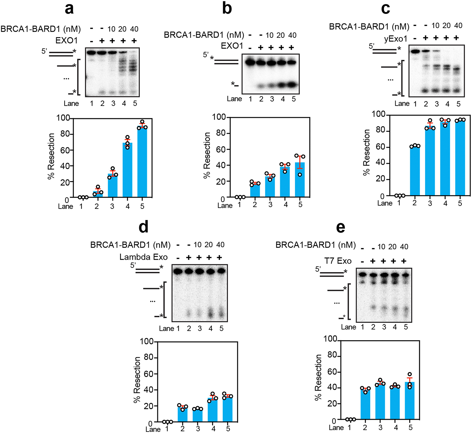
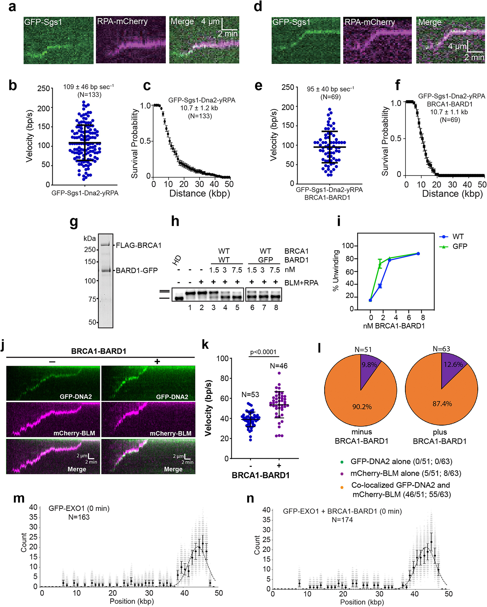
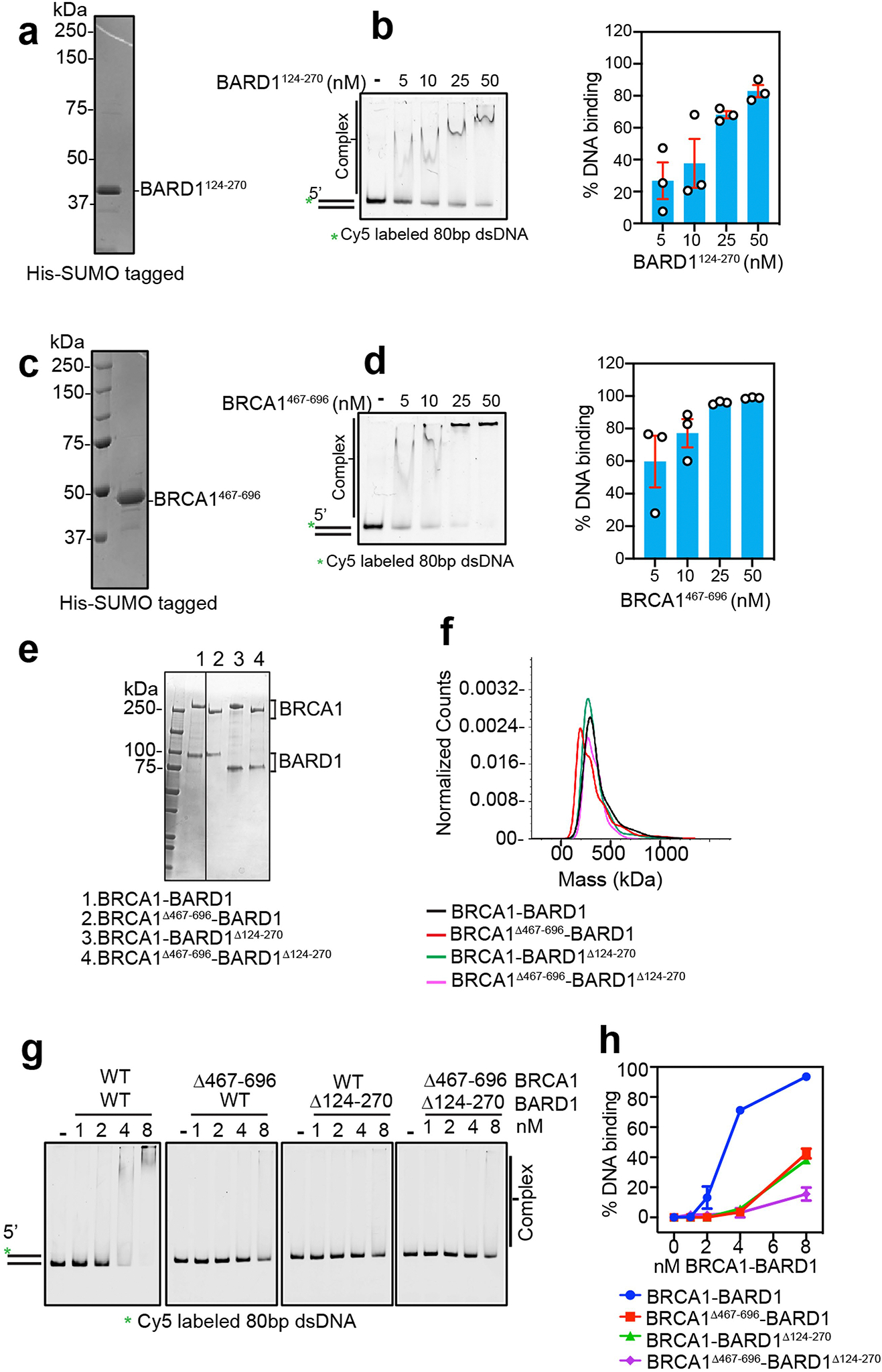
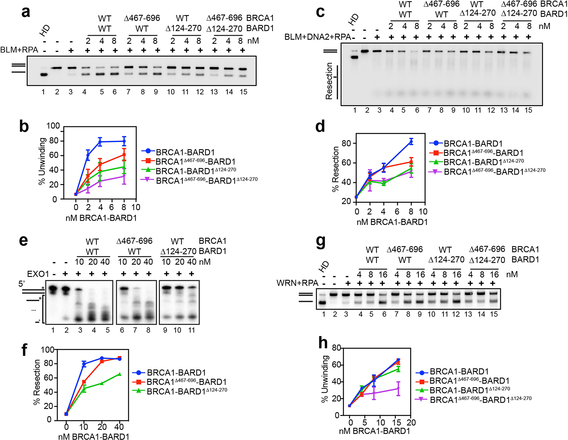
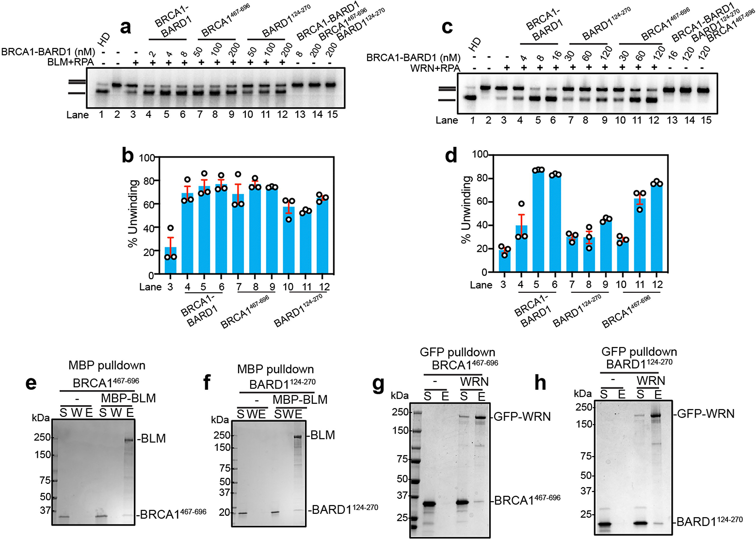
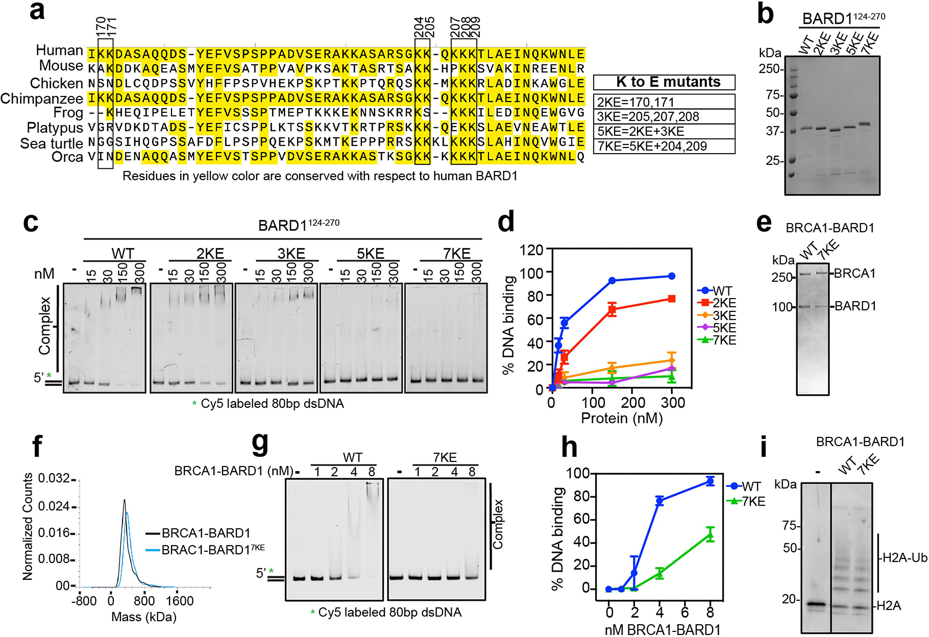
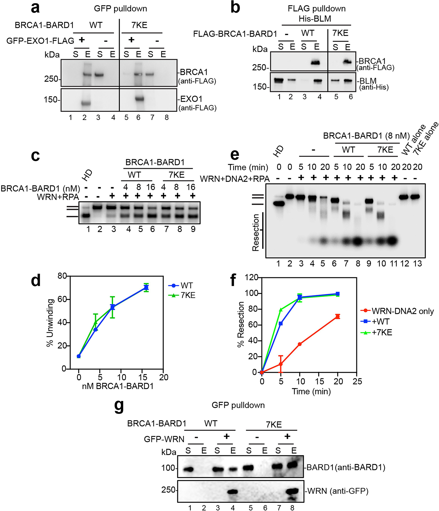
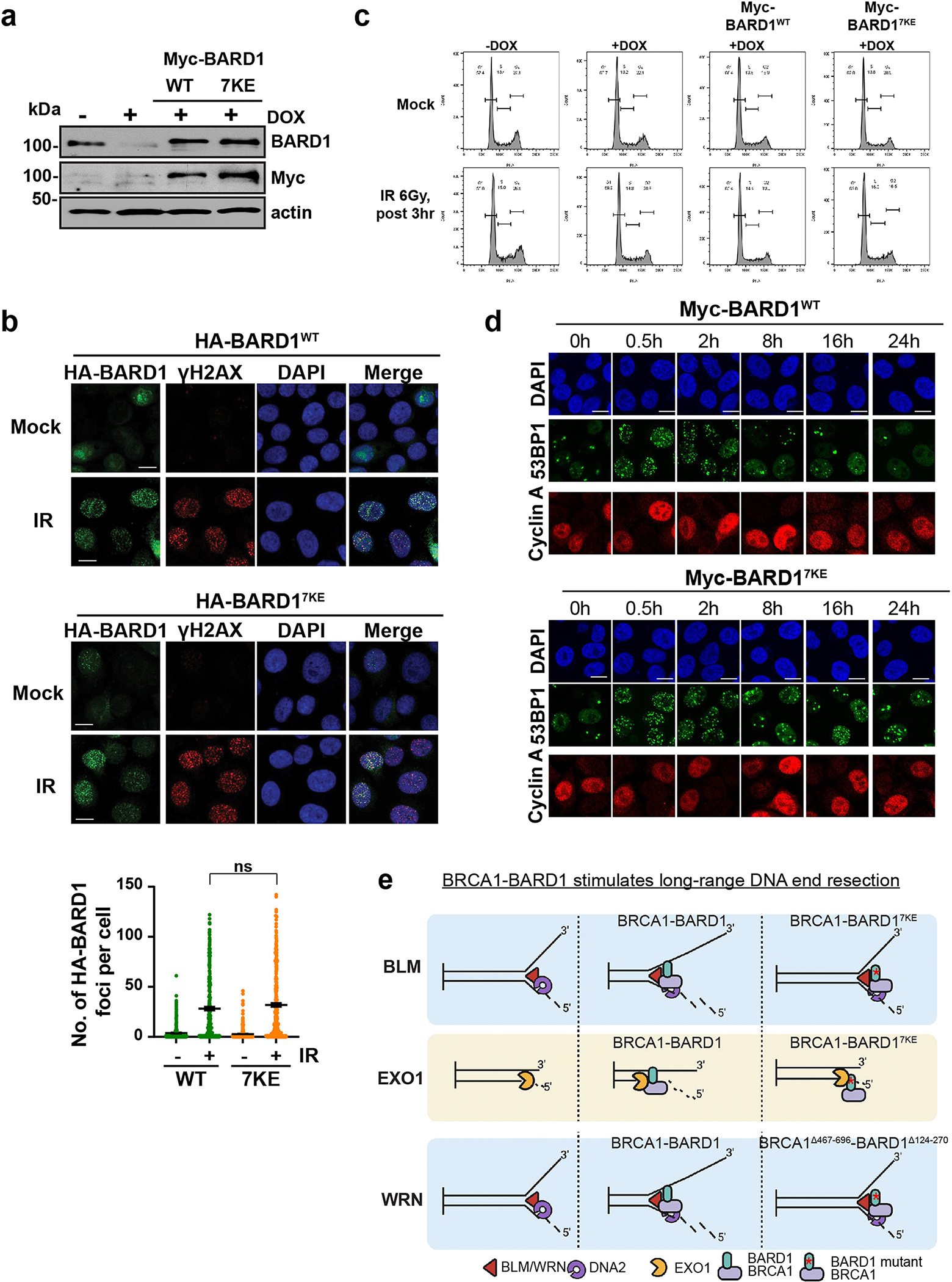
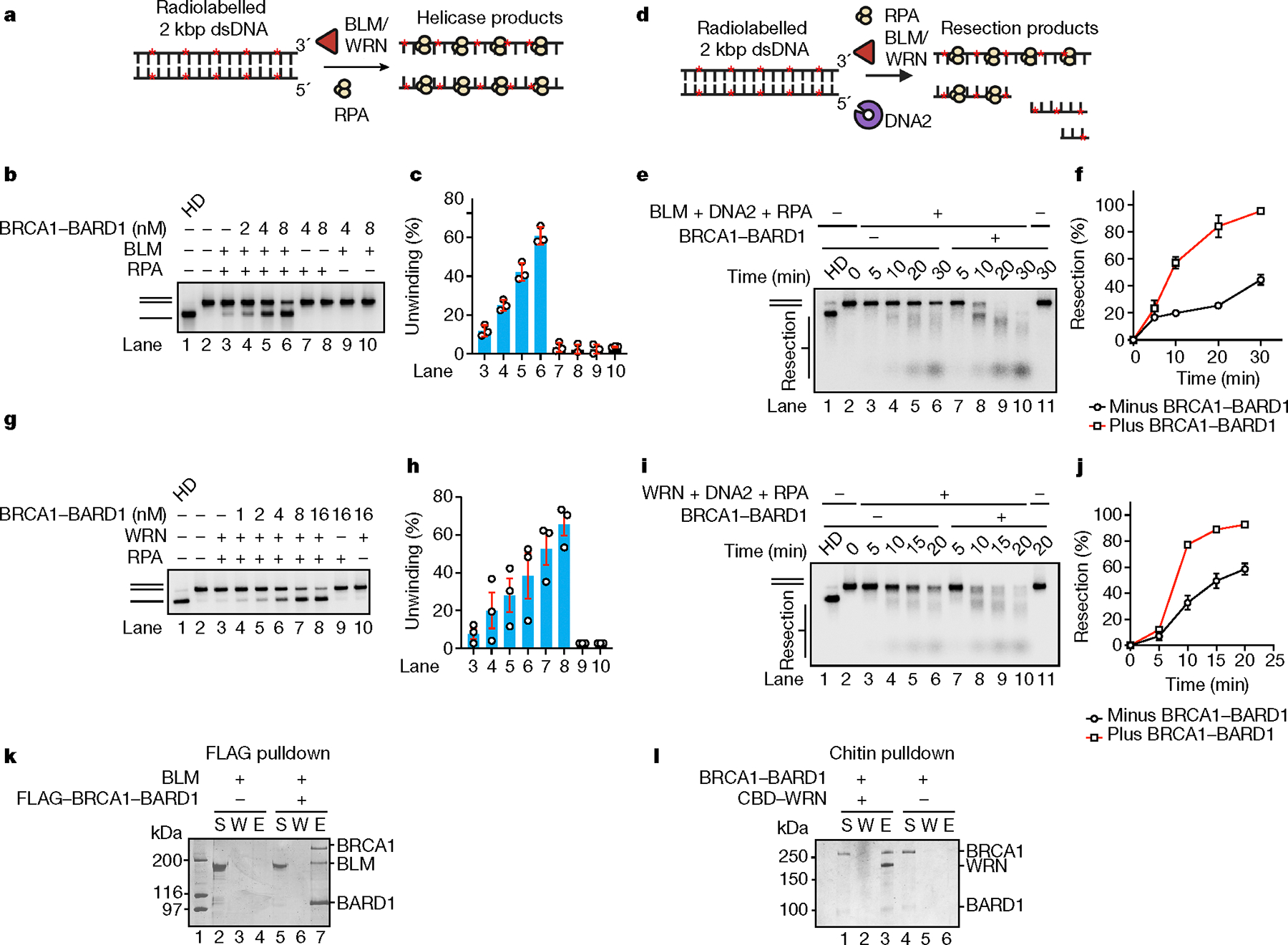
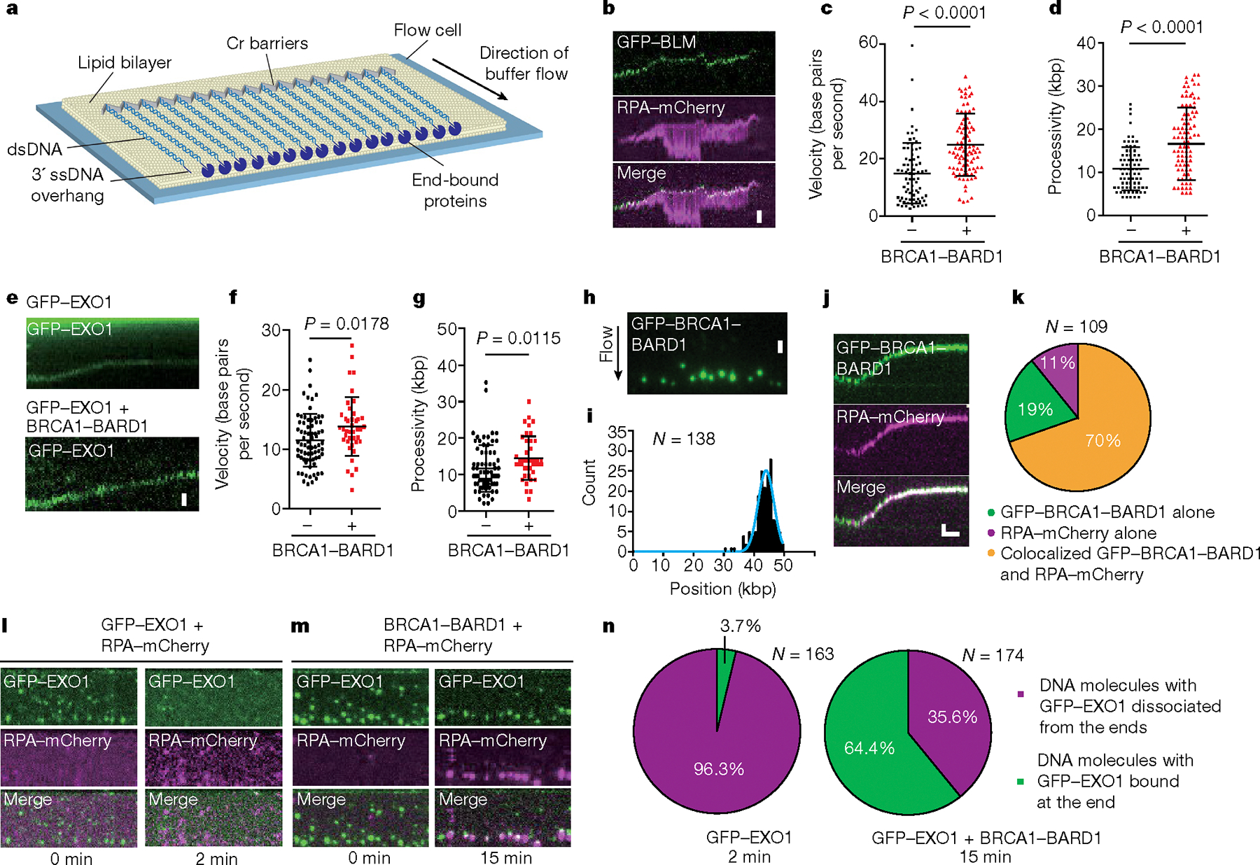
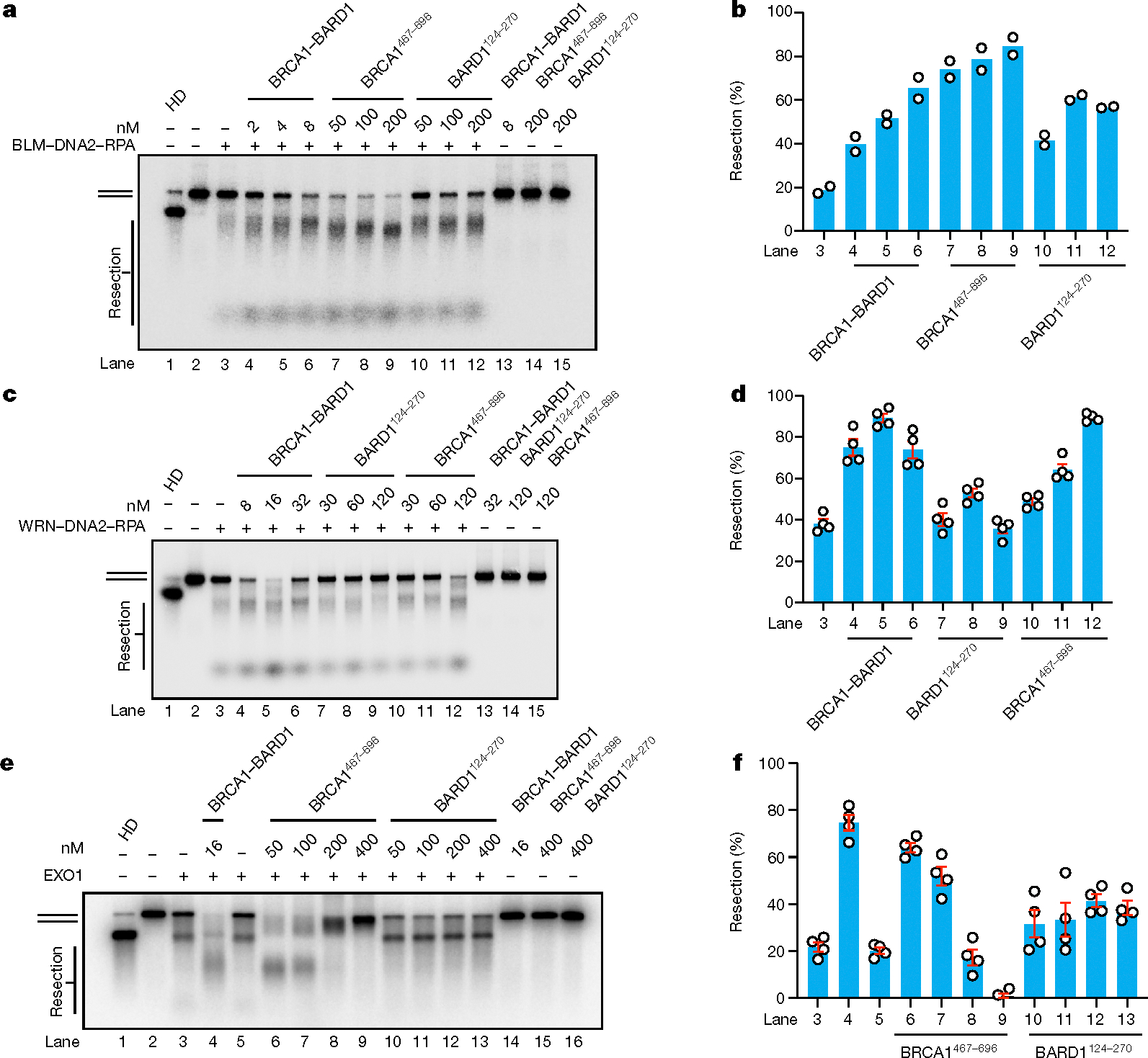
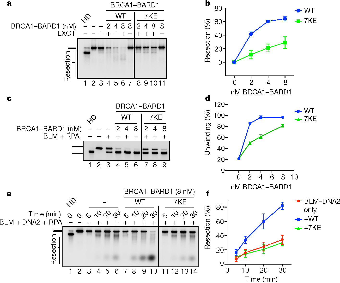
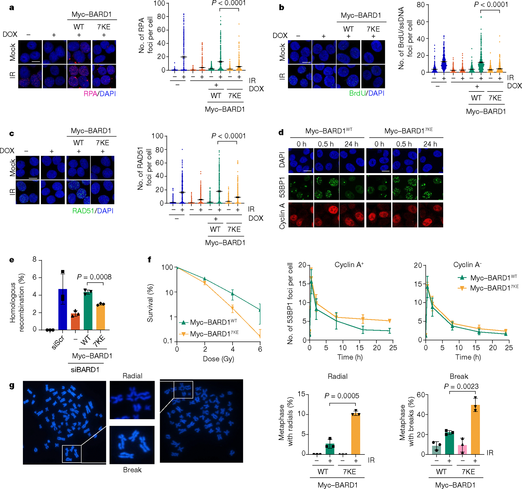
The licensing step of DNA double-strand break repair by homologous recombination entails resection of DNA ends to generate a single-stranded DNA template for assembly of the repair machinery consisting of the RAD51 recombinase and ancillary factors1. DNA end resection is mechanistically intricate and reliant on the tumour suppressor complex BRCA1-BARD1 (ref. 2). Specifically, three distinct nuclease entities-the 5'-3' exonuclease EXO1 and heterodimeric complexes of the DNA endonuclease DNA2, with either the BLM or WRN helicase-act in synergy to execute the end resection process3. A major question concerns whether BRCA1-BARD1 directly regulates end resection. Here, using highly purified protein factors, we provide evidence that BRCA1-BARD1 physically interacts with EXO1, BLM and WRN. Importantly, with reconstituted biochemical systems and a single-molecule analytical tool, we show that BRCA1-BARD1 upregulates the activity of all three resection pathways. We also demonstrate that BRCA1 and BARD1 harbour stand-alone modules that contribute to the overall functionality of BRCA1-BARD1. Moreover, analysis of a BARD1 mutant impaired in DNA binding shows the importance of this BARD1 attribute in end resection, both in vitro and in cells. Thus, BRCA1-BARD1 enhances the efficiency of all three long-range DNA end resection pathways during homologous recombination in human cells.


Conflict of interest statement

Competing interests The authors declare no competing interests.

Figures

Extended Data Fig. 1 |
Extended Data Fig. 1 |. Influence of BRCA1-BARD1 on BLM and DNA2 activities.
a, SDS-PAGE analysis of BRCA1-BARD1 purified as described. b, Mass photometry profile of BRCA1-BARD1, n = 3. c, Testing of BLM helicase mutant with BRCA1-BARD1. BRCA1-BARD1 was incubated with wild-type (8 nM) or the K695R (8 nM) variant of BLM and RPA (200 nM) with the 32P-labeled 2-kbp dsDNA substrate, n = 2. d, BRCA1-BARD1 (100 nM) was tested for its influence on ATP hydrolysis by BLM (10 nM). Following incubation of proteins with γ32P-ATP and unlabeled 2-kbp dsDNA (64 nM ends), reactions were analyzed by thin layer chromatography, n = 3. e, BRCA1-BARD1 was tested for its effect on the unwinding of a 2-kbp 32P-labeled dsDNA by Sgs1 (25 nM) and yRPA (200 nM) in a 20 min reaction. HD, heat-denatured DNA. Error bars show mean ± SEM; n = 3. f, BRCA1-BARD1 (20 nM) and DNA2 were incubated with 3′ Cy5-labeled Y DNA with and without RPA (20 nM) and then analyzed, n = 3. g, BRCA1-BARD1 (100 nM) was tested for its effect on ATP hydrolysis by DNA2 (100 nM). Reactions were analyzed following incubation of proteins with γ32P-ATP and unlabeled 2-kbp dsDNA (64 nM ends), n = 2. h, BRCA1-BARD1 (10 nM), BLM (1 nM), DNA2 (15 nM), RPA (200 nM) were incubated with the 32P-labeled 2-kbp dsDNA substrate. Reactions were subjected to heat denaturation prior to gel electrophoresis for 2 h followed by data quantification. Error bars show mean ± SEM; n = 3. i, dsDNA or j, ssDNA generated by heat denaturation of the dsDNA substrate was incubated with BLM (1 nM), DNA2 (15 nM), RPA (200 nM) with or without BRCA1-BARD1 (10 nM). Results were quantified and plotted. Error bars show mean ± SD; n = 3. In k, results from the 10 min and 15 min time points of experiments in panel i and j were graphed. n=number of independent experiments. Uncropped gel images are provided in Supplementary Fig. 1.
Extended Data Fig. 2 |
Extended Data Fig. 2 |. Characterization of WRN-DNA2 resection stimulation by BRCA1-BARD1.
a, His6-tagged BRCA1-BARD1 was tested for DNA2 interaction by affinity pulldown using Talon resin (specific for the His6 tag). The supernatant (S), wash (W) and SDS eluate (E) fractions were analyzed by SDS-PAGE and Coomassie blue staining, n = 2. b, c, Interaction between BRCA1-BARD1 and BLM fragments was assessed by affinity pulldown. Indicated GST-tagged BLM fragments immobilized on glutathione resin were incubated with BRCA1-BARD1. Proteins were eluted from the resin and analyzed by SDS-PAGE with Coomassie blue staining. S, supernatant and E, SDS eluate of resin, n = 2. d, Wild-type WRN (2 nM) or WRNK577A (2 nM) was tested with BRCA1-BARD1, RPA (200 nM), and the 2-kbp 32P-labeled dsDNA for helicase activity, n = 2. e, Effect of BRCA1-BARD1 (10 nM) on DNA end resection mediated by WRN (2 nM)-DNA2 (20 nM), RPA (200 nM), reaction mixtures were subjected to heat denaturation prior to gel electrophoresis for 2 h followed by data quantification. Error bars show mean ± SEM; n = 4. f, dsDNA or g, ssDNA generated by heat denaturation of the 2-kbp dsDNA substrate was incubated with WRN (2 nM), DNA2 (20 nM), RPA (200 nM) with or without BRCA1-BARD1 (10 nM) for the indicated times. Results from each dsDNA resection or ssDNA digestion experiment were quantified and plotted. Error bars show mean ± SD; n = 3. h, results from the 10 min and 15 min time points of experiments testing WRN-DNA2 from panel f and g were plotted as bar graphs. HD, heat-denatured DNA substrate. n=number of independent experiments. Uncropped gel images are provided in Supplementary Fig. 1.
Extended Data Fig. 3 |
Extended Data Fig. 3 |. Stimulation of EXO1 by BRCA1-BARD1.
a, BRCA1-BARD1 and EXO1 (5 nM) were incubated with 3′ 32P-labeled 80-mer dsDNA (2.5 nM) for 10 min and then analyzed. The results were quantified and plotted. Error bars show mean ± SEM; n = 3. b, Influence of BRCA1-BARD1 on removal of the 5′ nucleotide by EXO1 (5 nM). BRCA1-BARD1 and EXO1 were incubated with 5′ end labeled 30-mer dsDNA (2.5 nM) for 10 min and then analyzed. The results were quantified and plotted. Error bars show mean ± SEM; n = 3. c, BRCA1-BARD1 and yExo1 (5 nM) were incubated with 3′ 32P-labeled 80-mer dsDNA for 10 min and then analyzed. The results were quantified and plotted. Error bars show mean ± SEM; n = 3. d, e, BRCA1-BARD1 was tested with Lambda or T7 exonuclease (0.5 U) (NEB) using the 3′ 32P-labeled 80-mer dsDNA (2.5 nM) for 10 min and then analyzed. The results were quantified and plotted. Error bars show mean ± SEM; n = 3. n=number of independent experiments. Uncropped gel images are provided in Supplementary Fig. 1.
Extended Data Fig. 4 |
Extended Data Fig. 4 |. Single molecule analysis of Sgs1-Dna2.
a, Kymograph showing the movement of GFP-Sgs1 pre–bound to unlabeled dsDNA when chased with yeast Dna2, yeast RPA-mCherry (2 nM) and ATP with b, corresponding velocity distribution (N = 133;n = 3), and c, processivity plot (N = 133;n = 3). d, Kymograph showing the movement of GFP-Sgs1 pre-bound to unlabeled dsDNA when chased with yeast Dna2, yeast RPA-mCherry, BRCA1-BARD1 and ATP with e, corresponding velocity distribution (N = 69;n = 3) and f, processivity plot (N = 69;n = 3). g, Purified BRCA1-BARD1 with a C-terminal GFP tag on BARD1 was analyzed by SDS-PAGE. h, GFP-tagged BRCA1-BARD1 was incubated with BLM (5 nM), RPA (200 nM), and the 2-kbp 32P-labeled dsDNA substrate for 20 min. HD denotes heat-denatured DNA. i, Quantification of the results in panel h. Error bars show mean ± SEM from 3 experiments. j, Kymographs of DNA end resection by mCherry-BLM, GFP-DNA2, RPA and +/− BRCA1-BARD1. k, Velocity of resection reactions containing mCherry-BLM, GFP-DNA2, RPA and +/− BRCA1-BARD1. p < 0.0001; from two-tailed unpaired-t-test. l, Pie charts summarizing the localization pattern of mCherry-BLM and GFP-DNA2 from panel j. m, n, Binding distribution of GFP-EXO1 at 0 min with (N = 174; n = 3) and without BRCA1-BARD1 (N = 163; n = 3). Individual data points correspond to the dataset generated by bootstrap analysis; Error bars represent SD from the mean. N=number of DNA molecules analyzed; n=number of flow cell repeats. Uncropped gel images are provided in Supplementary Fig. 1.
Extended Data Fig. 5 |
Extended Data Fig. 5 |. BRCA1 and BARD1 DNA binding fragments and internal deletion mutants.
a, SDS-PAGE analysis of purified BARD1124–270. b, Testing of BARD1124–270 for DNA binding using Cy5-labeled 80 bp (3 nM) DNA as substrate. Results were quantified and plotted. Error bars show mean ± SEM; n = 3. c, SDS-PAGE analysis of purified BRCA1467–696. d, Testing of BRCA1467–696 for DNA binding using Cy5-labeled 80 bp (3 nM) DNA as substrate. Results were quantified and plotted. Error bars show mean ± SEM; n = 3. e, SDS-PAGE analysis of BRCA1-BARD1, BRCA1Δ467–696-BARD1, BRCA1-BARD1Δ124–270, and BRCA1Δ467–696-BARD1Δ124–270. f, Mass photometry profiles of wild type and mutant BRCA1-BARD1 species, n = 2. g, BRCA1-BARD1 and the indicated mutants were tested for DNA binding using Cy5-labeled 80 bp dsDNA (5 nM) as substrate. The reaction time was 10 min. h, Quantification of results from panel g, Error bars show mean ± SEM; n = 3. n=number of independent experiments. Uncropped gel images are provided in Supplementary Fig. 1.
Extended Data Fig. 6 |
Extended Data Fig. 6 |. Testing of BRCA1-BARD1 internal deletion mutants in DNA end resection.
a, Testing of BRCA1-BARD1 and the indicated mutants on the helicase activity of BLM (1 nM), RPA (200 nM) using the 32P-labeled 2-kbp dsDNA substrate (0.5 nM ends). The reaction time was 30 min. b, Quantification of results from panel a. Error bars show mean ± SEM; n = 3. c, Testing of BRCA1-BARD1 and the indicated mutants for their influence on DNA end resection mediated by BLM (1 nM), DNA2 (15 nM) and RPA (200 nM) using the 32P-labeled 2-kbp dsDNA substrate. The reaction time was 30 min. d, Quantification of results from panel c. Error bars show mean ± SEM; n = 3. e, Testing of BRCA1-BARD1 and the indicated mutants for their influence on DNA end resection mediated by EXO1 (5 nM) using the 3′ 32P-labeled 80-mer dsDNA (2.5 nM) substrate. f, Quantification of results from panel e. Error bars show mean ± SEM; n = 3. g, Testing of BRCA1-BARD1 and the indicated mutants on the helicase activity of WRN (2 nM), RPA (200 nM) using the 32P-labeled 2-kbp dsDNA substrate. The incubation time was 20 min. h, Quantification of results from panel g. Error bars show mean ± SEM; n = 5. n=number of independent experiments.
Extended Data Fig. 7 |
Extended Data Fig. 7 |. Testing of BRCA1 and BARD1 DNA binding domains on BLM and WRN helicase activity.
a, Testing of BRCA1467–696 and BARD1124–270 on the helicase activity of BLM (2 nM), RPA (200 nM) using the 32P-labeled 2-kbp dsDNA substrate. The reaction time was 30 min. b, Quantification of results from panel a. Error bars show mean ± SEM; n = 3. c, Testing of BRCA1467–696 and BARD1124–270 on the helicase activity of WRN (2 nM), RPA (200 nM) using the 32P-labeled 2-kbp dsDNA substrate. The incubation time was 20 min. d, Quantification of results from panel c. Error bars show mean ± SEM; n = 3. e, f, Interaction between BLM and BRCA1467–696 and BARD1124–270 was assessed by affinity pulldown (n = 2). MBP-BLM immobilized on amylose resin was incubated with BRCA1467–696 or BARD1124–270. Proteins were eluted from the resin and analyzed by SDS-PAGE with Coomassie blue staining. S, supernatant, W, wash and E, SDS eluate of resin. g, h, Interaction between GFP-WRN and BRCA1467–696 and BARD1124–270 was assessed by affinity pulldown assays (n = 2). GFP-WRN immobilized on anti-GFP resin was incubated with BRCA1467–696 and BARD1124–270. Proteins were eluted from the resin and analyzed by SDS-PAGE with Coomassie blue staining. S, supernatant, and E, SDS eluate of resin. n=number of independent experiments. Uncropped gel images are provided in Supplementary Fig. 1.
Extended Data Fig. 8 |
Extended Data Fig. 8 |. Construction and characterization of the BARD17KE DNA binding mutant.
a, Alignment of the human BARD1 DNA binding domain encompassing amino acid residues 124–270 with the equivalent region of the indicated BARD1 orthologues. Conserved residues with respect to human BARD1 are highlighted with yellow shade. Boxed lysine residues were changed in various combinations to glutamate to yield the 2KE, 3KE, 5KE, and 7KE mutants. b, SDS-PAGE analysis of BARD1WT and BARD1124–270 mutant species. c, Testing of the BARD1124–270 mutants for DNA binding using the Cy5-labeled 80 bp dsDNA (3 nM) substrate. d, Quantification of results from panel c. Error bars show mean ± SEM; n = 3. e, SDS-PAGE gel of purified BRCA1-BARD1 and BRCA1-BARD17KE. f, Profile of BRCA1-BARD1 and BRCA1-BARD17KE from mass photometry analysis, n = 2. g, Testing of BRCA1-BARD1 and BRCA1-BARD17KE using the a Cy5-labeled 80 bp dsDNA (5 nM) as substrate. h, Quantification of data from panel g. Error bars show mean ± SEM; n = 3. i, Testing BRCA1-BARD1 and BRCA1-BARD17KE for E3 ubiquitin ligase activity with histone H2A as substrate, n = 2. n=number of independent experiments. Uncropped gel images are provided in Supplementary Fig. 1.
Extended Data Fig. 9 |
Extended Data Fig. 9 |. Testing of BRCA1-BARD17KE with EXO1, WRN and WRN-DNA2.
a, BRCA1-BARD17KE was tested for interaction with EXO1. GFP-EXO1-FLAG and FLAG-BRCA1-BARD17KE. were incubated and EXO1 was captured using anti-GFP resin, n = 2. The supernatant and SDS eluate of the pulldown reaction were probed by immunoblotting for BRCA1 and EXO1 using anti-FLAG antibody. b, BRCA1-BARD17KE was tested for interaction with BLM, n = 2. FLAG-BRCA1-BARD17KE was preincubated with His6-BLM and captured using anti-FLAG resin. The supernatant and SDS eluate of the pulldown reaction were probed by immunoblotting for BRCA1 and BLM using anti-FLAG antibody and anti-His antibody. c, Testing of BRCA1-BARD1 and BRCA1-BARD17KE for their influence on the helicase activity of WRN (2 nM) with RPA (200 nM) and the 32P-labeled 2-kbp dsDNA substrate. The reaction time was 20 min. d, Quantification of results from panel c. Error bars show mean ± SEM; n = 3. e, Testing of BRCA1-BARD1 and BRCA1-BARD17KE DNA end resection mediated by WRN (2 nM), DNA2 (20 nM), and RPA (200 nM) using the 32P-labeled 2-kbp dsDNA substrate. f, Quantification of results from panel e. Error bars show mean ± SEM; n = 3. g, BRCA1-BARD17KE was tested for interaction with WRN, n = 2. BRCA1-BARD17KE was incubated with GFP-WRN and the latter was captured using anti-GFP resin. The supernatant and SDS eluate of the pulldown reaction were probed by immunoblotting for BARD1 and WRN using anti-BARD1 and anti-GFP antibody. n=number of independent experiments. Uncropped gel images are provided in Supplementary Fig. 1.
Extended Data Fig. 10 |
Extended Data Fig. 10 |. Cell biological characterization of BARD17KE cells.
a, Immunoblot analysis of lysates from HeLa cells with shRNA-mediated knockdown of BARD1 complemented with Myc-tagged BARD1WT or BARD17KE. b, Representative images of immunostaining of HeLa cells depleted of endogenous BARD1 by doxycycline treatment and expressing HA tagged BARD1WT or BARD17KE either mock-treated or exposed to ionizing radiation (6 Gy IR). Cells were pre-extracted and immunostained with anti-HA antibody (green), anti-γH2AX (red) and with DAPI (blue) to identify nuclei. Dot plot shows HA-BARD1 foci per cell (370, 349, 397 and 353 number of cells counted from left to right) from 3 independent experiments. Error bars show mean ± SEM;, ns=not significant. Scale bars, 10 μm. c, HeLa cells depleted of endogenous BARD1 by doxycycline treatment and expressing BARD1WT or BARD17KE were either mock-treated or exposed to ionizing radiation, and analyzed by single parameter flow cytometry after propidium iodide staining for DNA content (x-axis). See Supplementary fig. 2 for gating strategy. d, HeLa cells depleted of endogenous BARD1 and expressing BARD1 or BARD17KE were exposed to ionizing radiation and incubated for the indicated times. Cells were co-immunostained for 53BP1 foci (green) and cyclin A (to demarcate cells in S/G2, red). Nuclei were stained with DAPI (blue). e, Working model: our results demonstrate that BRCA1-BARD1 associates with DNA ends as well as with BLM, WRN, and EXO1 to accelerate long range resection. Mechanistically, we show that BRCA1-BARD1 translocates with the resection machinery and examination of various BRCA1-BARD1 mutants provides evidence that the DNA binding attribute of BRCA1-BARD1 is needed for efficient end resection by BLM-DNA2, WRN-DNA2, and EXO1. Schematic in e was created using BioRender (https://BioRender.com). Uncropped gel images are provided in Supplementary Fig. 1.
Fig. 1 |
Fig. 1 |. Enhancement of BLM/WRN–DNA2-mediated DNA end resection by BRCA1–BARD1.
a, Schematic of the DNA unwinding assay. b, The effect of BRCA1–BARD1 on unwinding of the 32P-labelled, 2 kbp dsDNA substrate (0.5 nM ends) by BLM (2 nM) and RPA (200 nM) was tested in a 30 min reaction. Lane 1, DNA was heat denatured (HD). c, Quantification of results from b. Error bars show mean ± s.e.m.; n = 3. d, Schematic of BLM/WRN–DNA2 resection assay. e, Resection of the 32P-labelled, 2 kbp dsDNA substrate was tested with BLM (2 nM), DNA2 (15 nM) and RPA (200 nM) with or without BRCA1–BARD1 (10 nM). f, Quantification of data in e. Error bars show mean ± s.e.m.; n = 3. g, The effect of BRCA1–BARD1 on unwinding of the 32P-labelled, 2 kbp dsDNA substrate by WRN (2 nM) and RPA (200 nM) was tested in a 20 min reaction. h, Quantification of results from g. Error bars show mean ± s.e.m.; n = 3. i, Resection of the 32P-labelled, 2 kbp dsDNA substrate was tested with WRN (2 nM), DNA2 (20 nM) and RPA (200 nM) with or without BRCA1–BARD1 (8 nM). j, Quantification of results from i. Error bars show mean ± s.e.m.; n = 3. k, The interaction between BRCA1–BARD1 and BLM was assessed by affinity pulldown (n = 2). FLAG-tagged BRCA1–BARD1 immobilized on anti-FLAG resin was incubated with His-tagged BLM, and proteins were eluted from the resin and analysed by SDS–polyacrylamide gel electrophoresis (SDS–PAGE) with Coomassie blue staining. l, Interaction between BRCA1–BARD1 and WRN was assessed by affinity pulldown (n = 2). WRN (protein tagged with a chitin-binding domain (CBD)) immobilized on chitin resin was incubated with BRCA1–BARD1, and proteins were eluted and analysed as in k. a,d, Red asterisks (*) denote 32P label. Uncropped gel images are provided in Supplementary Fig. 1. n, Number of independent experiments; E, SDS eluate of resin; S, supernatant; W, wash.
Fig. 2 |
Fig. 2 |. Enhancement of EXO1-mediated DNA end resection by BRCA1–BARD1.
a, Schematic of the EXO1 resection assay. b, EXO1 (2 nM) was tested with the 2 kbp, 32P-labelled substrate with and without the presence of BRCA1–BARD1 or RPA (100 nM); incubation time was 10 min. c, Quantification of data in b; error bars show mean ± s.e.m.; n = 3. d, The interaction between BRCA1–BARD1 and EXO1 was assessed by affinity pulldown (n = 2). GFP-tagged EXO1 immobilized on anti-GFP affinity resin was incubated with BRCA1–BARD1, and proteins were eluted from the resin and analysed by SDS–PAGE with Coomassie blue staining. e, Interaction between BRCA1–BARD1 and EXO1 fragments was assessed by affinity pulldown (n = 2). Either glutathione S-transferase (GST)-tagged EXO11–346 or EXO1347–846 was immobilized on glutathione Sepharose and then incubated with BRCA1–BARD1. Supernatant and SDS eluate were analysed by immunoblotting using either anti-FLAG antibody (specific for the FLAG tag on BRCA1) or anti-GST antibody (specific for the GST tag on EXO1 fragments). f, EXO11–346 (10 nM) was incubated with 3′ 32P-labelled (asterisks (*) denote 32P label), 80-mer dsDNA (2.5 nM) with or without BRCA1–BARD1 for 10 min, then analysed. g, Quantification of results from f. Error bars show mean ± s.e.m.; n = 3. a, Red asterisks (*) denote the 32P label. Uncropped gel images are provided in Supplementary Fig. 1. n, Number of independent experiments.
Fig. 3 |
Fig. 3 |. Single-molecule analysis of BRCA1–BARD1 in BLM- and EXO1-dependent resection.
a, Schematic of the DNA curtains analytical set-up. b, Kymographs of DNA resection by GFP–BLM, RPA–mCherry, DNA2 and BRCA1–BARD1. c,d, Velocity (N = 77, n = 3 and N = 93, n = 3) (c) and processivity of reactions (d) with GFP–BLM, RPA–mCherry and DNA2 with or without BRCA1–BARD1 (N = 77, n = 3 and N = 93, n = 3). e, Kymographs of DNA resection by GFP–EXO1 alone and GFP–EXO1 + BRCA1–BARD1. f,g, Velocity (N = 73, n = 3 and N = 41, n = 3) (f) and processivity of resection reactions (N = 73, n = 3 and N = 41, n = 3) (g). h, Wide-field TIRFM image showing association of GFP–BRCA1–BARD1 (green) with DNA ends. i, Binding distribution of GFP–BRCA1–BARD1 following bootstrap analysis (N = 138, n = 4). j, Kymograph showing DNA end resection by unlabelled BLM, GFP–BRCA1–BARD1 (green), unlabelled DNA2 and RPA–mCherry (magenta). k, Percentage of resecting DNA molecules with GFP–BRCA1–BARD1 and RPA–mCherry colocalization (N = 109, n = 4). l, Wide-field TIRFM image showing rapid loss of end-bound GFP–EXO1 in the absence of BRCA1–BARD1. m, Wide-field TIRFM image showing BRCA1–BARD1 mediating GFP–EXO1 retention on DNA in the presence of RPA–mCherry. n, Pie chart summarizing the occupancy of GFP–EXO1 at DNA ends in the absence (2 min time point; N = 163, n = 3) and presence (15 min time point; N = 174, n = 3) of BRCA1–BARD1. c,d,f,g, Error bars represent standard deviation of means, and P values are derived from two-tailed unpaired t-testing. Scale bars, 2 μm (b,e,h,j (vertical)), 3 min (j, horizontal). N, number of DNA molecules analysed; n, number of flow cell repeats.
Fig. 4 |
Fig. 4 |. Functionality of BRCA1467–696 and BARD1124–270 in DNA end resection.
a, Testing the influence of BRCA1467–696 and BARD1124–270 alongside BRCA1–BARD1 on DNA end resection mediated by BLM (2 nM), RPA (200 nM) and DNA2 (15 nM) using the 32P-labelled, 2 kbp dsDNA substrate in a 30 min reaction. b, Quantification of results from a (n = 2). c, Testing the influence of BRCA1467–696 and BARD1124–270 alongside BRCA1–BARD1 on DNA resection mediated by WRN (2 nM), RPA (200 nM) and DNA2 (20 nM) using the 32P-labelled, 2 kbp dsDNA substrate in a 20 min reaction. d, Quantification of results from c. Error bars show mean ± s.e.m.; n = 4. e, Testing the influence of BRCA1467–696 and BARD1124–270 alongside BRCA1–BARD1 on DNA end resection mediated by EXO1 (2 nM) using the 32P-labelled, 2 kbp dsDNA substrate. Reaction time was 10 min. f, Quantification of results from e. Error bars show mean ± s.e.m.; n = 4. Uncropped gel images are provided in Supplementary Fig. 1. n, Number of independent experiments.
Fig. 5 |
Fig. 5 |. Testing of BRCA1–BARD17KE DNA-binding mutant in DNA end resection.
a, EXO1 (2 nM) was incubated with the 2 kbp, 32P-labelled dsDNA substrate with or without the indicated BRCA1–BARD1 species for 10 min and then analysed. b, Quantification of results from a. Error bars show mean ± s.e.m.; n = 3. c, Assessment of BLM (2 nM) and RPA (200 nM) helicase activity using the 2 kbp, 32P-labelled dsDNA substrate for 30 min with or without the indicated BRCA1–BARD1 species. d, Quantification of results from c. Error bars show mean ± s.e.m.; n = 3. e, DNA end resection was examined with BLM (2 nM), RPA (200 nM) and DNA2 (15 nM) using the 2 kbp, 32P-labelled substrate with or without BRCA1–BARD1 or BRCA1–BARD17KE for the indicated times. f, Quantification of results from e. Error bars show mean ± s.e.m.; n = 3. Uncropped gel images are provided in Supplementary Fig. 1. n, Number of independent experiments; WT, wild type.
Fig. 6 |
Fig. 6 |. Cellular phenotypes of the BARD17KE mutant.
a, Representative images of mock-treated (Mock) and irradiated (IR) HeLa cells depleted of endogenous BARD1 by doxycycline treatment (DOX) and expressing BARD1WT or BARD17KE. Cells were immunostained with either anti-RPA antibody (red) or DAPI (blue) to identify nuclei. Error bars show mean ± s.e.m.; n = 3 (300 cells counted). b, Representative images of mock-treated and irradiated cells immunostained with anti-BrdU antibody (green) or DAPI (blue). Error bars show mean ± s.e.m.; n = 3 (300 cells counted). c, Representative images of mock-treated and irradiated cells immunostained with anti-RAD51 antibody (green) or DAPI (blue). Error bars show mean ± s.e.m.; n = 3 (431, 400, 404, 339, 386, 415, 418 and 373 cells counted (left to right)). d, Representative images of irradiated cells co-immunostained for 53BP1 foci (green) and cyclin A (red). Rates of DSB repair in S/G2 cells were determined by scoring 53BP1 foci in cyclin A-positive nuclei. The average number of 53BP1 foci remaining was plotted against the indicated time following ionizing radiation. Error bars show mean ± s.d.; n = 3. e, HR efficiency was measured by quantification of GFP expression in U2OS cells harbouring a DR–GFP reporter following transfection with an I-SceI plasmid. Cells were depleted of endogenous BARD1 using small interfering RNA (siBARD1) or a scrambled siRNA (siScr) and complemented with BARD1WT or BARD17KE. Error bars show mean ± s.d.; n = 3. f, Clonogenic survival of HeLa cells with knockdown of endogenous BARD1 and ectopic expression of BARD1WT or BARD17KE following exposure to the indicated doses of ionizing radiation. Error bars show mean ± s.d.; n = 3. g, Percentages of metaphase spreads with one or more radial chromosomes or breaks are plotted for irradiated HeLa cells with knockdown of endogenous BARD1 and ectopic expression of BARD1WT or BARD17KE. Error bars show mean ± s.d.; n = 3. In all cases, statistical analysis was performed using unpaired two-tailed Student’s t-test. P values are shown. Scale bars, 10 μm. n, Number of independent experiments.

Similar articles

  • CTC1-STN1-TEN1 controls DNA break repair pathway choice via DNA end resection blockade.
    Rogers CM, Kaur H, Swift ML, Raina VB, Zhou S, Kawale AS, Syed S, Kelly KG, Jasper AM, Salunkhe S, Kwon Y, Wang J, Shabestari AB, Daley JM, Sacks A, Gaczynska ME, Osmulski PA, Rawal Y, Tomimatsu N, Gayther SA, Lawrenson K, Burma S, Wasmuth EV, Olsen SK, Zhao W, Hromas R, Libich DS, Mazin AV, Zhou D, Greene EC, Chowdhury D, Sung P. Rogers CM, et al. Science. 2025 May 22;388(6749):881-888. doi: 10.1126/science.adt3034. Epub 2025 May 22. Science. 2025. PMID: 40403056
  • Mechanism of BRCA1-BARD1 function in DNA end resection and DNA protection.
    Ceppi I, Dello Stritto MR, Mütze M, Braunshier S, Mengoli V, Reginato G, Võ HMP, Jimeno S, Acharya A, Roy M, Sanchez A, Halder S, Howard SM, Guérois R, Huertas P, Noordermeer SM, Seidel R, Cejka P. Ceppi I, et al. Nature. 2024 Oct;634(8033):492-500. doi: 10.1038/s41586-024-07909-9. Epub 2024 Sep 11. Nature. 2024. PMID: 39261728 Free PMC article.
  • BRCA1-BARD1 promotes RAD51-mediated homologous DNA pairing.
    Zhao W, Steinfeld JB, Liang F, Chen X, Maranon DG, Jian Ma C, Kwon Y, Rao T, Wang W, Sheng C, Song X, Deng Y, Jimenez-Sainz J, Lu L, Jensen RB, Xiong Y, Kupfer GM, Wiese C, Greene EC, Sung P. Zhao W, et al. Nature. 2017 Oct 19;550(7676):360-365. doi: 10.1038/nature24060. Epub 2017 Oct 4. Nature. 2017. PMID: 28976962 Free PMC article.
  • Mechanisms and regulation of DNA end resection in the maintenance of genome stability.
    Ceccaldi R, Cejka P. Ceccaldi R, et al. Nat Rev Mol Cell Biol. 2025 Aug;26(8):586-599. doi: 10.1038/s41580-025-00841-4. Epub 2025 Mar 25. Nat Rev Mol Cell Biol. 2025. PMID: 40133633 Review.
  • BRCA1/BARD1 is a nucleosome reader and writer.
    Witus SR, Zhao W, Brzovic PS, Klevit RE. Witus SR, et al. Trends Biochem Sci. 2022 Jul;47(7):582-595. doi: 10.1016/j.tibs.2022.03.001. Epub 2022 Mar 26. Trends Biochem Sci. 2022. PMID: 35351360 Free PMC article. Review.

Cited by

References

    1. Daley JM, Gaines WA, Kwon Y & Sung P Regulation of DNA pairing in homologous recombination. Cold Spring Harb. Perspect. Biol. 6, a017954 (2014). - PMC - PubMed
    1. Tarsounas M & Sung P The antitumorigenic roles of BRCA1-BARD1 in DNA repair and replication. Nat. Rev. Mol. Cell Biol. 21, 284–299 (2020). - PMC - PubMed
    1. Cejka P & Symington LS DNA end resection: mechanism and control. Annu. Rev. Genet. 55, 285–307 (2021). - PubMed
    1. Prakash R, Zhang Y, Feng W & Jasin M Homologous recombination and human health: the roles of BRCA1, BRCA2, and associated proteins. Cold Spring Harb. Perspect. Biol. 7, a016600 (2015). - PMC - PubMed
    1. Sung P Catalysis of ATP-dependent homologous DNA pairing and strand exchange by yeast RAD51 protein. Science 265, 1241–1243 (1994). - PubMed

MeSH terms

LinkOut - more resources