. 2024 Sep;633(8031):840-847.
doi: 10.1038/s41586-024-07919-7. Epub 2024 Sep 11.

Drosophila are hosts to the first described parasitoid wasp of adult flies

Affiliations

Drosophila are hosts to the first described parasitoid wasp of adult flies

Logan D Moore et al. Nature. 2024 Sep.

Abstract

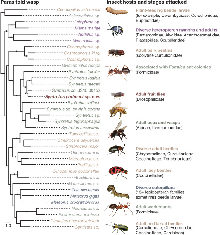
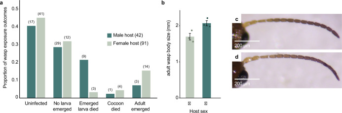
Parasitoid wasps are exceptionally diverse and use specialized adaptations capable of manipulating the physiology and behaviour of host organisms1. In more than two centuries since the first records of Drosophila-parasitizing wasps, nearly 200 described and provisional parasitoid species of drosophilids have been identified2. These include endoparasitoids and ectoparasitoids, as well as species attacking larval and pupal hosts3. Despite a deep history of research attention and remarkable biodiversity, a wasp species that attacks and develops inside the adult stage of a fly host has not been described previously. Here we report the discovery of a wasp species that infects the adult stage of fruit flies in the genus Drosophila, including one of the most deeply studied model organisms in biology, Drosophila melanogaster. Notably, this wasp can be easily collected from backyard fly baits and has a broad geographic distribution throughout the eastern USA. We document its life history and unique host interactions, including egg-laying into and larval emergence from adult flies, and provide protocols to raise wasps from wild-caught host flies. Our results emphasize the need for ongoing research investment in insect biodiversity and systematics. As parasitoid research continues to uncover unusual biology and supports fundamental mechanistic insights into immunity4, metabolism5, ecology6, evolution7-9 and behaviour10-12, we anticipate that this wasp's association with the laboratory model organism, D. melanogaster, will provide new research opportunities across the life sciences.


Conflict of interest statement

The authors declare no competing interests.

Figures

Fig. 1
Fig. 1. Adult Drosophila are parasitized by a previously undescribed species in the euphorine genus Syntretus.
A phylogram constructed from concatenated nuclear and mitochondrial gene sequences of select euphorine wasps (left), with images and descriptions of the host species (right). Nuclear loci are genes encoding carbamoylphosphate synthase domain protein (CAD), 18S rRNA and 28S rRNA. The mitochondrial locus is COI. Members of the sister taxa Cenocoelius and Asiacentistes are included as outgroups. The gene sequences of S.perlmani group with high support inside the genus Syntretus. Branches with Shimodaira–Hasegawa-like maximum likelihood support values ≥ 0.9 are labelled with a circle. Taxon labels are coloured by host order. GenBank sequence accession numbers are listed in the Supplementary Data. Host insect photographs are adapted with permission as follows (top to bottom, excluding the fruit fly): cerambycid larva, Gilles San Martin under a CC BY 2.0 licence; Nezara viridula, Bugwood.org, Robert and Lesley Ingram under a CC BY 3.0 licence; Pityogenes chalcographis, Gilles San Martin under a CC BY 2.0 licence; Formica sp., Bugwood.org, Joseph Berger under a CC BY 3.0 licence; Apis mellifera, Bugwood.org, David Cappaert under a CC BY 3.0 licence; T. carbonaria, Alison Bockoven; Disonycha triangularis, Bugwood.org, Joseph Berger under a CC BY 3.0 licence; Coleomegilla maculata, Bugwood.org, Whitney Cranshaw under a CC BY 3.0 licence; Helicoverpa armigera, Bugwood.org, Gyorgy Csoska under a CC BY 3.0 licence; Formica obscuriventris, Gary Alpert; Acalymma vittatum, Bugwood.org, G. J. Holmes under a CC BY 3.0 licence.
Fig. 2
Fig. 2. Life stages of S.perlmani.
a,b, The development of wasp larvae inside host flies (a) is accompanied by growth of wasp teratocytes (b, black arrows), which can be seen through the host abdominal cuticle and obstruct the view of the testes (b, white arrow). c, The second and following larval instars lack a head capsule and tail spike, and the final instar grows to nearly the length of the host fly (see Supplementary Video 2). d, Pupal development takes place within a white silken cocoon as is typical of euphorine wasps. e, Larval emergence is always from the abdomen and has been observed to occur between the second and third tergites (dorsolaterally) or laterally through a tear in the abdominal cuticle. f, The adult wasp (male shown) is small, yellowish brown and approximately 1.5 mm in length. Scale bars, 0.5 mm (ad) and 1 mm (e,f).
Fig. 3
Fig. 3. Life cycle, oviposition outcomes and geographic distribution of S.perlmani.
a, The life cycle of S.perlmani, including oviposition (1), early larval development (2), appearance of teratocytes (2′), late larval development and reduction in teratocyte number (3), larval emergence (4), cocoon formation and metamorphosis (5) and adult emergence (6). b, Duration of larval and cocoon stages for n = 24 and n = 22 wasps, respectively. Data are observations collected while rearing wasps from wild-caught and laboratory-infected flies. Centre line, median; box limits, upper and lower quartiles; whiskers, 1.5× quartile range. c, Global map showing the sampling locations of wild-caught D.melanogaster pools from which reads were sequenced. Green points indicate collection sites where S. perlmani mitochondrial DNA reads were absent and red triangles indicate collection sites where S.perlmani mitochondrial DNA reads were present. d, Map of the contiguous USA showing the locations where S.perlmani have been identified through field collections of D.affinis (dark green) or through in silico surveys of host DNA data (light green). Labels beside each state indicate the number of S. perlmani-infected D.affinis males caught and the total number collected in parentheses, or the number of DNA read sets positive for S. perlmani DNA reads and the total number analysed. Source Data
Fig. 4
Fig. 4. Morphology of S.perlmani.
a, Holotype lateral habitus. b, Holotype propodeum. c, Holotype forewing venation. Scale bars, 1 mm (a), 100 μm (b) and 400 μm (c).
Extended Data Fig. 1
Extended Data Fig. 1. Phylogenetic relationships in the genus Syntretus.
(a) and (b) Phylograms including members of the genus Syntretus constructed using concatenated (a) nuclear and (b) mitochondrial sequences. Nuclear loci are carbamoylphosphate synthase domain protein (CAD), 18S rRNA, and 28S rRNA. The mitochondrial locus is COI. Branches with SH-like support ≥0.9 are labeled with a circle. (c) A phylogram of the mitochondrial cytochrome oxidase subunit I (COI) locus of wasps attributed to the genus Syntretus showing the abundance and diversity of syntretine sequences in online databases. Included are all publicly available sequence entries on GenBank and the Barcode of Life Data System (BOLD). Species for which host records are known are shown as bold branches; those in light green are Syntretus trigonaphagus and the syntretine parasitoid of Apis cerana. The tree is rooted on the COI sequence of Myiocephalus boops.
Extended Data Fig. 2
Extended Data Fig. 2. Infection frequency of Syntretus perlmani.
Infection frequency of S. perlmani in wild-caught D. affinis males in Starkville, MS from April 2023 to February 2024. Fly abundance decreased into the late summer with increased temperature and reduced rainfall. Baits were placed in September, but visitation was low. Baits were not placed in November or December. One collection in late January yielded three males, one of which was infected, and is combined with the February data. Sample size (n) is shown in parentheses. Source Data
Extended Data Fig. 3
Extended Data Fig. 3. Parasitism success of Syntretus perlmani in laboratory exposures.
Percent success is shown categorized by outcome or stage achieved following exposure. Data is shown for all D. affinis and D. melanogaster exposures, including both host sexes. For larvae emerged, cocoons, and adults emerged, values calculated are percent of confirmed infections. Points are shaded by the position of the female wasp in the time of exposures, where the lightest color points correspond to data from the first trials, and the darkest points to the most recent. Results are shown for n = 5 female wasps.
Extended Data Fig. 4
Extended Data Fig. 4. Effects of host sex on Syntretus perlmani development.
(a) Exposure outcomes after male and female D. affinis were exposed to female S. perlmani wasps. Data excludes rearing attempts using coconut husk, as no adults were reared using this substrate. (b) For a cohort of 6 male offspring of an unmated female wasp (“Alice”), adult body size is shown. Wasps that developed in a male host (n = 3) were significantly smaller than those that developed in female hosts (n = 3; Welch’s t-test, t(3.77) = 3.23, p = 0.0349, 95% CI [0.044-0.702]; two-sided). Error bars display standard error of the mean. Additionally, from the same sample set, wasps that developed in (c) male hosts had antennae with fewer flagellomeres (12) than (d) wasps that developed in female hosts (13). Source Data
Extended Data Fig. 5
Extended Data Fig. 5. Melanotic encapsulation of Syntretus perlmani eggs in a resistant host species.
Photographs of the lateral or ventral abdomen are shown for a permissive host species, Drosophila acutilabella (a), and a resistant host species, Drosophila immigrans (b-d; representative images from one cohort of 15 infections). Arrows in (a) indicate small melanization spots associated with wasp oviposition. For both species, photographs were taken nine days post-oviposition by the same female wasp. The host pictured is female in (a-c) and male in (d).
Extended Data Fig. 6
Extended Data Fig. 6. Additional morphology of Syntretus perlmani.
(a) paratype lateral habitus, (b) paratype forewing and hindwing venation, (c) paratype head and antennomeres in profile, (d) paratype head, scape, and pedicel, (e) paratype ovipositor.

Similar articles

Cited by

  • Defensive tactics: lessons from Drosophila.
    Sadanandappa MK, Ahmad S, Mohanraj R, Ratnaparkhi M, Sathyanarayana SH. Sadanandappa MK, et al. Biol Open. 2024 Dec 15;13(12):bio061609. doi: 10.1242/bio.061609. Epub 2024 Dec 24. Biol Open. 2024. PMID: 39718046 Free PMC article. Review.

References

    1. Pennacchio, F. & Strand, M. R. Evolution of developmental strategies in parasitic hymenoptera. Annu. Rev. Entomol.51, 233–258 (2006). - PubMed
    1. Lue, C. H. et al. DROP: molecular voucher database for identification of Drosophila parasitoids. Mol. Ecol. Resour.21, 2437–2454 (2021). - PubMed
    1. Prevost, G. Parasitoids of Drosophila (Elsevier, 2009). - PubMed
    1. Rizki, R. M. & Rizki, T. M. Parasitoid virus-like particles destroy Drosophila cellular immunity. Proc. Natl Acad. Sci. USA87, 8388–8392 (1990). - PMC - PubMed
    1. Mrinalini et al. Parasitoid venom induces metabolic cascades in fly hosts. Metabolomics11, 350–366 (2015). - PMC - PubMed

LinkOut - more resources