. 2024 Sep;633(8031):887-894.
doi: 10.1038/s41586-024-07934-8. Epub 2024 Sep 11.

Self-organized tissue mechanics underlie embryonic regulation

Affiliations

Self-organized tissue mechanics underlie embryonic regulation

Paolo Caldarelli et al. Nature. 2024 Sep.

Abstract

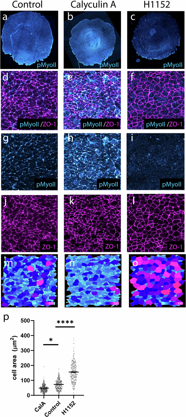
Early amniote development is highly self-organized, capable of adapting to interference through local and long-range cell-cell interactions. This process, called embryonic regulation1, has been well illustrated in experiments on avian embryos, in which subdividing the epiblast disk into different parts not only redirects cell fates to eventually form a complete and well-proportioned embryo at its original location, but also leads to the self-organization of additional, fully formed embryos2,3 in the other separated parts. The cellular interactions underlying embryonic self-organization are widely believed to be mediated by molecular signals, yet the identity of such signals is unclear. Here, by analysing intact and mechanically perturbed quail embryos, we show that the mechanical forces that drive embryogenesis self-organize, with contractility locally self-activating and the ensuing tension acting as a long-range inhibitor. This mechanical feedback governs the persistent pattern of tissue flows that shape the embryo4-6 and also steers the concomitant emergence of embryonic territories by modulating gene expression, ensuring the formation of a single embryo under normal conditions, yet allowing the emergence of multiple, well-proportioned embryos after perturbations. Thus, mechanical forces act at the core of embryonic self-organization, shaping both tissues and gene expression to robustly yet plastically canalize early development.


Conflict of interest statement

The authors declare no competing interests.

Figures

Fig. 1
Fig. 1. A model for self-organized force generation at the embryo margin.
a, Transmitted light picture of a stage XI quail embryo, depicting embryonic territories (left) and the position in millimetres and degrees along the margin used to quantify tissue motion (arrows, right). Ant., anterior; post. posterior. b, Trajectories depicting the tissue flows obtained by particle image velocimetry (PIV) analysis at t = 6–8 h after the onset of tissue motion. The overlay denotes active contractility (magenta, highest in the posterior) and the resulting tension (green, approximately uniform along the margin) in the anterior margin. c, Profile of velocity along the margin from n = 6 biologically independent embryos from a previous study at t = 4 h (0 mm is posterior; around ±3 mm is anterior). d, The time evolution of strain rates along the margin. The grey lines denote the evolution of angular positions. 0° is posterior, ±180° is anterior. e, Deformation of an initially square grid from an average of six embryos. f, The interplay between active contractility (magenta) and passive tension (green) proposed to regulate embryo formation through GDF1 expression. The solid arrows indicate local feedback arising from the response to tissue contraction versus stretching (see Extended Data Fig. 1). The dashed arrow indicates long-range tension propagation. g, Predicted profiles of contractility (magenta), tension (green) and velocity (black) at t = 4 h. h, Time evolution of strain rates along the margin. i, Global tissue deformation when self-organizing contractility is implemented in a in 2D fluid-mechanical model. Colours in d, h, e and i quantify contraction (red) and expansion (blue). Scale bar, 1 mm (a).
Fig. 2
Fig. 2. Tissue contractility modulates GDF1 expression in intact epiblasts.
af, Tissue deformation maps (a,e,i,m), time evolution of strain rates along the margin (b,f,j,n; 0 mm is posterior) and corresponding expression of GDF1 (c,g,k,o) and BRA (d,h,l,p) in control (ad; n = 32), calyculin-A-treated (eh; n = 18), H1152-treated (il; n = 13) and Ski-1-treated (mp; n = 6) biologically independent embryos. Colours in a, b, e, f, i, j, m and n quantify contraction (red) and expansion (blue). Scale bars, 1 mm (ac, de, g, h, ik, l, mo and p).
Fig. 3
Fig. 3. Self-organized tissue mechanics drives ectopic embryo formation after epiblast subdivision.
ae, Model predictions for the response to epiblast bisection in anterior halves. a, Sketch of epiblast bisection. be, Predicted contractility (magenta), tension (green) and velocity (black) profiles at t0 + 4 h (b), kymograph of margin strain rate (c; 0 mm is anterior) and deformation maps at 4 h (d) and 8 h (e) after bisection. f–w, UV-laser dissected anterior epiblast halves with (fk) or without (lw) epiboly and treated with H1152 (rw). The red dashed lines indicate the UV cut abrogating the epiboly process. f,l,r, A memGFP embryo at t0. g,m,s, Kymographs of the strain rates along the margin; 0 mm is anterior. h,n,t, Deformation maps at 10 h (h), 15 h (n) and 13 h (t) after epiblast bisection. i,o,u, SNAI2 expression in the same embryos fixed after live imaging (n = 8 out of 8 biologically independent embryos with epiboly; n = 5 out of 5 without epiboly (3 out of 5 show two ectopic primitive streaks, 2 out of 5 show one ectopic primitive streak); n = 4 out of 4 without epiboly and with H1152). j,k,p,q,v,w, GDF1 expression and the corresponding deformation maps at 4.5 h (j,k,p,q) and 6 h (v,w) after epiblast bisection. Scale bars, 1 mm (f, hk, l, nq, r and tw).
Fig. 4
Fig. 4. Mechanical feedback rescales embryonic territories.
a–p, Model predictions (ad and il) and experimental responses (eh and mp) to fixed and free boundary conditions in posterior epiblast halves. a,e,i,m, Sketches of the experiment (a,i) and memGFP posterior epiblast half after 3 h (e,m). b,f,j,n, Predicted and experimental profiles of contractility (magenta), tension (green) and velocity (in f and n, the black lines show the mean ± s.e.m. velocity profile from n = 19 control intact biologically independent embryos, shown in Extended Data Fig. 5b; the orange lines show n = 6 biologically independent embryos; and the blue lines show n = 13 biologically independent embryos) along the margin at t0 + 2 h. c,g,k,o, Kymographs of strain rates along the margin. d,h,l,p, Deformation maps at the end of the experiments and simulations (t0 + 3–4 h). q,r, GDF1, BRA and SOX3 mRNA in the embryos shown in eh and mp, respectively, fixed after live imaging. s, The size of the contracting domain in controls (black; n = 19 biologically independent embryos), attached halves (orange; n = 6 biologically independent embryos), smaller attached sectors (brown; n = 6 biologically independent embryos; see Extended Data Fig. 6a–c) and detached halves (blue; n = 13 biologically independent embryos) versus total margin length. The dotted lines show slopes of 0.37 for the controls and attached halves/sectors (R = 0.97), respectively, and 0.65 for detached halves (R = 0.90). t, Intensity profiles of the GDF1 mRNA signal along the margin in different conditions (colours are as described in s; see also Extended Data Fig. 5g for profiles of contraction, BRA and SOX3; the intensity profiles are normalized to the margin length). Data are mean ± s.e.m. u, GDF1 level versus contraction (Methods) in portions of the margin (the solid lines show the average for each condition; colours are as described in s). v, The relative size of the GDF1 domain versus the relative size of the contracted domain (Methods; R = 0.91; ***P = 0.0003, one-sided permutation test, n = 106 random permutations; colours are as described in s). Colours in c, d, g, h, k, l, o and p quantify contraction (red) and expansion (blue) as in Fig. 1. Scale bars, 1 mm (e, h, m, p, q and r). Source Data
Fig. 5
Fig. 5. Mechanical friction redirects tissue motion/axis formation, inducing ectopic embryo formation.
ah, Model predictions (ad) and the experimental response (el) to localized friction. a,e, Sketch of the experiment (a) and memGFP embryo with a hair on its ventral side (e). b,f, Profiles of contractility (magenta), tension (green) and velocity (black) along the margin at t0 + 4 h; 0 mm is posterior. c,g, Kymograph of margin strain rates; 0° is posterior. d,h, Deformation maps at 8 h (d) and 14 h (h) after hair deposition. n = 33 out of 34 biologically independent embryos. il, GDF1 (i; the arrowheads point to ectopic expression), BRA (j) and SOX3 (k) expression and the corresponding deformation map (l), 4.5 h after hair deposition (grey box). n = 15 biologically independent embryos. Colours in c, d, g, h and l quantify contraction (red) and expansion (blue) as in Fig. 1. Scale bars, 1 mm (e and hl).
Extended Data Fig. 1
Extended Data Fig. 1. Mechanical feedback underlying self-organization of force generation and tissue flows.
Contractile actomyosin cables (magenta segments in left panel) generate tension that propagates along the margin (green). An imbalance in contractility (lighter magenta segments in the anterior denote lower contractility, from lower cable density or activity) and therefore tension drives tissue flows across the embryonic disk (right panel) and differential contraction of the margin (red: contraction; blue: stretching). As depicted in the central plot, in the posterior contractility dominates over tension and the margin contracts, whereas in the anterior tension propagating from the posterior dominates over contractility and the margin extends. This is symbolized by equation (1), which takes different forms in the 1D linear and 2D nonlinear models (see Methods and Supplementary Discussion). Feedback occurs through the modulation of margin contractility by the strain rate, whereby stretching inhibits contractility, as schematized by the boxed panels (dotted lines depict cable breakdown under stretching) and described by equation (2); the transition from high to low contractility under increasing strain rate is described by the sigmoidal function f, plotted to the right of the equations.
Extended Data Fig. 2
Extended Data Fig. 2. Effect of Calyculin A and H1152 on Myosin II phosphorylation and apical cell area.
(a-c) Control (a), Calyculin A- (b) and H1152-treated (c) embryos immunostained using anti-pMyosin II antibodies (cyan). (d-l) Higher magnification in the posterior margin region showing pMyosin II (cyan) and ZO-1 (magenta) staining in control (d, g, j), Calyculin A- (e, h, k), and H1152- (f, i, l) treated embryos. (m-o) Colour-coded apical area of cells segmented using the ZO-1 staining. (p) Quantification of cell area; Calyculin A: n = 4 biologically independent embryos, 1914 cells; Control: n = 4 biologically independent embryos, 1059 cells; H1152: n = 3 biologically independent embryos, 629 cells; Mean ± s.e.m; *p = 0,0193, ****p < 0.0001; Nested one-way ANOVA. Source Data
Extended Data Fig. 3
Extended Data Fig. 3. Ski-1 treatment interferes with Gdf1 and Bra expression.
(a-i) Control (a-c, n = 10 biologically independent embryos) and Ski-1-treated embryos (d-i, n = 17 biologically independent embryos) showing overall decreased (d-f) and irregular (g-i) expression of Gdf1 (cyan/rainbow LUT) and Bra (magenta). Scale bar is 1 mm.
Extended Data Fig. 4
Extended Data Fig. 4. Formation of ectopic embryos in anterior parts bisected at varying angles.
(a-d) Experimental procedure for the evaluation of bisection angles on ectopic embryo formation in anterior parts. Epiblasts are bisected with an angle that is defined following the automated detection of the margin and anteroposterior axis using PIV analysis [(a); velocity fields, black arrows; margin and anteroposterior, in magenta; position of cut defined by the angle α, dotted red lines)]. Anterior epiblast parts are bisected using a UV-laser (b). The formation of ectopic axes is monitored by tissue deformation (c) and verified by (d) expression of Snai2. (e-x) Bisection experiments for different values of α (e, i, m, q, u, y) showing anterior epiblast parts at t0 (f, j, n, r, v, z), the deformation maps after 10 h (g, k, o, s, w, z’) and the corresponding Snai2 expression (d, h, l, p, t, x, z”). Scale bars, 1 mm. (e, n = 3/3; i, n = 6/6; m, n = 2/2; q, n = 2/2; u,y n = 5/5 biologically independent subdivided epiblasts).
Extended Data Fig. 5
Extended Data Fig. 5. Tissue motion and gene expression in intact epiblasts and posterior halves.
(a-f) Representative example of an intact epiblast whose developmental timing is precisely matched to posterior halves with free/attached boundary condition 3 h after bisection (t = 5 h ≈ t0 + 3 h, see Methods for details on the procedure). (a) Intact epiblast at t0. (b) Velocity profiles at the margin (n = 19; 0 mm is posterior). (c) Kymograph of strain rates at the margin, deformation map (d), and corresponding expression of Gdf1 (e), Brachyury and Sox3 (f) at t ≈ t0 + 3 h. (g) Mean ±SE profiles of normalized mRNA signal intensity and contraction at the margin in intact epiblasts (black, n = 19 biologically independent embryos) and posterior halves with attached (orange, n = 6 biologically independent embryos) and free (blue, n = 13 biologically independent embryos) edges. Scale bars, 1 mm.
Extended Data Fig. 6
Extended Data Fig. 6. Development of posterior parts with different sizes and at long times.
(a-c) Mechanical rescaling in a posterior sector <180° with reattached edges. Snapshot of a ~120° posterior sector following ablation (t0, a), deformation map at t0 + 4 h (b), and kymograph of strain rates along the margin (c; 0 mm is posterior; orange arrows indicate when edges reattach). (d-f) Long-term development of posterior half with repeated laser cuts to release border attachment. (d) Time series of a posterior epiblast half just after the 1st laser cut bisecting the epiblast (t0, left panel), just before the 2nd laser cut, as embryo borders reattach (t0 + 5 h, middle panel), and after the entire margin has converged and contributed to the forming primitive streak (t0 + 9 h, right panel). (b) Deformation map (t0 + 9 h). (f) Corresponding kymograph of strain rates at the margin (0 mm is posterior; orange arrows indicate when epiblast borders reattach; dotted red lines correspond to successive laser cuts). Scale bars, 1 mm.
Extended Data Fig. 7
Extended Data Fig. 7. Inhibition of obstacle-induced ectopic embryo formation by Ski-1 treatment.
(a-f) Ski-1-treated (a-c, n = 7 biologically independent embryos) and control (d-f, n = 10 biologically independent embryos) embryos on which a hair has been deposited. (a, d) Deformation maps. (b, e) Kymograph of strain rates at the margin. (c, f) Gdf1 (cyan) and Bra (magenta) expression. Red arrows point to sites of ectopic contraction anterior to the hair.
Extended Data Fig. 8
Extended Data Fig. 8. Asymmetric perturbations probe the range of tension propagation.
(a-h) Long-range inhibition of ectopic contraction in the presence of an obstacle interfering with tension propagation one side of the margin in experiments (a-d; n = 5/7 biologically independent embryos) and simulations (e-h). Notice the propagation past the anterior and all the way to the obstacle of motion towards the posterior (velocity fields at t0 + 8 h in b,f) and stretching (deformation maps at t0 + 8 h in c, g, overlaid with initially circular contours from a,e’, and kymographs of strain rate along the margin in d,h). In simulations, this outcome is obtained with parameter values that increase the range of tension propagation by 20% compared to our main parameter set (cf. Supplementary Methods Bc). (i-p) Occasionally in experiments (i-l; n = 2/7 biologically independent embryos), and with our main parameter set in simulations (m-p), motion towards the posterior does not propagate all the way to the obstacle and an ectopic contraction develops anterior to the obstacle, as seen in the velocity fields (j,n), deformation maps (k,o), and kymographs of strain rate along the margin (l,p). Magenta arrows in the second (resp. third) column show the orientation of the presumptive axis at t0 (resp. t0 + 8 h), highlighting a rotation of the axis induced by the obstacle.

Similar articles

Cited by

References

    1. Driesch, H. The Science and Philosophy of the Organism the Gifford Lectures Delivered Before the University of Aberdeen in the Year 1907–1908 (Adam and Charles Black, 1908).
    1. Lutz, H. Sur la Production Expérimentale de la Polyembryonie et de la Monstruosité Double Chez les Oiseau (Université de Strasbourg, 1949).
    1. Spratt, N. T. & Haas, H. Integrative mechanisms in development of the early chick blastoderm. I. Regulative potentiality of separated parts. J. Exp. Zool.145, 97–137 (1960).
    1. Gräper, L. Die primitiventwicklung des hühnchens nach stereokinematographischen untersuchungen, kontrolliert durch vitale farbmarkierung und verglichen mit der entwicklung anderer wirbeltiere. Dev. Genes Evol.116, 382–429 (1929). - PubMed
    1. Wetzel, R. Untersuchungen am Hühnchen. Die entwicklung des keims während der ersten beiden bruttage. Wilhelm Roux Arch. Entwickl. Mech. Org.119, 188–321 (1929). - PubMed

MeSH terms

LinkOut - more resources