. 2024 Sep 11;22(1):834.
doi: 10.1186/s12967-024-05636-3.

USP14 inhibition promotes DNA damage repair and represses ovarian granulosa cell senescence in premature ovarian insufficiency

Affiliations

USP14 inhibition promotes DNA damage repair and represses ovarian granulosa cell senescence in premature ovarian insufficiency

Lin-Zi Ma et al. J Transl Med. .

Abstract

Background: Premature ovarian insufficiency (POI) is a condition characterized by a substantial decline or loss of ovarian function in women before the age of 40. However, the pathogenesis of POI remains to be further elucidated, and specific targeted drugs which could delay or reverse ovarian reserve decline are urgently needed. Abnormal DNA damage repair (DDR) and cell senescence in granulosa cells are pathogenic mechanisms of POI. Ubiquitin-specific protease 14 (USP14) is a key enzyme that regulates the deubiquitylation of DDR-related proteins, but whether USP14 participates in the pathogenesis of POI remains unclear.

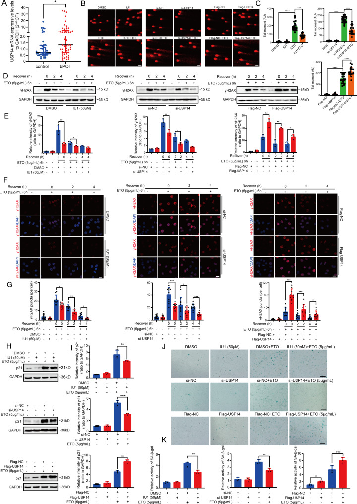
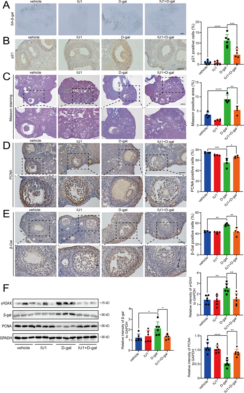
Methods: We measured USP14 mRNA expression in granulosa cells from biochemical POI (bPOI) patients. In KGN cells, we used IU1 and siRNA-USP14 to specifically inhibit USP14 and constructed a cell line stably overexpressing USP14 to examine its effects on DDR function and cellular senescence in granulosa cells. Next, we explored the therapeutic potential of IU1 in POI mouse models induced by D-galactose.

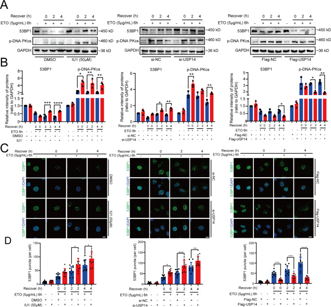
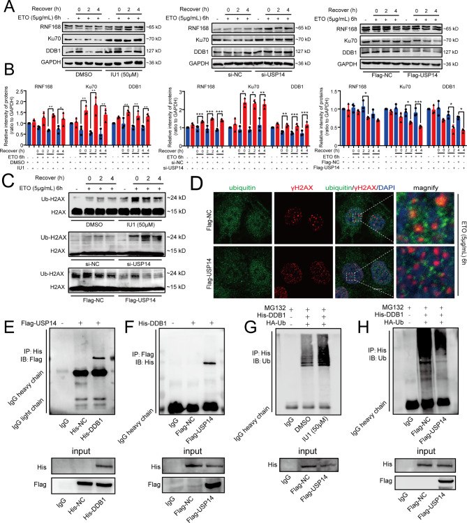
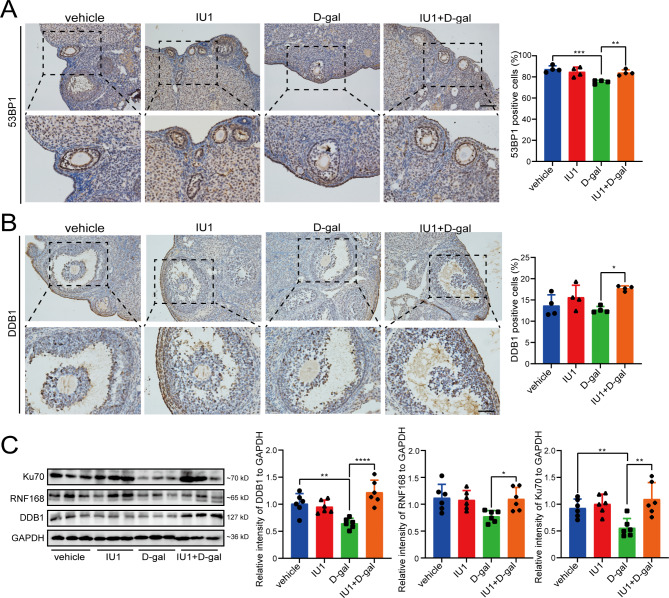
Results: USP14 expression in the granulosa cells of bPOI patients was significantly upregulated. In KGN cells, IU1 treatment and siUSP14 transfection decreased etoposide-induced DNA damage levels, promoted DDR function, and inhibited cell senescence. USP14 overexpression increased DNA damage, impaired DDR function, and promoted cell senescence. Moreover, IU1 treatment and siUSP14 transfection increased nonhomologous end joining (NHEJ), upregulated RNF168, Ku70, and DDB1, and increased ubiquitinated DDB1 levels in KGN cells. Conversely, USP14 overexpression had the opposite effects. Intraperitoneal IU1 injection alleviated etoposide-induced DNA damage in granulosa cells, ameliorated the D-galactose-induced POI phenotype, promoted DDR, and inhibited cell senescence in ovarian granulosa cells in vivo.

Conclusions: Upregulated USP14 in ovarian granulosa cells may play a role in POI pathogenesis, and targeting USP14 may be a potential POI treatment strategy. Our study provides new insights into the pathogenesis of POI and a novel POI treatment strategy.

Keywords: Cell senescence; DNA damage repair; Granulosa cells; Premature ovarian insufficiency; Ubiquitin-specific protease 14.


Conflict of interest statement

The authors declare that they have no conflict of interest.

Figures

Fig. 1
Fig. 1
USP14 affects DNA damage repair and aggravates the senescence of KGN cells. A The mRNA expression levels of USP14 in granulosa cells from bPOI and control patients were tested by qRT‒PCR. Gene expression was normalized relative to GAPDH (mean ± SD). B, C Comet assay of KGN cells stimulated with 5 µg/mL ETO for 6 h with or without 24 h pretreatment with 50 µM IU1, transfected with si-NC or si-USP14, and overexpressing Flag-NC or Flag-USP14 before ETO induction (bar = 50 μm). D, E Western blot of γH2AX in KGN cells at 0 h, 2 h and 4 h after ETO induction for 6 h with or without pretreatment with 50 µM IU1 for 24 h, transfected with si-NC or si-USP14, and overexpressing Flag-NC or Flag-USP14. F, G Immunofluorescence assay of γH2AX in KGN cells at 0 h, 2 h and 4 h after ETO induction for 6 h with or without pretreatment with 50 µM IU1 for 24 h, transfected with si-NC or si-USP14, and overexpressing Flag-NC or Flag-USP14 (bar = 10 μm). H, I Western blot analysis of p21 in KGN cells after ETO induction for 12 h with or without pretreatment with 50 µM IU1 for 24 h, transfected with si-NC or si-USP14, and overexpressing Flag-NC or Flag-USP14. J, K SA-β-Gal staining of KGN cells after ETO induction for 12 h with or without pretreatment with 50 µM IU1 for 24 h, transfection with si-NC or si-USP14, or Flag-NC or Flag-USP14 overexpression. (bar = 50 μm). All the data are presented as the means ± SDs of at least three independent experiments; *P < 0.05, **P < 0.01, ***P < 0.001, ****P < 0.0001
Fig. 2
Fig. 2
USP14 impairs the NHEJ pathway in KGN cells. A, B Western blot analysis of 53BP1 and p-DNA-PKcs in KGN cells at 0 h, 2 h and 4 h after ETO induction for 6 h with or without pretreatment with 50 µM IU1 for 24 h, transfection with si-NC or si-USP14, and Flag-NC or Flag-USP14 overexpression. C, D Immunofluorescence assay of 53BP1 in KGN cells at 0 h, 2 h and 4 h after ETO induction for 6 h with or without pretreatment with 50 µM IU1 for 24 h, transfection with si-NC or si-USP14, and overexpression of Flag-NC or Flag-USP14. (bar = 10 μm). All the data are presented as the means ± SDs of at least three independent experiments; *P < 0.05, **P < 0.01, ***P < 0.001,  ****P<0.0001
Fig. 3
Fig. 3
USP14 regulates the expression of RNF168, Ku70, DDB1 and ubiquitination of DDB1. A, B Western blot analysis of RNF168, Ku70 and DDB1 in KGN cells at 0 h, 2 h and 4 h after ETO induction for 6 h with or without pretreatment with 50 µM IU1 for 24 h, transfection with si-NC or si-USP14, and overexpression of Flag-NC or Flag-USP14. C Western blot analysis of H2AX monoubiquitination in KGN cells at 0 h, 2 h and 4 h after ETO induction for 6 h with or without pretreatment with 50 µM IU1 for 24 h, transfection with si-NC or si-USP14, and overexpression of Flag-NC or Flag-USP14. D Immunofluorescence assay showing the colocalization of γH2AX and ubiquitin in KGN cells overexpressing USP14 after stimulation with 5 µg/mL ETO for 6 h (scale bar = 5 μm). E Co-IP assay of Flag expression in HEK 293T cells overexpressing Flag-USP14 and His-DDB1 by IP with the His antibody. F Co-IP assay of His expression in HEK 293T cells overexpressing Flag-USP14 and His-DDB1 by IP with an anti-Flag antibody. G Co-IP assay of Ub expression in HEK 293T cells overexpressing His-DDB1 and HA-Ub with or without pretreatment with 50 µM IU1 for 24 h by IP with the His antibody. H Co-IP assay of Ub expression in HEK 293T cells overexpressing Flag-USP14, His-DDB1 and HA-Ub by IP with the His antibody. All data are presented as the means ± SDs of at least three independent experiments; *P < 0.05, **P < 0.01, ***P < 0.001
Fig. 4
Fig. 4
Treatment with IU1 ameliorates the decreased ovary reserve in D-gal-induced POI model mice. A Representative HE staining of mouse ovary sections from different groups (bar = 500 μm, 100 μm; n = 6). B Counts of primordial follicles, primary follicles, secondary follicles, antral follicles, atretic follicles and corpus luteum in every mouse ovary (n = 6). C-E ELISAs of serum FSH, estradiol and AMH levels in mice from each group (n = 6). All data are presented as the means ± SDs; *P < 0.05, **P < 0.01, ***P < 0.001, ****P < 0.0001
Fig. 5
Fig. 5
IU1 alleviates D-gal-induced DNA damage in mouse ovarian granulosa cells. A TUNEL staining of DNA damage in mouse ovarian granulosa cells. (bar = 100 μm; n = 6) B Immunofluorescence assay of γH2AX in mouse ovarian granulosa cells (bar = 100 μm; n = 6). C Immunofluorescence assay of and cleaved PARP in mouse ovarian granulosa cells (bar = 100 μm; n = 6). All data are presented as the means ± SDs; *P < 0.05, **P < 0.01, ***P < 0.001
Fig. 6
Fig. 6
IU1 improves D-gal-induced ovarian senescence in mice. A SA-β-gal staining of mouse ovary sections from different groups. (bar = 100 μm; n = 4) B IHC assay of p21-positive granulosa cells in mouse ovarian granulosa cells. (bar = 100 μm; n = 6) C Masson staining for ovarian fibrosis in mouse ovaries (bar = 100 μm; n = 4) D IHC assay of PCNA in mouse ovarian granulosa cells. (bar = 100 μm; n = 4) E IHC assay of β-gal in mouse ovarian granulosa cells. (bar = 100 μm; n = 4) F Western blot analysis of γH2AX, β-gal, and PCNA in mouse ovaries from different groups. GAPDH was used as a loading control (n = 6). All data are presented as the means ± SDs, *P < 0.05, **P < 0.01, ***P < 0.001, ****P < 0.0001.
Fig. 7
Fig. 7
IU1 affects the expression of 53BP1, Ku70, RNF168 and DDB1 in mouse ovaries. A IHC assay of 53BP1 in mouse ovarian granulosa cells. (bar = 100 μm; n = 4) B IHC assay of DDB1 in mouse ovarian granulosa cells. (bar = 50 μm; n = 4) C Western blot analysis of Ku70, RNF168 and DDB1 in mouse ovaries from different groups. GAPDH was used as a loading control (n = 6). All data are represented as the mean ± SD, *P < 0.05, **P < 0.01, ***P < 0.001, ****P < 0.0001
Fig. 8
Fig. 8
Schematic illustration of the mechanism by which USP14 regulates DNA damage repair and cell senescence of granulosa cells in POI. In healthy granulosa cells, modest amounts of USP14 maintain the ubiquitination of RNF168, Ku70 and DDB1 thereby regulating the NHEJ pathway and leading to normal DDR. In granulosa cells with POI, the expression of USP14 was significantly upregulated, resulting in impaired DDR function and cellular senescence via decreases in the expression and ubiquitination of RNF168, Ku70 and DDB1. By targeted inhibition of USP14 using IU1, the dysfunction of granulosa cells was significantly reversed

References

    1. Jiao X, Zhang H, Ke H, Zhang J, Cheng L, Liu Y, Qin Y, Chen ZJ. Premature ovarian insufficiency: phenotypic characterization within different etiologies. J Clin Endocrinol Metab. 2017;102(7):2281–90. 10.1210/jc.2016-3960 - DOI - PubMed
    1. Huang QY, Chen SR, Chen JM, Shi QY, Lin S. Therapeutic options for premature ovarian insufficiency: an updated review. Reprod Biol Endocrinol. 2022;20(1):28. 10.1186/s12958-022-00892-8 - DOI - PMC - PubMed
    1. Gonfloni S, Jodice C, Gustavino B, Valentini E. DNA damage stress response and follicle activation: signaling routes of mammalian Ovarian Reserve. Int J Mol Sci. 2022;23(22):14379. 10.3390/ijms232214379 - DOI - PMC - PubMed
    1. Yang Q, Mumusoglu S, Qin Y, Sun Y, Hsueh AJ. A kaleidoscopic view of ovarian genes associated with premature ovarian insufficiency and senescence. FASEB J. 2021;35(8):e21753. 10.1096/fj.202100756R - DOI - PubMed
    1. Wang X, Zhang X, Dang Y, Li D, Lu G, Chan WY, Leung PCK, Zhao S, Qin Y, Chen ZJ. Long noncoding RNA HCP5 participates in premature ovarian insufficiency by transcriptionally regulating MSH5 and DNA damage repair via YB1. Nucleic Acids Res. 2020;48(9):4480–91. 10.1093/nar/gkaa127 - DOI - PMC - PubMed

Substances