. 2024 Sep 11;57(1):65.
doi: 10.1186/s40659-024-00538-6.

The effect of CGRP and SP and the cell signaling dialogue between sensory neurons and endothelial cells

Affiliations

The effect of CGRP and SP and the cell signaling dialogue between sensory neurons and endothelial cells

Alice Leroux et al. Biol Res. .

Abstract

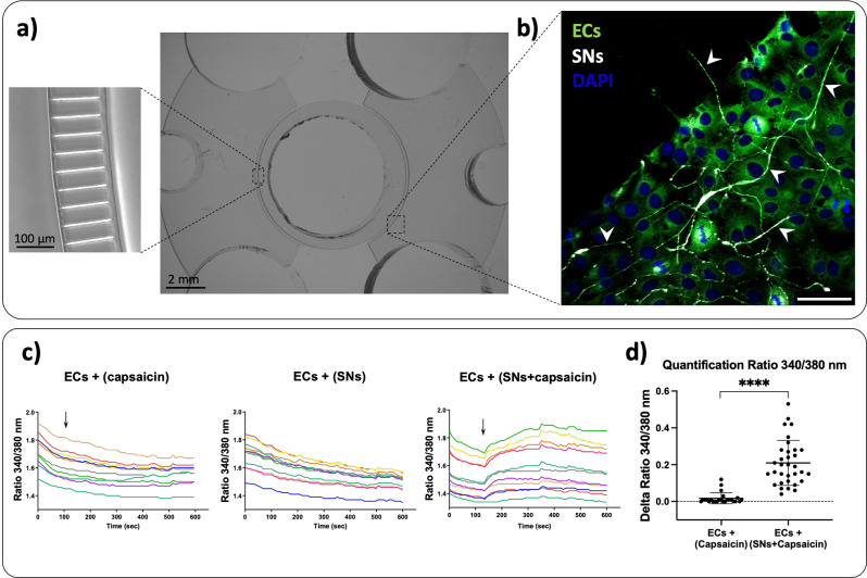
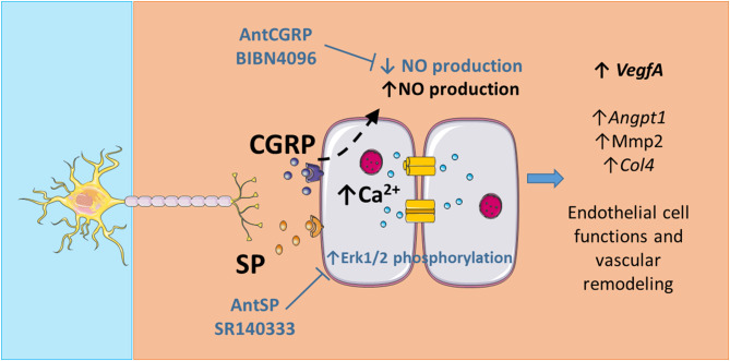
Increasing evidences demonstrate the role of sensory innervation in bone metabolism, remodeling and repair, however neurovascular coupling in bone is rarely studied. Using microfluidic devices as an indirect co-culture model to mimic in vitro the physiological scenario of innervation, our group demonstrated that sensory neurons (SNs) were able to regulate the extracellular matrix remodeling by endothelial cells (ECs), in particular through sensory neuropeptides, i.e. calcitonin gene-related peptide (CGRP) and substance P (SP). Nonetheless, still little is known about the cell signaling pathways and mechanism of action in neurovascular coupling. Here, in order to characterize the communication between SNs and ECs at molecular level, we evaluated the effect of SNs and the neuropeptides CGRP and SP on ECs. We focused on different pathways known to play a role on endothelial functions: calcium signaling, p38 and Erk1/2; the control of signal propagation through Cx43; and endothelial functions through the production of nitric oxide (NO). The effect of SNs was evaluated on ECs Ca2+ influx, the expression of Cx43, endothelial nitric oxide synthase (eNOS) and nitric oxide (NO) production, p38, ERK1/2 as well as their phosphorylated forms. In addition, the role of CGRP and SP were either analyzed using respective antagonists in the co-culture model, or by adding directly on the ECs monocultures. We show that capsaicin-stimulated SNs induce increased Ca2+ influx in ECs. SNs stimulate the increase of NO production in ECs, probably involving a decrease in the inhibitory eNOS T495 phosphorylation site. The neuropeptide CGRP, produced by SNs, seems to be one of the mediators of this effect in ECs since NO production is decreased in the presence of CGRP antagonist in the co-culture of ECs and SNs, and increased when ECs are stimulated with synthetic CGRP. Taken together, our results suggest that SNs play an important role in the control of the endothelial cell functions through CGRP production and NO signaling pathway.

Keywords: Angiogenesis; CGRP; Calcium signaling; Innervation; Nitric oxide; Substance P.


Conflict of interest statement

The authors declare that they have no relevant financial or non-financial interests to disclose.

Figures

Fig. 1
Fig. 1
Representative images of the co-culture between ECs and SNs in microfluidic devices and intracellular Ca2+ levels in endothelial cells (ECs) in the presence or absence of sensory neurons (SNs). (a) Microfluidic devices composed of 3 compartments: one central compartment containing SNs surrounded by two lateral compartments containing ECs. Central and lateral compartments are separated by microchannels with precise dimensions that permit axons to pass through, as characterized in our previous study [3]. (b) Confocal microscopy representative image of the co-culture between SNs and ECs at day 7. SNs are shown in white (β-III tubulin) and ECs in green (von Willebrand factor). The white arrowheads show neurites emitted by the SNs passing through microchannels and within ECs culture. Scale bar = 50 μm. (c) Representative traces of a region of interest (ROI) around ECs from a random region in a microfluidic device showing the intracellular calcium levels in ECs cultured alone having capsaicin added in the central compartment (ECs + (capsaicin)), ECs culture in the presence of SNs without capsaicin stimulation (ECs + (SNs)) and ECs co-culture with SNs with capsaicin stimulation (ECs + (SNs + capsaicin)). Each line represents an endothelial cell previously defined as a ROI using the Metafluor software. The values of calcium levels are expressed as Ratio (340/380 nm) as a function of time. The black arrow represents the addition of capsaicin in the SNs compartment. These figures are representative of three independent experiments. (d) Variation of intracellular calcium levels in ECs quantified by Delta Ratio 340/380 nm as the maximum value of the 340/380 nm ratio measured in ECs after capsaicin addition in the central compartment minus the value of the 340/380 nm ratio in ECs at the moment of capsaicin addition. Data are represented as n = 24–42 ROIs from three independent experiments. n = 8–14 ROIs analyzed per microfluidic device. Mean values ± SD analyzed by Student’s t test, **** p < 0.0001
Fig. 2
Fig. 2
Effect of (a-d) the co-culture of ECs with SNs and (e-h) CGRP and SP antagonists on Cx43 expression and phosphorylation in endothelial cells (ECs). (a) Gene expression of Cx43 by ECs cultured in presence (grey bars) or absence (white bars) of SNs after 4 and 7 days. (b) Representative western blot of Cx43 protein expression by ECs cultured in presence (+) and absence (-) of SNs after 4 and 7 days of culture. Black arrowheads represent phosphorylated forms of Cx43 (pCx43) with slightly higher molecular weight. (c) Total Cx43 protein expressed by ECs cultured in presence (grey bars) or absence (white bars) of SNs after 4 and 7 days relative to EC alone Gapdh at day 4. (d) Phosphorylated and non-phosphorylated forms of Cx43 were quantified separately and the Cx43 phosphorylated ratio (pCx43/Cx43) was evaluated relative to ECs alone at day 4. (a-d) n = 6 independent experiments. (e) Cx43 gene expression in the presence of AntCGRP, AntSP and vehicle after 7 days of co-culture between ECs and SNs in the presence of each antagonist and vehicle. (f) Representative western blot of Cx43 protein expression relative to Gapdh after 7 days of co-culture between ECs and SNs in the presence of each antagonist and vehicle. Black arrowheads represent pCx43 form. (g) Total Cx43 protein expression relative to Gapdh in ECs co-cultured with SNs for 7 days in the presence of each antagonist relative to the vehicle. (h) Phosphorylated and non-phosphorylated forms of Cx43 in ECs cultured with SNs for 7 days in the presence of each antagonist and vehicle. The Cx43 phosphorylated ratio (pCx43/Cx43) was quantified relative to the vehicle. (e-h) n = 3 independent experiments. All graphs represent mean values ± SD for both gene and protein evaluation using ANOVA test followed by Bonferroni (a, c and d) or Tukey (e, g and h) as post-hoc, * p < 0.05. Uncropped western blots images are shown in Supplementary Fig. 2
Fig. 3
Fig. 3
Effect of (a-c) the co-culture of ECs with SNs and (d-f) CGRP and SP on eNOS expression and nitrite production. (a) Representative western blot of eNOS protein expression by ECs cultured in presence (+) and absence (-) of SNs after 4 and 7 days of culture. The phosphorylation of T495 site was assessed using specific antibody (p-eNOS) (Table 2). (b) eNOS protein expression relative to Gapdh in ECs cultured in presence (grey bars) or absence (white bars) of SNs relative to ECs alone at day 4. (c) Phosphorylation ratio of the T495 site (p-eNOS/eNOS) relative to ECs alone at day 4. (d) Quantification of NO degradation products was performed at day 7 in the ECs culture medium in the presence or absence of SNs. (e) Quantification of NO degradation products was performed at day 7 in the ECs culture medium supplemented with CGRP and SP antagonists in the presence or absence of SNs. The vehicle group is considered as control. (f) Effect of the two neuropeptides CGRP (100 nM) and SP (1 µM) separately on nitrite production by ECs cultured alone for 7 days. ECs medium supplemented with the vehicle is considered as control (n = 4 independent experiments). The graphs represent mean values ± SD using ANOVA test followed by Bonferroni (b, c) or Tukey (e, f) as post-hoc or Student’s t test (d), * p < 0.05, ** p < 0.01, **** p < 0.0001. Uncropped western blots images are shown in Supplementary Fig. 3
Fig. 4
Fig. 4
Effect of (a-c) the co-culture of ECs with SNs and (d-f) CGRP and SP antagonists on p38 expression. (a) Representative western blot of p38 and its phosphorylated form (p-p38) expression by ECs cultured in presence (+) and absence (-) of SNs after 4 and 7 days of culture. P-p38 expression was assessed using specific antibody (Table 2). (b) p38 expression relative to Gapdh by ECs cultured in presence (grey bars) or absence (white bars) of SNs, after 4 and 7 days relative to ECs alone at day 4. (c) Phosphorylated and non-phosphorylated forms of p38 were quantified separately and the phosphorylated ratio (p-p38/p38) was evaluated relative to ECs alone at day 4. (d) Representative western blot of p38 and p-p38 expression in EC when co-cultured with SNs for 7 days in the presence of CGRP and SP antagonists (AntCGRP and AntSP, respectively). The vehicle group is used as control. (e) Total p38 expression relative to Gapdh in ECs co-cultured with SNs for 7 days in the presence of each antagonist relative to the vehicle. (f) Phosphorylation ratio (p-p38/p38) in the presence of each antagonist relative to the vehicle (n = 4 independent experiments). The graphs represent mean values ± SD using ANOVA test followed by Bonferroni (b, c) or Tukey (e, f) as post-hoc. Uncropped western blots images are shown in Supplementary Fig. 4
Fig. 5
Fig. 5
Effect of (a-c) the co-culture of ECs with SNs, (d-f) CGRP and SP antagonists, and (g-i) the neuropeptides CGRP and SP on Erk1/2 expression and phosphorylation. (a) Representative western blot of Erk1/2 and its phosphorylated form (p-Erk1/2) expression by ECs cultured in presence (+) and absence (-) of SNs after 4 and 7 days of culture. P-Erk1/2 expression was assessed using specific antibody (Table 2). (b) Erk1/2 expression relative to Gapdh by ECs cultured in presence (grey bars) or absence (white bars) of SNs, after 4 and 7 days relative to ECs alone at day 4. (c) Phosphorylated and non-phosphorylated forms of Erk1/2 were quantified separately and the phosphorylated ratio (p-Erk1/2-to-Erk1/2) was evaluated relative to ECs alone at day 4. (a-c) n = 4 independent experiments. (d) Representative western blot of Erk1/2 and p-Erk1/2 expression in presence of CGRP or SP antagonists (AntCGRP and AntSP, respectively) and vehicle. (e) Erk1/2 expression relative to Gapdh by ECs co-cultured with SNs in the presence of each antagonist relative to the vehicle. (f) Erk1/2 phosphorylation ratio (p-Erk1/2-to-Erk1/2) in the presence of each antagonist relative to the vehicle. (d-f) n = 3 independent experiments. (g) Representative western blot of Erk1/2 and p-Erk1/2 expression in ECs cultured in well plates in the presence of 100 nM CGRP, 1 µM SP, and vehicle. (h) Erk1/2 expression relative to Gapdh in ECs alone cultured in the presence of 100 nM CGRP, 1 µM SP relative to the vehicle. (i) Erk1/2 phosphorylation ratio in the presence of 100 nM CGRP, 1 µM SP relative to the vehicle. (g-i) n = 3 independent experiments. The graphs represent mean values ± SD using ANOVA test followed by Bonferroni (b, c) or Tukey (e, f) as post-hoc, * p < 0.05. Uncropped western blots images are shown in Supplementary Fig. 5
Fig. 6
Fig. 6
Putative role and mechanisms of action of sensory neurons (SN) on endothelial cells (ECs). Capsaicin-stimulated SNs induce Ca2+ influx in ECs. The presence of AntSP (SR140333) increases phosphorylation of Erk1/2 and a tendency on Cx43 downregulation (p = 0.07) is also observed. SNs induce an increase of NO production in ECs, and the neuropeptide CGRP seems to be the mediator of this effect since NO production is decreased in the presence of AntCGRP (BIBN4096) in the co-culture of ECs and SNs and increased when ECs are stimulated with synthetic CGRP. Considering our previous work showing that SNs regulates endothelial cell functions and vascular remodeling through the upregulation of VegfA, Angpt1, Mmp2 and Col4 [3] and the effect of VEGFA on NO production [14], SNs seem to play an important role in the control of ECs angiogenic functions

References

    1. Tomlinson RE, Clemens TL, Maes C. Vascular and nerve interactions. Principles of Bone Biology. Elsevier; 2020. pp. 205–18.
    1. Marrella A, Lee TY, Lee DH, et al. Engineering vascularized and innervated bone biomaterials for improved skeletal tissue regeneration. Mater Today. 2018;21:362–76. 10.1016/j.mattod.2017.10.005.10.1016/j.mattod.2017.10.005 - DOI - PMC - PubMed
    1. Leroux A, Paiva dos Santos B, Leng J, et al. Sensory neurons from dorsal root ganglia regulate endothelial cell function in extracellular matrix remodelling. Cell Commun Signal. 2020;18:162. 10.1186/s12964-020-00656-0. 10.1186/s12964-020-00656-0 - DOI - PMC - PubMed
    1. Qin Q, Lee S, Patel N, et al. Neurovascular coupling in bone regeneration. Exp Mol Med. 2022;54:1844–9. 10.1038/s12276-022-00899-6. 10.1038/s12276-022-00899-6 - DOI - PMC - PubMed
    1. Li Y, Fraser D, Mereness J, et al. Tissue Engineered Neurovascularization Strategies for Craniofacial tissue regeneration. ACS Appl Bio Mater. 2022;5:20–39. 10.1021/acsabm.1c00979. 10.1021/acsabm.1c00979 - DOI - PMC - PubMed

MeSH terms

LinkOut - more resources