Review
. 2024 Aug 13;27(9):110710.
doi: 10.1016/j.isci.2024.110710. eCollection 2024 Sep 20.

The mitochondrial stress signaling tunes immunity from a view of systemic tumor microenvironment and ecosystem

Affiliations
Review

The mitochondrial stress signaling tunes immunity from a view of systemic tumor microenvironment and ecosystem

Cheng-Liang Kuo et al. iScience. .

Abstract

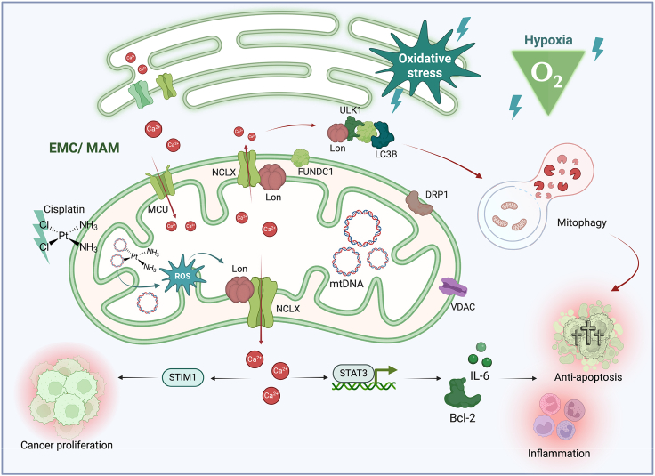
Mitochondria play important roles in cell fate, calcium signaling, mitophagy, and the signaling through reactive oxygen species (ROS). Recently, mitochondria are considered as a signaling organelle in the cell and communicate with other organelles to constitute the mitochondrial information processing system (MIPS) that transduce input-to-output biological information. The success in immunotherapy, a concept of systemic therapy, has been proved to be dependent on paracrine interactions within the tumor microenvironment (TME) and distant organs including microbiota and immune components. We will adopt a broader view from the concept of TME to tumor micro- and macroenvironment (TM 2 E) or tumor-organ ecosystem (TOE). In this review, we will discuss the role of mitochondrial signaling by mitochondrial ROS, calcium flux, metabolites, mtDNA, vesicle transportation, and mitochondria-derived peptide in the TME and TOE, in particular immune regulation and effective cancer immunotherapy.

Keywords: Cancer; Immune response; Microenvironment.


Conflict of interest statement

The authors have no conflicts of interest to declare.

Figures

None
Graphical abstract
Figure 1
Figure 1
The model of mitochondrial signal transduction in the tumor microenvironment (TME) Mitochondria in the cytoplasm are positioned at the interface between incoming signals from the outside extracellular environment and the inside compartment of cells. As a mitochondrial information processing system (MIPS), all mitochondria have the ability to perform input sensing, integration, and output signaling that contribute to cellular and organismal adaptation. In this scenario, mitochondria serve as central hubs that integrate input signals to release processed signals into the environment after a complicate information processing. Upon cancer cells stimulated by environmental stresses (blue colored), mitochondria respond and generate integrating signals, such as mitochondrial reactive oxygen species (mtROS), metabolites, mitochondrial DNA (mtDNA), Ca2+, and MDVs, and coordinate the signals under stresses (purple colored). These integration and processing subsequently lead to the release of downstream molecules as input signals (red colored) that then alter the TME. ER, endoplasmic reticulum; EMC, ER-mitochondria contact site; MDVs, mitochondria-derived vesicles; EVs, extracellular vesicles. The scheme was created with BioRender.com.
Figure 2
Figure 2
Mitochondria synthesize and release signals evolved to alter cellular functions in the tumor microenvironment (TME) Schematic illustration of major mitochondrial pathways modulating cancer malignancy under stressed TME. (A) Mitochondria are key hubs for ROS generation. Under stresses, the mitochondrial chaperone protein Lon coordinates with the ETC complex I subunit, NDUFS8, and PYCR1, leading to the production of mtROS. This phenomenon triggers pathways related to anti-apoptosis, angiogenesis, epithelial-mesenchymal transition (EMT)/metastasis, and immunosuppression in cancer cells. (B) Oncometabolites may be secreted into the TME and potentially exert negative effects on the peripheral immune system. Lactate, a glycolysis product enriched under the Warburg effect, has been found to boost mitochondrial activity via oxidative phosphorylation (OXPHOS). The shift suggests that mitochondrial metabolism has some defects in TCA cycle and is taken over by glycolysis in cancer metabolic pathways. (C) The generation of mtROS impacts mtDNA causing fragments that are released into the cytosol and induce cellular PD-L1 expression. This process further drives immunosuppressive pathways associated with tumorigenesis. mtPQC, mitochondrial protein quality control; PYCR1, pyrroline-5-carboxylate reductase 1; NDUFS8, NADH-dehydrogenase ubiquinone iron-sulfur protein 8; NF-κB, nuclear factor kappa-light-chain enhancer of activated B cells; STAT, signal transducer and activator of transcription; HIF-1ɑ, hypoxia-induced factor 1 alpha; AMPK, adenosine-monophosphate-activated protein kinase; NADPH, nicotinamide adenine dinucleotide phosphate; STING, stimulator of interferon gene; TBK, TANK-binding kinase; IFN-γ, interferon gamma; PD-L1, programmed cell death receptor ligand 1; TLR9, Toll-like receptor 9; ATP, adenosine triphosphate; PI3K, phosphoinositide 3-kinase; IL-1β, interleukin-1β; IL-6, interleukin-6; M2 TAMs, M2 tumor-associated macrophages; SUCNR1, succinate receptor 1; PDH, pyruvate dehydrogenase; TCA cycle, tricarboxylic acid cycle; GM-CSF, granulocyte-macrophage colony-stimulating factor; PBMCs, peripheral blood mononuclear cells. The scheme was created with BioRender.com.
Figure 3
Figure 3
The scheme of the interaction and communication between mitochondria and ER regulate mitophagy and cell survival under stresses Mitochondrial machinery that crosstalks with organelles boosts cancer malignancies under stress condition in the TME. Upon ROS and hypoxia condition, mitochondrial chaperone Lon promotes FUNDC1-ULK1-mediated mitophagy at the EMC/MAM site via forming the complex, which is dependent on the binding with mitochondrial Na+/Ca2+ exchanger (NCLX). Cisplatin treatment and ROS cause mtDNA damages and further induce Lon protein expression that is a mtDNA-binding protein. Mitochondrial Lon activates NCLX to release mitochondrial calcium (Ca2+) to the cytosol. Cytosolic Ca2+ thereby stimulates the STAT3 signal pathway. Activated STAT3 translocates to nucleus to activate IL-6 and Bcl-2 expression that increases the survival of cancer cells leading to cisplatin resistance. EMC/MAM, endoplasmic reticulum-mitochondria contact site/mitochondria-associated membrane; Ca2+, calcium ion; ULK1, Unc-51-like kinase 1; LC3B, microtubule-associated proteins 1A/1B light chain 3B; MCU, mitochondrial calcium uniporter; NCLX, mitochondrial sodium calcium exchanger; FUNDC1, FUN14 domain containing 1; DRP1, dynamin-related protein 1; VDAC, voltage dependent anion channel 1; STIM1, stromal interaction molecule 1; STAT3, signal transducer and activator of transcription 3; Bcl-2, B-cell lymphoma 2; IL-6, interleukin-6. The scheme was created with BioRender.com.
Figure 4
Figure 4
The scheme of mitochondrial stress signaling regulates inflammation and immunity to promote tumorigenesis from a view of the tumor microenvironment (TME) to the tumor-organ ecosystem (TOE) Mitochondria are the major cellular source of ROS generation due to the metabolic process. Chaperone Lon binds with NDFUS8 in the complex I of electron transport chain and with PYCR1 reductase to upregulate mitochondrial ROS (mtROS) generation that promotes cell proliferation and inflammation. mtROS cause the oxidative damage on mtDNA and induce IFN signaling that upregulates PD-L1 expression to inhibit T cell activation. Under ROS stress, cancer cells produce NF-κB-dependent inflammatory cytokines, TGFβ, VEGF-A, IL-6, and IL-10, to cause the immunosuppressive state of macrophages, dendritic cells (DC), and T cells (Treg). In addition, Lon upregulation by ROS and hypoxia induces the secretion of extracellular vehicles (EVs) that carry mtDNA and PD-L1. Therefore, mtROS cause an immunosuppressive TME to promote immunoescape, survival, and EMT/metastasis of cancer cells. Recently, the emerging data indicated that the TME view is too narrow to accurately reflect the complexities of the systemic networks in tumor. Since we consider cancer as a systemic disease, cancer progression and metastasis is driven by paracrine interactions within the TME and by distant organs including microbiota in the tumor-organ ecosystem (TOE).

References

    1. Stowe D.F., Camara A.K.S. Mitochondrial reactive oxygen species production in excitable cells: modulators of mitochondrial and cell function. Antioxidants Redox Signal. 2009;11:1373–1414. doi: 10.1089/ARS.2008.2331. - DOI - PMC - PubMed
    1. Holmström K.M., Finkel T. Cellular mechanisms and physiological consequences of redox-dependent signalling. Nat. Rev. Mol. Cell Biol. 2014;15:411–421. doi: 10.1038/nrm3801. - DOI - PubMed
    1. Murphy M.P. How mitochondria produce reactive oxygen species. Biochem. J. 2009;417:1–13. doi: 10.1042/BJ20081386. - DOI - PMC - PubMed
    1. Margulis L. Symposia of the Society for Experimental Biology; 1975. Symbiotic Theory of the Origin of Eukaryotic Organelles; Criteria for Proof; pp. 21–38. - PubMed
    1. Roger A.J., Muñoz-Gómez S.A., Kamikawa R. The Origin and Diversification of Mitochondria. Curr. Biol. 2017;27 doi: 10.1016/j.cub.2017.09.015. R1177-r1192. - DOI - PubMed

LinkOut - more resources