. 2024 Jul 31;27(9):110631.
doi: 10.1016/j.isci.2024.110631. eCollection 2024 Sep 20.

Maternal high-fat diet alters Tet-mediated epigenetic regulation during heart development

Affiliations

Maternal high-fat diet alters Tet-mediated epigenetic regulation during heart development

Yuhan Yang et al. iScience. .

Abstract

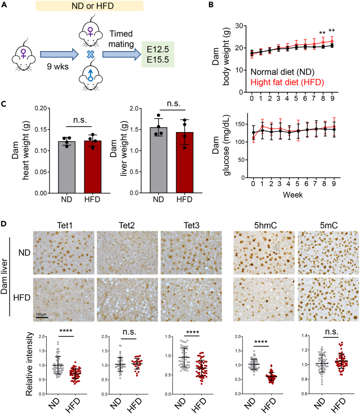
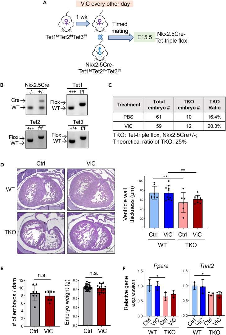
Imbalanced dietary intake, such as a high-fat diet (HFD) during pregnancy, has been associated with adverse offspring outcomes. Metabolic stress from imbalanced food intake alters the function of epigenetic regulators, resulting in abnormal transcriptional outputs in embryos to cause congenital disorders. We report herein that maternal HFD exposure causes metabolic changes in pregnant mice and non-compaction cardiomyopathy (NCC) in E15.5 embryos, accompanied by decreased 5-hydroxymethylcytosine (5hmC) levels and altered chromatin accessibility in embryonic heart tissues. Remarkably, maternal vitamin C supplementation mitigates these detrimental effects, likely by restoring iron, a cofactor for Tet enzymes, in a reduced state. Using a genetic approach, we further demonstrated that the cardioprotective benefits of vitamin C under HFD conditions are attributable to enhanced Tet activity. Our results highlight an interaction between maternal diet, specifically HFD or vitamin C, and epigenetic modifications during early heart development, emphasizing the importance of balanced maternal nutrition for healthy embryonic development.

Keywords: Developmental biology; Diet; Epigenetics; Model organism; Pregnancy.


Conflict of interest statement

The authors declare no competing interests.

Figures

None
Graphical abstract
Figure 1
Figure 1
Maternal HFD alters Tet activities in the liver of dams (A) Experimental timeline for normal diet (ND) or high-fat diet (HFD) feeding of timed mated female mice. (B) Measurements of the body weight (top) and blood glucose levels (bottom) for female mice fed with ND or HFD over a nine-week period (n = 11 mice/group; mean ± SD); ∗∗p < 0.01 (two-sided unpaired Student’s t test). (C) Weight measurement of heart and liver collected from dams fed with ND or HFD for 9 weeks (n = 4 mice/group; mean ± SD). n.s., not significant. (D) (Top) Representative images showing immunohistochemistry (IHC) staining of the indicated Tet1-3 proteins and DNA modifications (5hmC or 5mC) in liver tissues collected from dams (n = 4–5) exposed to ND or HFD for 9 weeks. Scale bar, 100 μm. (Bottom) Quantifications of the relative intensities of the corresponding markers (n = 50–60 cells; mean ± SD); ∗∗∗∗p < 0.0001 (two-sided unpaired Student’s t test).
Figure 2
Figure 2
Maternal HFD suppresses TET protein expression and causes ventricular non-compaction cardiomyopathy (NCC) in embryos (A) Representative H&E-stained sections (Left) and quantification of ventricular area and ventricular wall thickness (Right) from E15.5 embryonic heart tissues following maternal ND or HFD (n = 3–6 embryos per group, mean ± SD). ∗p < 0.05 (two-sided unpaired Student’s t test). Scale bar, 200 μm (4x) or 100 μm (20x). (B) Representative IHC images (top) and quantification (bottom) of Tet1, Tet3 and 5hmC levels in the ventricular area of E15.5 embryonic hearts from the ND and HFD groups (n = 50–60 cells/group; mean ± SD. ∗p < 0.05, ∗∗∗∗p < 0.0001 (two-sided unpaired Student’s t test). Scale bar, 100 μm. (C) Western blot analysis of Tet1 in heart tissues collected from E15.5 embryos exposed to maternal ND or HFD (n = 2 embryos/group). Tubulin was used as a loading control. (D) 5hmC dotblot analysis on DNA samples from heart tissues collected from E12.5 and E15.5 embryos exposed to maternal ND or HFD (n = 2 embryos/group). Methylene blue (ME-Blue) staining is used as loading control. (E) Genomic distribution of differentially enriched ATAC-seq peaks in heart tissues collected from E15.5 embryos following maternal ND or HFD exposure. (F) GREAT analysis of functionally annotated differentially enriched ATAC-seq peaks from cardiac tissues of E15.5 embryos under maternal ND or HFD. (G) Genome browser views of ATAC-seq data at the Ppara and Tnnt2 loci in heart tissues collected from E15.5 embryos exposed to maternal ND (blue) or HFD (red). (H) hMeDIP analysis of 5hmC enrichment at the promoters of Ppara and Tnnt2 in heart tissues collected from E15.5 embryos exposed to maternal ND or HFD. IgG is used as a negative control (n = 4 embryos, mean ± SD; ∗p < 0.05, ∗∗p < 0.01; two-sided unpaired Student’s t test). (I) Real-time qPCR analysis on Ppara and Tnnt2 expression in heart tissues collected from E15.5 embryos exposed to maternal ND or HFD (n = 3–4 embryos; mean ± SD). ∗p < 0.05 (two-sided unpaired Student’s t test).
Figure 3
Figure 3
Vitamin C ameliorates maternal HFD-induced glucose intolerance and ventricle wall thinning in embryos (A) Experimental timeline of maternal vitamin C treatment under ND or HDF conditions. (B) Changes in body weight of dams on ND or HFD, with and without vitamin C (ViC) treatment (n = 7–8 dams per group; mean ± SD). ∗∗∗p < 0.001 (two-sided unpaired Student’s t test). (C) Embryos weights in maternal ND or HFD groups, with or without vitamin C (ViC) treatment (n = 17–23 embryos per group; mean ± SD; ∗∗∗∗p < 0.0001, ∗∗p < 0.01, ∗p < 0.05; two-sided unpaired Student’s t test). (D) Glucose tolerance test (GTT) results from dams on ND or HFD, with and vitamin C (ViC) treatment (n = 3–7 dams/group; mean ± SD). ∗∗∗p < 0.001 (two-sided unpaired Student’s t test). (E and F) Quantification of levels of reduced (Fe2+), oxidized (Fe3+), and total iron in placenta (E, left), embryonic heart tissue (E, middle), embryonic liver (E, right), and maternal serum (F), under maternal ND or HFD conditions, with and without vitamin C treatment (n = 3–5/group; mean ± SD). ∗∗p < 0.01, ∗∗∗p < 0.001 (two-sided unpaired Student’s t test). (G) Representative H&E images (left) and quantification (right) of ventricular wall thickness of embryonic heart tissues at E15.5 from dams on ND and HFD, with or without vitamin C treatment (n = 4–10 embryos/group; mean ± SD. ∗∗∗p < 0.001, ∗∗∗∗p < 0.0001 (two-sided unpaired Student’s t test). Scale bar, 100 μm. (H) Representative IHC images (left) and quantification (right) of 5hmC levels in the trabecular area of embryonic heart tissue at E15.5 from dams on HFD, with or without vitamin C treatment (n = 35–45 cells/group; mean ± SD). ∗∗∗∗p < 0.0001 (two-sided unpaired Student’s t test). Scale bar, 40 μm. (I) Real-time qPCR analysis of Ppara expression in heart tissues collected from E15.5 embryos exposed to maternal HFD with and without vitamin C treatment (n = 3–4 embryos; mean ± SD). ∗∗p < 0.001 (two-sided unpaired Student’s t test).
Figure 4
Figure 4
Tet proteins contribute to vitamin C-mediated cardioprotection during development in maternal HFD-exposed embryos (A) Schematic of the experimental setup for administering vitamin C to myocardium-specific Tet-TKO mouse models under a normal diet (ND). (B) Genotyping results confirm cardiac specific deletion of Tet genes in embryos. (C) Total count of Tet-TKO embryos obtained from dams treated with PBS or vitamin C. (D) Representative H&E-stained sections of embryonic hearts (left) and quantification of ventricular wall thickness of heart tissues (right) collected from E15.5 Tet-TKO embryos, with or without vitamin C treatment (n = 6–10 embryos/group; mean ± SD. ∗∗p < 0.01 (two-sided unpaired Student’s t test). Scale bar, 200 μm. (E) Quantification of the number and weight of embryos collected from Tet-TKO dams treated with or without vitamin C (n = 7 dams/group, n = 20–29 embryos/group; mean ± SD). n.s., no statistical significance (two-sided unpaired Student’s t test). (F) Real-time qPCR analysis on Ppara and Tnnt2 expression in WT and Tet-TKO heart tissues collected from E15.5 embryos treated with or without vitamin C (n = 3 embryos; mean ± SD; ∗p < 0.05; two-sided unpaired Student’s t test).

Similar articles

Cited by

References

    1. Urbonaite G., Knyzeliene A., Bunn F.S., Smalskys A., Neniskyte U. The impact of maternal high-fat diet on offspring neurodevelopment. Front. Neurosci. 2022;16 doi: 10.3389/fnins.2022.909762. - DOI - PMC - PubMed
    1. Fernandes D.J., Spring S., Roy A.R., Qiu L.R., Yee Y., Nieman B.J., Lerch J.P., Palmert M.R. Exposure to maternal high-fat diet induces extensive changes in the brain of adult offspring. Transl. Psychiatry. 2021;11:149. doi: 10.1038/s41398-021-01274-1. - DOI - PMC - PubMed
    1. Zhang Q., Xiao X., Zheng J., Li M., Yu M., Ping F., Wang T., Wang X. A Maternal High-Fat Diet Induces DNA Methylation Changes That Contribute to Glucose Intolerance in Offspring. Front. Endocrinol. 2019;10:871. doi: 10.3389/fendo.2019.00871. - DOI - PMC - PubMed
    1. Keleher M.R., Zaidi R., Hicks L., Shah S., Xing X., Li D., Wang T., Cheverud J.M. A high-fat diet alters genome-wide DNA methylation and gene expression in SM/J mice. BMC Genom. 2018;19:888. doi: 10.1186/s12864-018-5327-0. - DOI - PMC - PubMed
    1. Mdaki K.S., Larsen T.D., Wachal A.L., Schimelpfenig M.D., Weaver L.J., Dooyema S.D.R., Louwagie E.J., Baack M.L. Maternal high-fat diet impairs cardiac function in offspring of diabetic pregnancy through metabolic stress and mitochondrial dysfunction. Am. J. Physiol. Heart Circ. Physiol. 2016;310:H681–H692. doi: 10.1152/ajpheart.00795.2015. - DOI - PMC - PubMed