. 2024 Aug 20;10(16):e36386.
doi: 10.1016/j.heliyon.2024.e36386. eCollection 2024 Aug 30.

HL-60 cells as a valuable model to study LPS-induced neutrophil extracellular traps release

Affiliations

HL-60 cells as a valuable model to study LPS-induced neutrophil extracellular traps release

Sonya J Malavez-Cajigas et al. Heliyon. .

Abstract

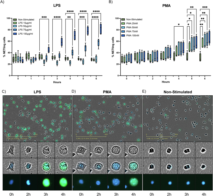
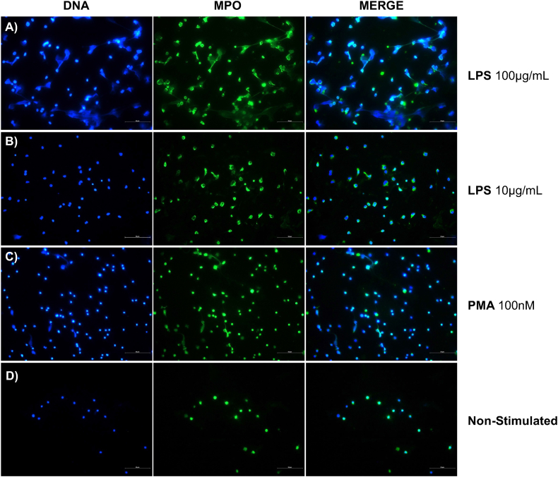
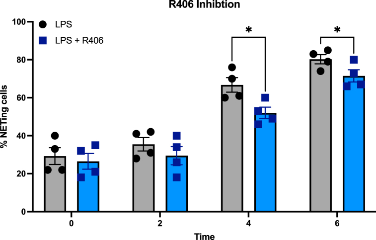
Neutrophil Extracellular Traps (NETs) present a paradoxical role in infectious diseases, contributing to both immunity and pathogenesis. The complex nature of this process necessitates further characterization to elucidate its clinical implications. However, studying NETs faces challenges with primary neutrophils due to their heterogeneity, short lifespan, and lack of adequate cryopreservation. Researchers often turn to alternative models, such as differentiated HL-60 cells (dHL-60). This study explored LPS-induced NETs formation in dHL-60 cells, revealing significant responses to LPS from Pseudomonas aeruginosa, although significantly lower than primary neutrophils. Moreover, Spleen Tyrosine Kinase (SYK) inhibition with R406, the active metabolite of the drug Fostamatinib, previously demonstrated to suppress NETs in primary neutrophils, effectively reduced NETs release in dHL-60 cells. dHL-60 cells, offering easier manipulation, consistent availability, and no donor variability in functional responses, possess characteristics suitable for high-throughput studies evaluating NETosis. Overall, dHL-60 cells may be a valuable in vitro model for deciphering the molecular mechanisms of NETosis in response to LPS, contributing to our available tools for understanding this complex immune process.

Keywords: HL-60; Human leukemia cell line; Lipopolysaccharide; Neutrophil Extracellular Traps; Neutrophils; SYK inhibitor.


Conflict of interest statement

The authors declare that they have no known competing financial interests or personal relationships that could have appeared to influence the work reported in this paper.

Figures

Fig. 1
Fig. 1
LPS induces NETosis comparable to PMA in dHL-60 cells. The percent of dHL-60 cells undergoing NETosis (n = 4) after stimulation with A) Different concentrations of LPS from P. aeruginosa and B) Different concentrations of PMA, compared to non-stimulated dHL-60 cells. Representative images (20×) showing nuclei (blue fluorescence) and DNA release labeling (green fluorescence) at 6 h after stimulation with C) LPS (100 μg/mL), D) PMA (100 nM) or E) non-stimulated cells are shown. NETs are quantified by total fluorescence object area (μm2/image) using Incucyte NETosis assay. Analysis was performed using two-way ANOVA with multiple comparisons to assess differences across concentrations versus non-stimulated cells. *, P < 0.05; **, P < 0.01; ***, P < 0.001; ****, P < 0.0001. Data are graphed as box and whisker plots showing the max and min values.
Fig. 2
Fig. 2
Visualization of NETs MPO-DNA co-localization. Immunofluorescence staining was performed using antibodies targeting Myeloperoxidase (MPO), shown in green (FITC). DNA was counterstained with DAPI (4′,6-diamidino-2-phenylindole), shown in blue. This experiment was performed with dHL-60 cells stimulated with Lipopolysaccharide (LPS) from P. aeruginosa at A) 100 μg/mL and B) 10 μg/mL, C) PMA at 100 nM or D) non-stimulated. Cells were fixed after 2 h of stimulation. Representative images depict NETs structures with MPO-DNA co-localization. Images were captured at a magnification of 40×, and scale bar was set at 50 μm.
Fig. 3
Fig. 3
R406 reduces LPS-induced NETosis in dHL-60 cells. NETs formation (n = 4) in dHL-60 cells after stimulation with LPS (100 μg/mL) P. aeruginosa alone or with a 30-min preincubation with R406. NETosis is showed as a percent of cells undergoing NETosis, per described in the methods section. NETs release is quantified by total green object area (μm2/image) using Incucyte NETosis assay. Analysis was performed with multiple paired t-tests. *, P < 0.05; **, P < 0.01; ***, P < 0.001. Data are graphed in columns showing the mean ± SEM.

References

    1. Delgado-Rizo V., et al. Neutrophil extracellular traps and its implications in inflammation: an overview. Front. Immunol. 2017;8 - PMC - PubMed
    1. Hidalgo A., et al. Neutrophil extracellular traps: from physiology to pathology. Cardiovasc. Res. 2022;118(13):2737–2753. - PMC - PubMed
    1. Papayannopoulos V. Neutrophil extracellular traps in immunity and disease. Nat. Rev. Immunol. 2018;18(2):134–147. - PubMed
    1. Stoimenou M., et al. Methods for the assessment of NET formation: from neutrophil biology to translational research. Int. J. Mol. Sci. 2022;23(24) - PMC - PubMed
    1. Silvestre-Roig C., Hidalgo A., Soehnlein O. Neutrophil heterogeneity: implications for homeostasis and pathogenesis. Blood. 2016;127(18):2173–2181. - PubMed

LinkOut - more resources