. 2024 Aug 23;10(17):e36377.
doi: 10.1016/j.heliyon.2024.e36377. eCollection 2024 Sep 15.

Construction and drug screening of Co-culture system using extrahepatic cholangiocarcinoma organoids and tumor-associated macrophages

Affiliations

Construction and drug screening of Co-culture system using extrahepatic cholangiocarcinoma organoids and tumor-associated macrophages

Yinghao Guo et al. Heliyon. .

Abstract

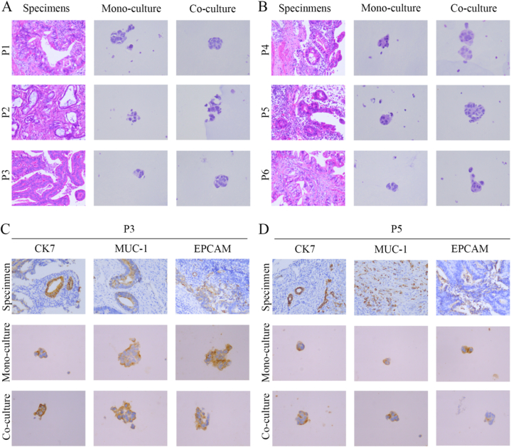
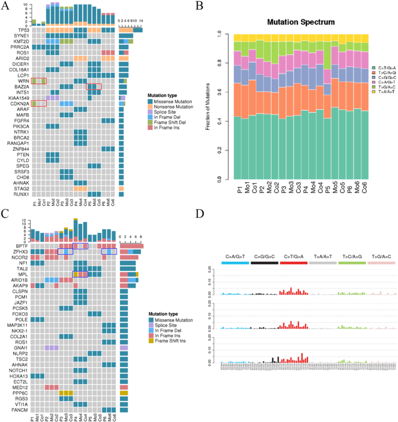
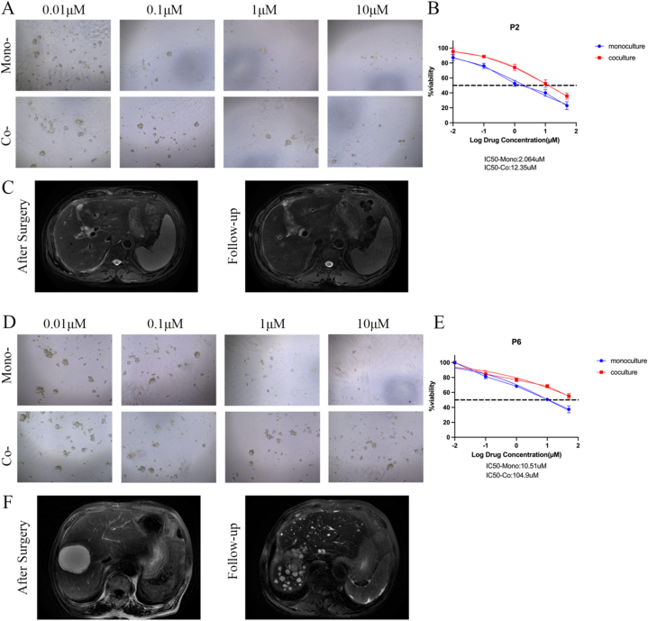
Patient-derived organoids (PDOs) have been proposed as a novel in vitro tumor model that can be applied to tumor research and drug screening. However, current tumor organoid models lack components of the tumor microenvironment, particularly tumor-associated macrophages(TAMs).We collected peripheral blood and tumor samples from 6 patients with extrahepatic cholangiocarcinoma(eCCA). Monocytes were induced into TAMs by cytokine and conditioned medium, and then co-cultured with tumor organoids. Our comprehensive analysis and comparison of histopathology and genomics results confirmed that this co-culture model can better capture intra- and inter-tumor heterogeneity retain the specific mutations of the original tumor. Drug sensitivity data in vitro revealed that gemcitabine and cisplatin are effective drugs for cholangiocarcinoma, but TAMs in the tumor microenvironment promote organoids growth and chemotherapy resistance. In conclusion, our organoid model of cholangiocarcinoma co-cultured with TAMs can not only shorten the model construction cycle, but also preserve the heterogeneity of original tumors to improve the accuracy of drug screening, and can also be applied to the researches of TAMs and tumors.

Keywords: Extrahepatic cholangiocarcinoma; Genetic profiles; Organoids; Therapy resistance; Tumor-associated macrophages.


Conflict of interest statement

The authors declare that they have no known competing financial interests or personal relationships that could have appeared to influence the work reported in this paper.

Figures

Fig. 1
Fig. 1
Construction of extrahepatic cholangiocarcinoma organoids and TAMs co-culture system. (A) Study design used to create co-culture system from eCCA patients. (B) Morphology of eCCA organoids with TAMs in bright field. Organoids had a round structure (red arrow), and TAMs had a spindle structure. (magnification, 100×).
Fig. 2
Fig. 2
Histopathological characterization comparing eCCA organoids from mono-culture and co-culture models and corresponding specimens. (A–B) Representative H&E staining of eCCA specimens and organoids (specimen magnification, 200×; organoid magnification, 400×). (C–D) Representative immunohistochemistry staining for CK7, MUC-1, and EPCAM in eCCA specimens and organoids (specimen magnification, 200×; organoid magnification, 400×).
Fig. 3
Fig. 3
General genetic alterations in two original specimens and organoids. (A) Number of SNP in different regions of the genome (left) and number of different types of SNP in the coding region (right) from patients 4. (B) Number of INDEL in different regions of the genome (left) and number of different types of INDEL in the coding region (right) from patients 4. (C) Number of SNP in different regions of the genome (left) and number of different types of SNP in the coding region (right) from patients 6. (D) Number of INDEL in different regions of the genome (left) and number of different types of INDEL in the coding region (right) from patients 6.
Fig. 4
Fig. 4
Detailed genetic profiles of six original specimens and organoids. (A) Representative driver genes of the specimens and organoids are presented. The horizontal coordinate is the sample (P: specimens; Mo: monocultured organoids; Co: co-cultured organoids); the vertical coordinate is the gene; the top is the number of gene mutations in each sample; the right is the number of mutations in each gene in these samples presented. (B) Representative mutation spectrum in the specimens and organoids are presented. (C) Representative cancer predisposition genes in the specimens and organoids are presented. (D) Mutation characteristics of the specimens and organoids are presented.
Fig. 5
Fig. 5
Organoids response to gemcitabine and cisplatin. (A) Representative images showing patient 2 organoids from the two models response to gemcitabine (magnification, 100×). (B) Dose-response curves of organoids chemosensitivity to gemcitabine. Data are expressed as means ± SD. (C) Representative MR Images of patient 2 during follow-up. (D) Representative images showing patient 6 organoids from the two models response to cisplatin (magnification, 100×). (E) Dose-response curves of organoids chemosensitivity to cisplatin. Data are expressed as means ± SD. (F) Representative MR Images of patient 6 during follow-up.
Fig. 6
Fig. 6
Organoids response to 5-fluorouracil and paclitaxel. (A) Representative images showing patient 6 organoids from the two models response to 5-fluorouracil(magnification, 100×). (B–D) Dose-response curves of organoids chemosensitivity to 5-fluorouracil. Data are expressed as means ± SD. (E) Representative images showing patient 8 organoids from the two models response to paclitaxel(magnification, 100×). (F–H) Dose-response curves of organoids chemosensitivity to paclitaxel. Data are expressed as means ± SD.
Fig. 7
Fig. 7
TAMs promotes the growth of eCCA organoids in vitro and in vivo. (A) Representative images of eCCA organoids in mono-culture and co-culture(magnification, 40×). (B) Diameters of eCCA organoids cultured with or without TAMs(n = 6, 3 organoids for each well randomly were measured, **P < 0.01, ***P < 0.001). Data are expressed as means ± SD. (C) Cell viability of eCCA organoids cultured with or without TAMs(n = 6 experimental settings with 3 biological replicates for each; **P < 0.01, ***P < 0.001). Data are expressed as means ± SD. (D) Representative images of the tumors from mono-transplantation and co-transplantation. (E–F) Weights and volumes of tumors from mono-transplantation and co-transplantation(n = 5, **P < 0.01, ***P < 0.001). Data are expressed as means ± SD. (G) Representative H&E staining of tumors from specimens, mono-transplantation, and co-transplantation. (H) Representative immunohistochemistry staining for CK7, MUC-1, and EPCAM in tumors from specimens, mono-transplantation and co-transplantation (magnification, 200×).

Similar articles

Cited by

References

    1. Bertuccio P., Malvezzi M., Carioli G., Hashim D., Boffetta P., El-Serag H.B., La Vecchia C., Negri E. Global trends in mortality from intrahepatic and extrahepatic cholangiocarcinoma. J. Hepatol. 2019;71(1):104–114. - PubMed
    1. Fabris L., Perugorria M.J., Mertens J., Björkström N.K., Cramer T., Lleo A., Solinas A., Sänger H., Lukacs-Kornek V., Moncsek A., Siebenhüner A., Strazzabosco M. The tumour microenvironment and immune milieu of cholangiocarcinoma. Liver Int. 2019;39(Suppl 1):63–78. - PMC - PubMed
    1. Sun D., Luo T., Dong P., Zhang N., Chen J., Zhang S., Liu L., Dong L., Zhang S. CD86+/CD206+ tumor-associated macrophages predict prognosis of patients with intrahepatic cholangiocarcinoma. PeerJ. 2020;8 - PMC - PubMed
    1. Tacke F. Targeting hepatic macrophages to treat liver diseases. J. Hepatol. 2017;66(6):1300–1312. - PubMed
    1. Kong L., Zhou Y., Bu H., Lv T., Shi Y., Yang J. Deletion of interleukin-6 in monocytes/macrophages suppresses the initiation of hepatocellular carcinoma in mice. J. Exp. Clin. Cancer Res. 2016;35(1):131. - PMC - PubMed

LinkOut - more resources