Review
. 2024 Aug 31;14(4):698-724.
doi: 10.21037/cdt-24-52. Epub 2024 Jul 31.

Technical principles, benefits, challenges, and applications of photon counting computed tomography in coronary imaging: a narrative review

Affiliations
Review

Technical principles, benefits, challenges, and applications of photon counting computed tomography in coronary imaging: a narrative review

Antonella Meloni et al. Cardiovasc Diagn Ther. .

Abstract

Background and objective: The introduction of photon-counting computed tomography (PCCT) represents the most recent groundbreaking advancement in clinical computed tomography (CT). PCCT has the potential to overcome the limitations of traditional CT and to provide new quantitative imaging information. This narrative review aims to summarize the technical principles, benefits, and challenges of PCCT and to provide a concise yet comprehensive summary of the applications of PCCT in the domain of coronary imaging.

Methods: A review of PubMed, Scopus, and Google Scholar was performed until October 2023 by using relevant keywords. Articles in English were considered.

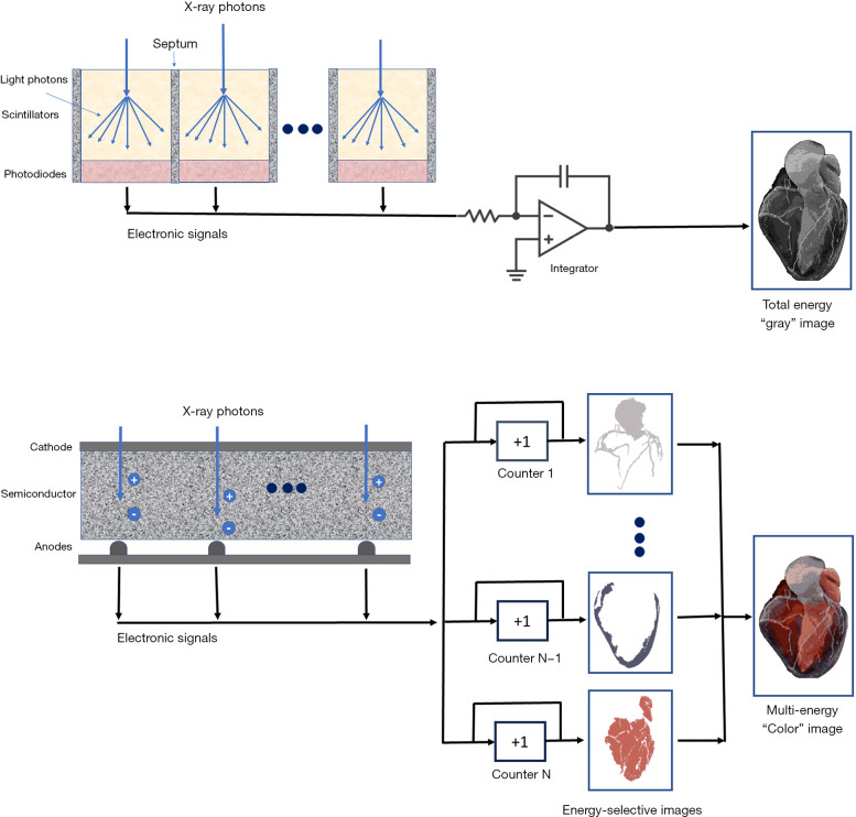
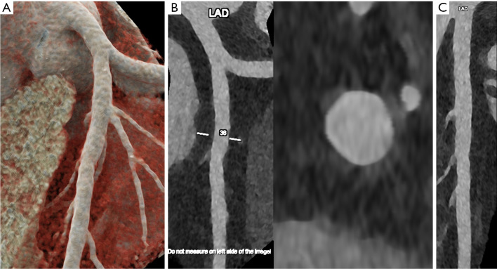
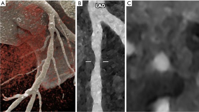
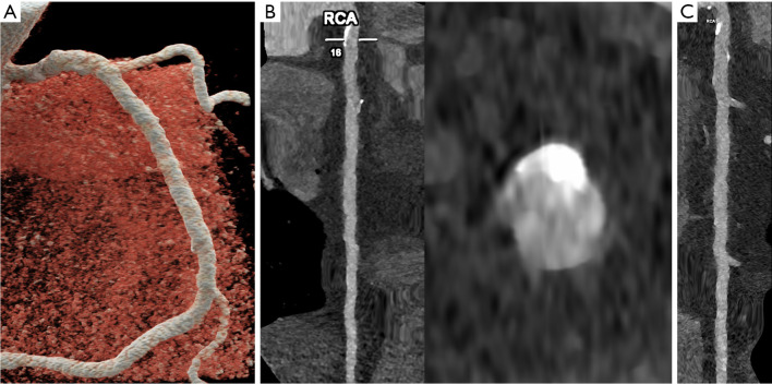
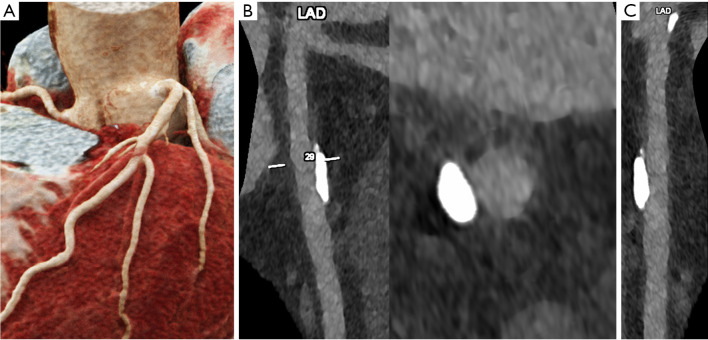
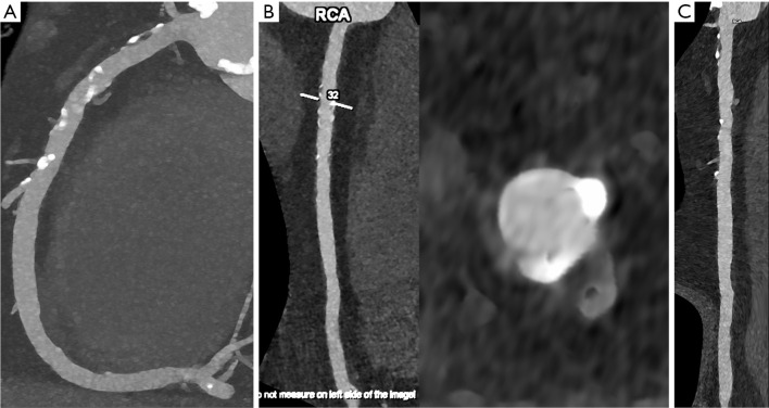
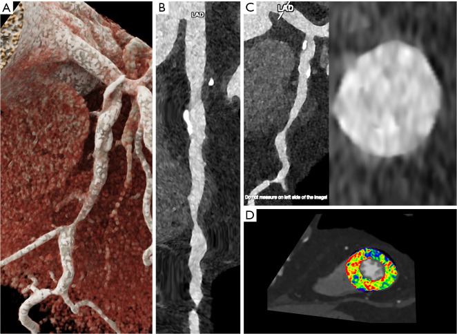
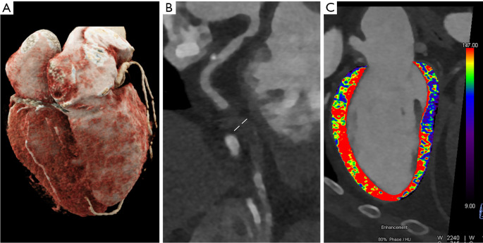
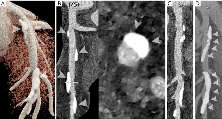
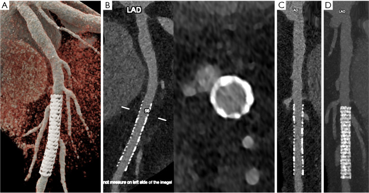
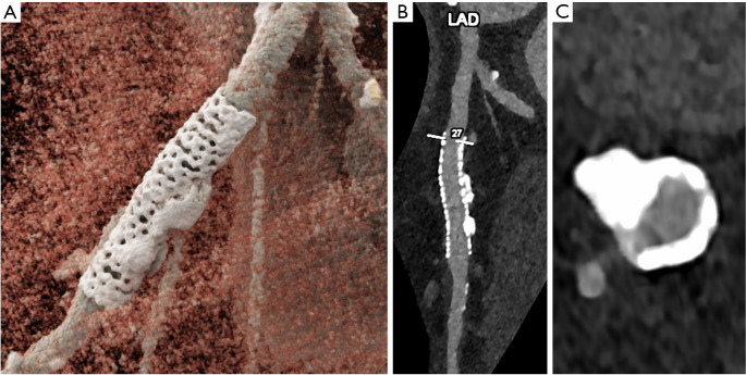
Key content and findings: The main advantages of PCCT over traditional CT are enhanced spatial resolution, improved signal and contrast characteristics, diminished electronic noise and image artifacts, lower radiation exposure, and multi-energy capability with enhanced material discrimination. These key characteristics have made room for improved assessment of plaque volume and severity of stenosis, more precise assessment of coronary artery calcifications, also preserved in the case of a reduced radiation dose, improved assessment of plaque composition, possibility to provide details regarding the biological processes occurring within the plaque, enhanced quality and accuracy of coronary stent imaging, and improved radiomic analyses.

Conclusions: PCCT can significantly impact diagnostic and clinical pathways and improve the management of patients with coronary artery diseases (CADs).

Keywords: Photon-counting detectors; computed tomography angiography (CT angiography); coronary arteries.


Conflict of interest statement

Conflicts of Interest: All authors have completed the ICMJE uniform disclosure form (available at https://cdt.amegroups.com/article/view/10.21037/cdt-24-52/coif). F.C. and L.S. serve as unpaid editorial board members of Cardiovascular Diagnosis and Therapy from September 2023 to August 2025. The other authors have no conflicts of interest to declare.

Figures

Figure 1
Figure 1
Schematic representation of an energy integrating detector (top) and of a photon-counting detector that directly converts X-rays into electrical signals (bottom). In the photon-counting detector, the interaction between the incident X-ray photon and the semiconductor generates a cloud of positive and negative charges, which pull away from each other under the influence of the electric field. As the electrons reach the anodes, they generate short current pulses, which are converted into voltage pulses. The detector’s electronics analyzes the pulses by comparing their amplitudes (proportional to the photon’s energy) with predefined threshold levels.
Figure 2
Figure 2
Cardiac/coronary PCCT examples of normal coronary arteries. The figure shows a proximal left coronary artery with 3D cinematic rendering (A), longitudinal MPR and axial cross-section of LAD (B), and longitudinal stretched MPR (C). In this case, coronary arteries are normal (no calcium and no non-calcified atherosclerosis). The scan was performed on a commercial whole-body Dual Source Photon Counting CT scanner (Naeotom Alpha, Siemens Healthineers) with 0.2 mm slice thickness, 0.1 mm reconstruction increment, FOV 140 mm, and IQ level 55. The scan is performed with retrospective ECG gating with tube current modulation and images are displayed with a resolution matrix of 1,024×1,024 pixels on the source axial reconstructions with a kernel filtering of Bv60 (vascular kernel medium-sharp) and with maximum intensity of QIR 4. The actual displayed resolution is 0.1 mm (100 microns). PCCT, photon-counting computed tomography; 3D, three-dimensional; MPR, multiplanar reconstructions; LAD, left anterior descending coronary artery; CT, computed tomography; FOV, field of view; IQ, image quality; ECG, electrocardiogram; QIR, quantum iterative reconstruction.
Figure 3
Figure 3
Cardiac/coronary PCCT examples of non-obstructive CAD. In the figure, a proximal left coronary artery is shown with 3D cinematic rendering (A), longitudinal MPR (B), and axial cross-section of LAD (C). In this case, proximal LAD shows predominantly non-calcified atherosclerosis with some positive remodelling. The scan was performed on a commercial whole-body Dual Source Photon Counting CT scanner (Naeotom Alpha, Siemens Healthineers) with 0.2 mm slice thickness, 0.1 mm reconstruction increment, FOV 140 mm, and IQ level 55. The scan is performed with retrospective ECG gating with tube current modulation and images are displayed with a resolution matrix of 1024×1,024 pixels on the source axial reconstructions with a kernel filtering of Bv60 (vascular kernel medium-sharp) and with maximum intensity of QIR 4. The actual displayed resolution is 0.1 mm (100 microns). PCCT, photon-counting computed tomography; CAD, coronary artery disease; 3D, three-dimensional; MPR, multiplanar reconstructions; LAD, left anterior descending coronary artery; CT, computed tomography; FOV, field of view; IQ, image quality; ECG, electrocardiogram; QIR, quantum iterative reconstruction.
Figure 4
Figure 4
Cardiac/coronary PCCT examples of non-obstructive CAD. In the figure, a complete RCA is shown with 3D cinematic rendering (A), longitudinal MPR and axial cross-section of LAD (B), and longitudinal stretched MPR (C). In this case, proximal RCA shows calcified plaques that do not affect the lumen diameter. The scan was performed on a commercial whole-body Dual Source Photon Counting CT scanner (Naeotom Alpha, Siemens Healthineers) with 0.2 mm slice thickness, 0.1 mm reconstruction increment, FOV 140 mm, and IQ level 55. The scan is performed with retrospective ECG gating with tube current modulation and images are displayed with a resolution matrix of 1,024×1,024 pixels on the source axial reconstructions with a kernel filtering of Bv60 (vascular kernel medium-sharp) and with maximum intensity of QIR 4. The actual displayed resolution is 0.1 mm (100 microns). PCCT, photon-counting computed tomography; CAD, coronary artery disease; RCA, right coronary artery; 3D, three-dimensional; MPR, multiplanar reconstructions; LAD, left anterior descending coronary artery; CT, computed tomography; FOV, field of view; IQ, image quality; ECG, electrocardiogram; QIR, quantum iterative reconstruction.
Figure 5
Figure 5
Cardiac/coronary PCCT examples of non-obstructive CAD. In the figure, a proximal left coronary artery is shown with 3D cinematic rendering (A), longitudinal MPR and axial cross-section of LAD (B), and longitudinal stretched MPR (C). In this case, proximal LAD shows a severely calcified plaque that does not affect the lumen diameter; even though the plaque is large, it appears to stay across the coronary artery wall without impacting significantly the lumen diameter. The scan was performed on a commercial whole-body Dual Source Photon Counting CT scanner (Naeotom Alpha, Siemens Healthineers) with 0.2 mm slice thickness, 0.1 mm reconstruction increment, FOV 140 mm, and IQ level 55. The scan is performed with retrospective ECG gating with tube current modulation and images are displayed with a resolution matrix of 1,024×1,024 pixels on the source axial reconstructions with a kernel filtering of Bv60 (vascular kernel medium-sharp) and with maximum intensity of QIR 4. The actual displayed resolution is 0.1 mm (100 microns). PCCT, photon-counting computed tomography; CAD, coronary artery disease; 3D, three-dimensional; MPR, multiplanar reconstructions; LAD, left anterior descending coronary artery; CT, computed tomography; FOV, field of view; IQ, image quality; ECG, electrocardiogram; QIR, quantum iterative reconstruction.
Figure 6
Figure 6
Cardiac/coronary PCCT examples of non-obstructive CAD. In the figure, a complete RCA is shown with 3D cinematic rendering (A), longitudinal MPR and axial cross-section of LAD (B), and longitudinal stretched MPR (C). In this case, proximal RCA shows diffuse calcified plaques from the ostium down to crux that do not affect the lumen diameter. The scan was performed on a commercial whole-body Dual Source Photon Counting CT scanner (Naeotom Alpha, Siemens Healthineers) with 0.2 mm slice thickness, 0.1 mm reconstruction increment, FOV 140 mm, and IQ level 55. The scan is performed with retrospective ECG gating with tube current modulation and images are displayed with a resolution matrix of 1,024×1,024 pixels on the source axial reconstructions with a kernel filtering of Bv60 (vascular kernel medium-sharp) and with maximum intensity of QIR 4. The actual displayed resolution is 0.1 mm (100 microns). PCCT, photon-counting computed tomography; CAD, coronary artery disease; RCA, right coronary artery; 3D, three-dimensional; MPR, multiplanar reconstructions; LAD, left anterior descending coronary artery; CT, computed tomography; FOV, field of view; IQ, image quality; ECG, electrocardiogram; QIR, quantum iterative reconstruction.
Figure 7
Figure 7
Cardiac/coronary PCCT examples of obstructive CAD. In the figure, a proximal left coronary artery is shown with 3D cinematic rendering (A), longitudinal MPR and axial cross-section of LAD (B), longitudinal stretched MPR (C), and short axis view of the apical segments of the left ventricle first pass rest perfusion map (D). In this case, proximal and middle LAD are severely diseased with predominantly non calcified plaques with positive remodelling and serial significant obstructions of the coronary lumen; the first pass static rest perfusion map shows a perfusion delay (D) at the level of the anterolateral wall of the left ventricle in middle-apical segments (blue colored overlay). The scan was performed on a commercial whole-body Dual Source Photon Counting CT scanner (Naeotom Alpha, Siemens Healthineers) with 0.2 mm slice thickness, 0.1 mm reconstruction increment, FOV 140 mm, and IQ level 55. The scan is performed with retrospective ECG gating with tube current modulation and images are displayed with a resolution matrix of 1,024×1,024 pixels on the source axial reconstructions with a kernel filtering of Bv60 (vascular kernel medium-sharp) and with maximum intensity of QIR 4. The actual displayed resolution is 0.1 mm (100 microns). PCCT, photon-counting computed tomography; CAD, coronary artery disease; 3D, three-dimensional; MPR, multiplanar reconstructions; LAD, left anterior descending coronary artery; CT, computed tomography; FOV, field of view; IQ, image quality; ECG, electrocardiogram; QIR, quantum iterative reconstruction.
Figure 8
Figure 8
Cardiac/coronary PCCT examples of obstructive CAD. In the figure, a dominant circumflex coronary artery is shown with 3D cinematic rendering (A), longitudinal MPR (B), and two-chamber long axis view of the left ventricle first pass rest perfusion map (C). In this case the middle-distal left circumflex is occluded from a non-calcified plaque; the first pass static rest perfusion map shows a large perfusion delay (C) at the level of the inferior wall of the left ventricle (blue colored overlay). The scan was performed on a commercial whole-body Dual Source Photon Counting CT scanner (Naeotom Alpha, Siemens Healthineers) with 0.2 mm slice thickness, 0.1 mm reconstruction increment, FOV 140 mm, and IQ level 55. The scan is performed with retrospective ECG gating with tube current modulation and images are displayed with a resolution matrix of 1,024×1,024 pixels on the source axial reconstructions with a kernel filtering of Bv60 (vascular kernel medium-sharp) and with maximum intensity of QIR 4. The actual displayed resolution is 0.1 mm (100 microns). PCCT, photon-counting computed tomography; CAD, coronary artery disease; 3D, three-dimensional; MPR, multiplanar reconstructions; LAD, left anterior descending coronary artery; CT, computed tomography; FOV, field of view; IQ, image quality; ECG, electrocardiogram; QIR, quantum iterative reconstruction.
Figure 9
Figure 9
Cardiac/coronary PCCT examples of non-obstructive CAD. In the figure, a proximal left coronary artery is shown with 3D cinematic rendering (A), longitudinal MPR and axial cross-section of LAD (B), longitudinal stretched MPR (C), and longitudinal stretched MIP (D). In this case, proximal-middle LAD shows massively/bulky calcified plaques that, even though very thick and dense, do not significantly affect the lumen diameter (A-D; arrowheads); the Agatston score of the displayed LAD is above 1,000. Technically, the presence of severe calcifications should be approached as the presence of coronary stents. The scan was performed on a commercial whole-body Dual Source Photon Counting CT scanner (Naeotom Alpha, Siemens Healthineers) with 0.2 mm slice thickness, 0.1 mm reconstruction increment, FOV 140 mm, and IQ level 55. The scan is performed with retrospective ECG gating with diastolic tube current modulation and images are displayed with a resolution matrix of 1,024×1,024 pixels on the source axial reconstructions with a kernel filtering of Bv72 (vascular kernel sharp) and with maximum intensity of QIR 4. The actual displayed resolution is 0.1 mm (100 microns). PCCT, photon-counting computed tomography; CAD, coronary artery disease; 3D, three-dimensional; MPR, multiplanar reconstructions; MIP, maximum intensity projection; LAD, left anterior descending coronary artery; CT, computed tomography; FOV, field of view; IQ, image quality; ECG, electrocardiogram; QIR, quantum iterative reconstruction.
Figure 10
Figure 10
Cardiac/coronary PCCT examples of coronary artery stent. In the figure, it is shown a stent in the mid left anterior descending coronary artery with 3D cinematic rendering (A), longitudinal MPR and axial cross section of coronary lumen within the stent segment (B), stretched MPR (C) and stretched MIP (D). In this case, the stent is perfectly visualized (even the metal stent struts) with perfect visualization of the in-stent lumen. The scan was performed on a commercial whole-body Dual Source Photon Counting CT scanner (Naeotom Alpha, Siemens Healthineers) with 0.2 mm slice thickness, 0.1 mm reconstruction increment, FOV 140 mm, and IQ level 55. The scan is performed with retrospective ECG gating with tube current modulation and images are displayed with a resolution matrix of 1,024×1,024 pixels on the source axial reconstructions with a kernel filtering of Bv72 (vascular kernel sharp) and with maximum intensity of QIR 4. The actual displayed resolution is 0.1 mm (100 microns). PCCT, photon-counting computed tomography; 3D, three-dimensional; MPR, multiplanar reconstructions; LAD, left anterior descending coronary artery; CT, computed tomography; FOV, field of view; IQ, image quality; ECG, electrocardiogram; QIR, quantum iterative reconstruction.
Figure 11
Figure 11
Cardiac/coronary PCCT examples of coronary artery stent. In the figure, it is shown a stent in the proximal left anterior descending coronary artery with 3D cinematic rendering (A), longitudinal MPR (B) and axial cross section of coronary lumen within the stent segment (C). In this case, even though there are some calcifications around the stent, the stent is perfectly visualized (even the metal stent struts) with perfect visualization of the in-stent lumen. The scan was performed on a commercial whole-body Dual Source Photon Counting CT scanner (Naeotom Alpha, Siemens Healthineers) with 0.2 mm slice thickness, 0.1 mm reconstruction increment, FOV 140 mm, and IQ level 55. The scan is performed with retrospective ECG gating with tube current modulation and images are reconstructed and displayed with a resolution matrix of 1,024×1,024 pixels on the source axial reconstructions with a kernel filtering of Bv72 (vascular kernel sharp) and with maximum intensity of QIR 4. The actual displayed resolution is 0.1 mm (100 microns). PCCT, photon-counting computed tomography; 3D, three-dimensional; MPR, multiplanar reconstructions; LAD, left anterior descending coronary artery; CT, computed tomography; FOV, field of view; IQ, image quality; ECG, electrocardiogram; QIR, quantum iterative reconstruction.

Similar articles

Cited by

References

    1. Brown JC, Gerhardt TE, Kwon E. Risk Factors for Coronary Artery Disease. In: StatPearls. Treasure Island (FL): StatPearls Publishing; January 23, 2023.
    1. Serruys PW, Kotoku N, Nørgaard BL, et al. Computed tomographic angiography in coronary artery disease. EuroIntervention 2023;18:e1307-27. 10.4244/EIJ-D-22-00776 - DOI - PMC - PubMed
    1. Met R, Bipat S, Legemate DA, et al. Diagnostic performance of computed tomography angiography in peripheral arterial disease: a systematic review and meta-analysis. JAMA 2009;301:415-24. 10.1001/jama.301.4.415 - DOI - PubMed
    1. Abdulla J, Abildstrom SZ, Gotzsche O, et al. 64-multislice detector computed tomography coronary angiography as potential alternative to conventional coronary angiography: a systematic review and meta-analysis. Eur Heart J 2007;28:3042-50. 10.1093/eurheartj/ehm466 - DOI - PubMed
    1. Budoff MJ, Dowe D, Jollis JG, et al. Diagnostic performance of 64-multidetector row coronary computed tomographic angiography for evaluation of coronary artery stenosis in individuals without known coronary artery disease: results from the prospective multicenter ACCURACY (Assessment by Coronary Computed Tomographic Angiography of Individuals Undergoing Invasive Coronary Angiography) trial. J Am Coll Cardiol 2008;52:1724-32. 10.1016/j.jacc.2008.07.031 - DOI - PubMed

LinkOut - more resources