Review
. 2024 Jul 5;5(8):561-572.
doi: 10.1016/j.hroo.2024.07.001. eCollection 2024 Aug.

Optimizing ventricular tachycardia ablation through imaging-based assessment of arrhythmic substrate: A comprehensive review and roadmap for the future

Affiliations
Review

Optimizing ventricular tachycardia ablation through imaging-based assessment of arrhythmic substrate: A comprehensive review and roadmap for the future

Janneke C Burger et al. Heart Rhythm O2. .

Abstract

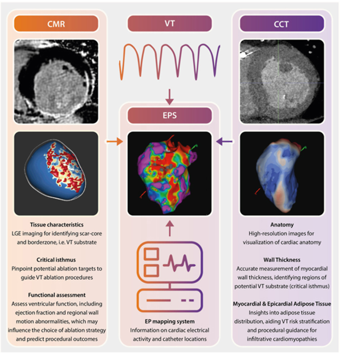
Ventricular tachycardia (VT) is a life-threatening heart rhythm and has long posed a complex challenge in the field of cardiology. Recent developments in advanced imaging modalities have aimed to improve comprehension of underlying arrhythmic substrate for VT. To this extent, high-resolution cardiac magnetic resonance (CMR) and cardiac computed tomography (CCT) have emerged as tools for accurately visualizing and characterizing scar tissue, fibrosis, and other critical structural abnormalities within the heart, providing novel insights into VT triggers and substrate. However, clinical implementation of knowledge derived from these advanced imaging techniques in improving VT treatment and guiding invasive therapeutic strategies continues to pose significant challenges. A pivotal concern lies in the absence of standardized imaging protocols and analysis methodologies, resulting in a large variance in data quality and consistency. Furthermore, the clinical significance and outcomes associated with VT substrate characterization through CMR and CCT remain dynamic and subject to ongoing evolution. This highlights the need for refinement of these techniques before their reliable integration into routine patient care can be realized. The primary objectives of this study are twofold: firstly, to provide a comprehensive overview of the studies conducted over the last 15 years, summarizing the current available literature on imaging-based assessment of VT substrate. Secondly, to critically analyze and evaluate the selected studies, with the aim of providing valuable insights that can inform current clinical practice and future research.

Keywords: Arrhythmogenic substrate; Cardiac computed tomography; Cardiac magnetic resonance imaging; Image integration; Ventricular tachycardia.


Figures

None
Graphical abstract
Figure 1
Figure 1
Role of magnetic resonance imaging–derived scarmaps in guiding ventricular tachycardia (VT) ablation. A: Electroanatomic map (EAM) (left) and cardiac magnetic resonance–derived scarmap (right) of a patient with ischemic cardiomyopathy where ablation of the conduction channel (CC) rendered the VT noninducible. B: Comparable overview with EAM (left) and scarmap (right) of a patient with nonischemic cardiomyopathy where activation mapping was not feasible due to hemodynamic instability during VT. Fourt different VT foci were identified using targeted mapping in regions with border zone and CCs. The CC, which traversed through a large midwall septal substrate, was ablated, resulting in noninducibility of the clinical VT.
Figure 2
Figure 2
Clinical utility of wideband late gadolinium enhancement (LGE) for artifact suppression. Left: Conventional long- and short-axis LGE images with a large susceptibility artifact from the can of an implantable cardioverter-defibrillator on the left ventricular anterior wall. Right: Wide-band LGE images in the same patient with resolution of the artifact providing diagnostic quality images.
Figure 3
Figure 3
Cardiac computer tomography (CCT)-derived wall thickness. Wall thickness analysis from computer tomographic imaging in a patient with ischemic cardiomyopathy demonstrating a conduction channel (blue area; normal wall thickness) surrounded by wall thinning (red areas). EAM = electroanatomic mapping.
Figure 4
Figure 4
Recommendations for a standardized image acquisition and analysis workflow before ventricular tachycardia (VT) ablation. A concise overview of recommended imaging-strategies for optimizing preprocedural imaging in patients undergoing VT ablation. If available, 3-dimensional late gadolinium enhanced (3D-LGE) imaging should be preferred over 2-dimensional (2D)-LGE. Scarmaps, processed using commercially available software, can significantly facilitate procedural guidance. If no cardiac magnetic resonance (CMR) imaging is available, late enhancement cardiac computed tomography (LE-CCT) or CT angiography (CTA) should be acquired before VT ablation. LE analysis or wall thickness in conjunction with intramyocardial fat can be used to generate comparable scarmaps. Integration of CMR or CT-derived scarmaps with the electroanatomical system should be performed by using standardized landmarks such as the pulmonary artery (PA) or aorta. These recommendations aim to enhance the accuracy and efficiency of substrate identification, ultimately improving the precision and efficacy of VT ablation procedures. ARVC = arrhythmogenic right ventricular cardiomyopathy; EAVM = electroanatomic voltage mapping; FWHM = full-width half-maximum.

References

    1. Sultan A., Futyma P., Metzner A., et al. Management of ventricular tachycardias: insights on centre settings, procedural workflow, endpoints, and implementation of guidelines-results from an EHRA survey. Europace. 2024;26 - PMC - PubMed
    1. Samuel M., Elsokkari I., Sapp J.L. Ventricular tachycardia burden and mortality: association or causality? Can J Cardiol. 2022;38:454–464. - PubMed
    1. Roca-Luque I., Mont-Girbau L. Cardiac magnetic resonance for ventricular tachycardia ablation and risk stratification. Front Cardiovasc Med. 2021;8 - PMC - PubMed
    1. Samuel M., Healey J.S., Nault I., et al. Ventricular tachycardia and ICD therapy burden with catheter ablation versus escalated antiarrhythmic drug therapy. JACC Clin Electrophysiol. 2023;9 - PubMed
    1. Garcia Garcia J., Arya A., Dinov B., et al. Impact of repeat ablation of ventricular tachycardia in patients with structural heart disease. Europace. 2023;26 - PMC - PubMed

LinkOut - more resources