

Comparative Histopathologic Analysis of Inner Ear Damage in Meningitis: Otogenic Versus Meningogenic Routes
- PMID: 39263886
- PMCID: PMC11725716
- DOI: 10.1002/lary.31759
Comparative Histopathologic Analysis of Inner Ear Damage in Meningitis: Otogenic Versus Meningogenic Routes
Abstract
Objective: To distinguish the patterns of inner ear changes between meningogenic and otogenic routes in meningitis cases. Our hypothesis is that pinpointing distinct patterns linked to each route could aid in the development of diagnostic strategies and targeted therapies.
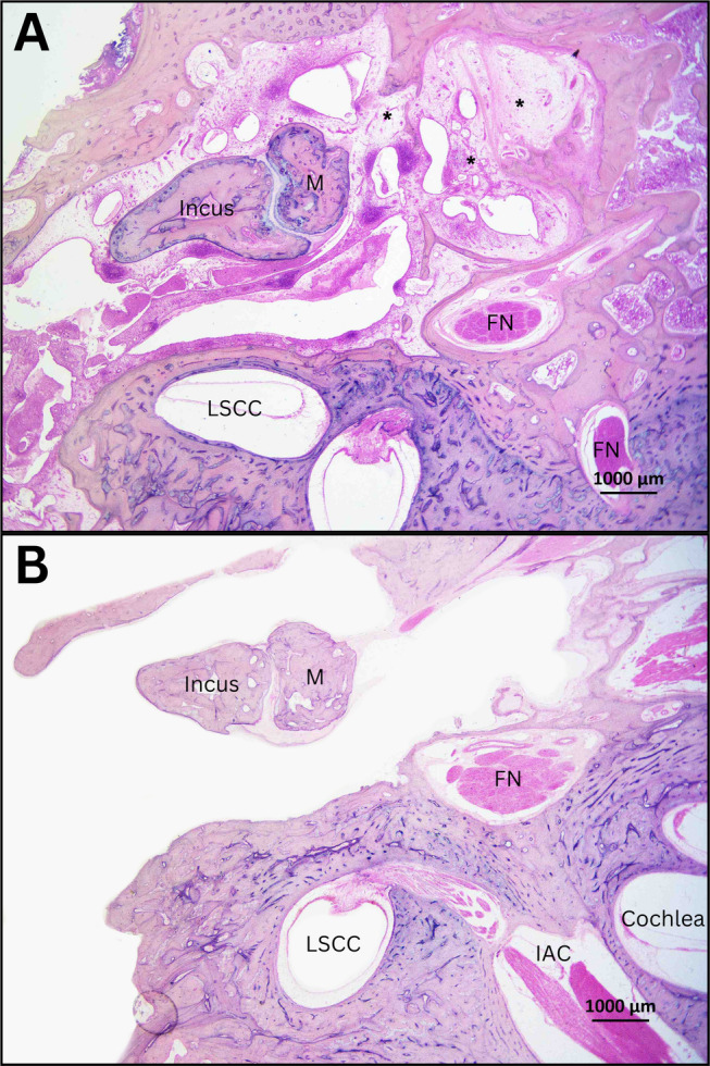
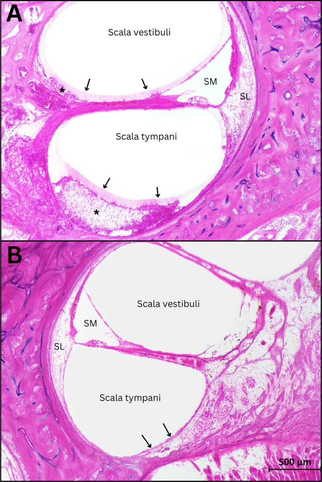
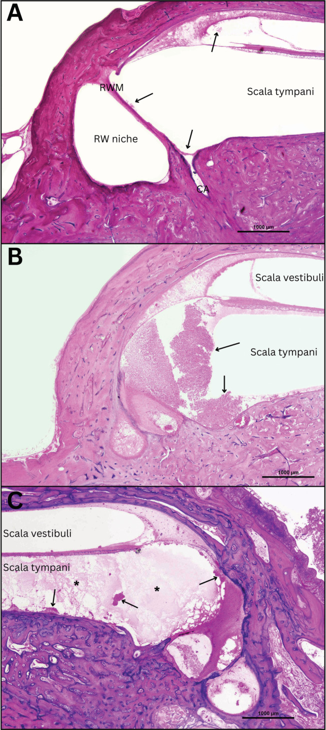
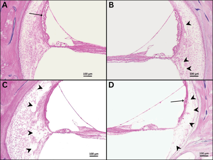
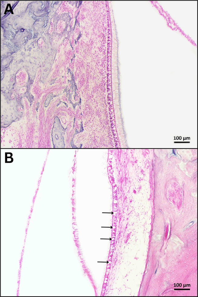
Methods: Temporal bones (TBs) from patients with a history of meningitis and histopathological evidence of labyrinthitis were divided into two groups (otogenic and meningogenic). Inner ear histopathological examination was performed to identify qualitative and semi-quantitative changes. This assessment encompassed inflammation patterns, indications of early ossification, hair cell loss, and alterations in the lateral wall, round window membrane, cochlear aqueduct and vestibular aqueduct.
Results: Thirty-six TBs were included in the study (otogenic, 21; meningogenic, 15). Generalized labyrinthitis was more common in otogenic cases (100% vs. 53%, p < 0.001). Early signs of cochlear ossification were exclusively observed in otogenic cases (9 TBs). The spiral ligament of otogenic cases has shown a uniform loss of fibrocytes across all cochlear turns, while meningogenic cases showed more severe loss in the apical turn. Otogenic cases exhibited a higher prevalence of severe inflammation of the cochlear aqueduct and endolymphatic sac. Meningogenic cases showed more severe loss of vestibular hair cells in the otolithic organs.
Conclusion: Otogenic cases displayed a higher prevalence of changes in the spiral ligament and signs of early ossification, whereas meningogenic cases were associated with a higher degree of vestibular damage. Our findings emphasize the importance of considering the infection route and its implications for timely diagnosis and development of pathology-oriented treatment strategies.
Level of evidence: NA Laryngoscope, 135:864-872, 2025.
Keywords: human temporal bone; labyrinthitis; meningitis; otopathology.
© 2024 The Author(s). The Laryngoscope published by Wiley Periodicals LLC on behalf of The American Laryngological, Rhinological and Otological Society, Inc.
Figures
References
-
- Worsøe L, Cayé‐Thomasen P, Brandt CT, Thomsen J, Østergaard C. Factors associated with the occurrence of hearing loss after pneumococcal meningitis. Clin Infect Dis. 2010;51(8):917‐924. - PubMed
-
- Lucas MJ, Brouwer MC, van de Beek D. Neurological sequelae of bacterial meningitis. J Infect. 2016;73(1):18‐27. - PubMed
-
- Edmond K, Clark A, Korczak VS, Sanderson C, Griffiths UK, Rudan I. Global and regional risk of disabling sequelae from bacterial meningitis: a systematic review and meta‐analysis. Lancet Infect Dis. 2010;10(5):317‐328. - PubMed
Publication types
MeSH terms
Grants and funding
LinkOut - more resources
Full Text Sources
Medical
Research Materials
