. 2024 Aug-Sep;72(8-9):495-515.
doi: 10.1369/00221554241274878. Epub 2024 Sep 12.

Renal Mast Cell-Specific Proteases in the Pathogenesis of Tubulointerstitial Fibrosis

Affiliations

Renal Mast Cell-Specific Proteases in the Pathogenesis of Tubulointerstitial Fibrosis

Dmitrii Atiakshin et al. J Histochem Cytochem. 2024 Aug-Sep.

Abstract

Chronic kidney disease is detected in 8-15% of the world's population. Along with fibrotic changes, it can lead to a complete loss of organ function. Therefore, a better understanding of the onset of the pathological process is required. To address this issue, we examined the interaction between mast cells (MCs) and cells in fibrous and intact regions, focusing on the role of MC proteases such as tryptase, chymase, and carboxypeptidase A3 (CPA3). MCs appear to be involved in the development of inflammatory and fibrotic changes through the targeted secretion of tryptase, chymase, and CPA3 to the vascular endothelium, nephron epithelium, interstitial cells, and components of intercellular substances. Protease-based phenotyping of renal MCs showed that tryptase-positive MCs were the most common phenotype at all anatomic sites. The infiltration of MC in different anatomic sites of the kidney with an associated release of protease content was accompanied by a loss of contact between the epithelium and the basement membrane, indicating the active participation of MCs in the formation and development of fibrogenic niches in the kidney. These findings may contribute to the development of novel strategies for the treatment of tubulointerstitial fibrosis.

Keywords: carboxypeptidase A3; chymase; fibrogenic niche; kidney fibrosis; mast cells; specific tissue microenvironment; tryptase.


Figures
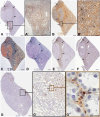
Figure 1.
Figure 1.
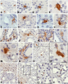
Features of the mast cell (MC) association with elements of the kidney stroma in nephrofibrosis. Technique: (A, E–G) immunohistochemical staining of MC tryptase (A, G), α-smooth muscle actin (α-SMA; E), and vimentin (F); (B) silver impregnation; (C) Heidenhain’s Azan staining; (D) Alcian blue and periodic acid Schiff staining. (A) Selective localization of tryptase-positive MCs in the kidney with high protease secretion activity (arrowed). (A′) Enlarged fragment. (A, B–D) Formation of distinct tryptase-positive inductive fields, coinciding in localization with areas of high content of reticular fibers and collagen fibrillogenesis (B, B′, arrowed), as well as collagen fibers and an amorphous component of the extracellular matrix of the connective tissue (C, D, arrowed). (E, F) Colocalization of intraorganic expression of α-SMA (E) and vimentin (F) with MC histotopography (arrowed). (G) Participation of MCs in the formation of a profibrogenic niche with a tryptase-inductive zone in the unaltered kidney parenchyma (G′,G′′ arrowed). Bars A′, B′, G′ = 50 µm; bar G′′ = 5 µm; bars A–G = 500 µm.
Figure 2.
Figure 2.
Histo- and cytotopography of mast cell tryptase under the formation of fibrotic changes in the kidney. (A) Close mast cell localization to the renal corpuscle in the cortex. (B–D) Target tryptase secretion by mast cells to the basement epithelial membrane of the proximal convoluted tubule of the nephron (arrowed) and interstitium cells (double arrowed). (E) Tryptase secretion to the basement membrane of the epithelium of the distal convoluted tubule of the nephron and the structures of the interstitium (arrowed). (F) Tryptase secretion into the extracellular matrix of the kidney interstitium in the cortex at the border of the distal and proximal convoluted tubules of the nephron (arrowed). (G) Interaction of mast cells with stromal cells in the renal cortex (arrowed), including fibroblast (double arrowed). (H–N) Options for the formation of profibrogenic niches by mast cells in the interstitium of the kidney with local tryptase accumulation in the extracellular matrix (arrowed). (O) Target tryptase secretion to the stromal cell with penetration into the nuclear structures (arrowed). (P–R) Tryptase-positive nuclei in the cells of a specific tissue microenvironment of profibrogenic renal niches. The positivity of the nucleus to tryptase, mainly in the cells of the interstitium (arrowed), is highlighted. O′ - enlarged fragment O. Q′ - enlarged fragment Q. Bars A–R = 10 µm.
Figure 3.
Figure 3.
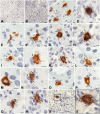
Carboxypeptidase A3 (CPA3)-positive mast cells (MCs) in the kidney in nephrofibrosis. (A, B) High content of MCs in the kidney area with fibrotic changes. (C) MCs form groups in the paracrine zone near the fibroblast (arrowed). (D) Colocalization of MCs with several stromal cells in the fibrosis site, active secretion of CPA3. (E–G) Secretion of CPA3 to the epithelium of the nephron (arrowed) in fibrous areas of the renal medulla. (H) Simultaneous effect of CPA3 on the nephron epithelium and interstitium cells (arrowed). (I) MC participation in the epithelial–mesenchymal transformation of nephron cells (presumably arrowed). (J) MC colocalization with myofibroblast of the interstitium of the kidney (presumably arrowed). (K–S) Morphological equivalents of MC participation in the formation of profibrogenic niches with CPA3 secretion (arrowed) to the targets of a specific tissue microenvironment, including the epithelium of the proximal and distal convoluted tubules (K–M), interstitium cells (N–R), and leaves of the Shumlyansky–Bowman’s capsule (S). Bars A and B = 50 µm; bars C–S and S′ = 5 µm.
Figure 4.
Figure 4.
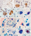
Mast cells (MCs) in the formation of profibrotic changes in the kidney. Techniques: (A–E) immunohistochemical staining of MC chymase; (F, G, K–N) staining with toluidine blue; (H–J) Giemsa staining; (O) Picro Mallory staining. (A) Accumulation of chymase in mature fibrous tissue (arrowed) at the border with the renal cortex. (B–D) Target chymase secretion to the basement membrane of the epithelium of the proximal (B, C, arrowed) and distal (D, arrowed) parts of the kidney nephron. (E–H) Directed MC degranulation to fibroblasts in the zone of fibrotic changes (arrowed). (I) MCs at the periphery of a profibrogenic niche with active secretory activity (arrowed). (J) Colocalization of an MC and eosinophil in the fibrous tissue. (K–M) Various variants of MC secretory activity through autonomous secretory granule excretion into the extracellular matrix (arrowed), resulting in a decreased number of granules in the cytoplasm (L) and the formation of areas of a specific tissue microenvironment filled with secretory granules (M). (N and O) Variants of MC colocalization with a fibrous component of the extracellular matrix in the kidney area with fibrotic changes. Bar A = 50 µm; bars B–O = 5 µm.
Figure 5.
Figure 5.
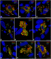
Profile of specific mast cell proteases during the formation of profibrogenic niches in the kidney stroma. (A) Active entry of tryptase and carboxypeptidase A3 (CPA3) to the targets of the extracellular matrix of the specific tissue microenvironment of the tissue niche in the composition of tunneling nanotubules (arrowed) and secretory granules (double arrowed). (B) Entry of tryptase and CPA3 into the profibrogenic niche as part of secretory granules (arrowed). (C, D) Dissemination of secretory granules with specific mast cell proteases in the interstitium (arrowed) adjacent to the stroma cells. (E) Contacting areas of the mast cell cytoplasm filled with large secretory granules, with predominant localization of tryptase and chymase along the periphery of the granules. (F, G) Mast cells in the state of secretion of specific proteases to the epitheliocytes of the nephron tubules (arrowed) of the renal medulla. (H, I) Active secretion of tryptase and CPA3 to interstitial cells of the fibrous part of the kidney (arrowed), including fibroblasts (presumably double arrowed). Bars A–I = 5 µm.
Figure 6.
Figure 6.
Histotopography of mast cells (MCs) in the interstitium of the cortex and medulla of the kidney with fibrosis (in percentage of the total number of MCs, staining technique—monoplex immunohistochemical). Abbreviation: CPA3, carboxypeptidase A3.
Figure 7.
Figure 7.
The content of α-smooth muscle actin (α-SMA)- and vimentin-positive cells and structures in the kidney (in percentage of the total number on the analysis area, staining technique—monoplex immunohistochemical).
Figure 8.
Figure 8.
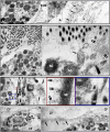
Mast cells (MCs) in the extracellular matrix (ECM) remodeling during the formation of fibrogenic niches in the tissue microenvironment of the kidneys. Electronic micrographs. (A) MC in the ECM mesophase with tactoid formation (arrowed) and the initial stages of collagen microfibril formation (double arrowed). A′—magnified fragment of A. (B) MC degranulation with specific positioning of secretome components (arrowed) and collagen fibrillogenesis initiation with microfibril formation (double arrowed). (C) Ultrastructural signs of the MC granule secretory material involved in collagen fibrillogenesis with ECM tactoid nucleation, specific positioning of collagen protofibrils (arrowed), and thickening of microfibrils (double arrowed). (D) Grouping of collagen microfibrils into target-specific bundles with developing features of the microfibril spatial distribution in the peripheral region of the collagen fiber (arrowed). Bars A–D = 1 µm. Abbreviation: CF, collagen fibers; MF, microfibril; SG, secretory granule.
Figure 9.
Figure 9.
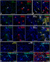
Spatial mapping and profile of specific mast cell (MC) proteases in the stromal and immune landscapes of fibrogenic niches. Technique: sequential multiplex immunohistochemical detection of molecular targets. (A, B) Sequential staining of tryptase and transforming growth factor-beta (TGF-β). High TGF-β expression in MCs adjacent to the nephron epithelium (arrowed) in the presence of MCs with moderate TGF-β expression. (C, D) Sequential staining of tryptase, chymase, carboxypeptidase A3 (CPA3), and CD31. The predominant phenotype of MCs, TryptaseHigh, Chymasehigh, and CPA3High, contact with CD31+ cells (arrowed). (E) Sequential staining of chymase, α-smooth muscle actin (α-SMA), CD163, CD68, CD38, and chymase. The most frequent MC colocalization with α-SMA-positive stromal cells of fibrous niches. Bars A–E = 50 µm.

References

    1. GBD Chronic Kidney Disease Collaboration. Global, regional, and national burden of chronic kidney disease, 1990-2017: a systematic analysis for the Global Burden of Disease Study 2017. Lancet. 2020;395(10225):709–33. doi: 10.1016/S0140-6736(20)30045-3. - DOI - PMC - PubMed
    1. Chen TK, Knicely DH, Grams ME. Chronic kidney disease diagnosis and management: a review. JAMA. 2019;322(13):1294–304. doi: 10.1001/jama.2019.14745. - DOI - PMC - PubMed
    1. Sun YB, Qu X, Caruana G, Li J. The origin of renal fibroblasts/myofibroblasts and the signals that trigger fibrosis. Differentiation. 2016;92(3):102–07. doi: 10.1016/j.diff.2016.05.008. - DOI - PubMed
    1. Hwang SH, Lee YM, Choi Y, Son HE, Ryu JY, Na KY, Chin HJ, Jeon NL, Kim S. Role of human primary renal fibroblast in TGF-β1-mediated fibrosis-mimicking devices. Int J Mol Sci. 2021;22(19):10758. doi: 10.3390/ijms221910758. - DOI - PMC - PubMed
    1. Liu L, Wang Y, Yan R, Liang L, Zhou X, Liu H, Zhang X, Mao Y, Peng W, Xiao Y, Zhang F, Liu L, Shi M, Guo B. BMP-7 inhibits renal fibrosis in diabetic nephropathy via miR-21 downregulation. Life Sci. 2019;238:116957. doi: 10.1016/j.lfs.2019.116957. - DOI - PubMed

Publication types

LinkOut - more resources