. 2024 Sep 25;44(9):BSR20240826.
doi: 10.1042/BSR20240826.

Loss of cardiomyocyte-specific adhesion G-protein-coupled receptor G1 (ADGRG1/GPR56) promotes pressure overload-induced heart failure

Affiliations

Loss of cardiomyocyte-specific adhesion G-protein-coupled receptor G1 (ADGRG1/GPR56) promotes pressure overload-induced heart failure

Jeanette Einspahr et al. Biosci Rep. .

Abstract

Adhesion G-protein-coupled receptors (AGPCRs), containing large N-terminal ligand-binding domains for environmental mechano-sensing, have been increasingly recognized to play important roles in numerous physiologic and pathologic processes. However, their impact on the heart, which undergoes dynamic mechanical alterations in healthy and failing states, remains understudied. ADGRG1 (formerly known as GPR56) is widely expressed, including in skeletal muscle where it was previously shown to mediate mechanical overload-induced muscle hypertrophy; thus, we hypothesized that it could impact the development of cardiac dysfunction and remodeling in response to pressure overload. In this study, we generated a cardiomyocyte (CM)-specific ADGRG1 knockout mouse model, which, although not initially displaying features of cardiac dysfunction, does develop increased systolic and diastolic LV volumes and internal diameters over time. Notably, when challenged with chronic pressure overload, CM-specific ADGRG1 deletion accelerates cardiac dysfunction, concurrent with blunted CM hypertrophy, enhanced cardiac inflammation and increased mortality, suggesting that ADGRG1 plays an important role in the early adaptation to chronic cardiac stress. Altogether, the present study provides an important proof-of-concept that targeting CM-expressed AGPCRs may offer a new avenue for regulating the development of heart failure.

Keywords: ADGRG1; Adhesion GPCR; GPR56; cardiomyocytes; heart failure; pressure overload.


Conflict of interest statement

The authors declare that there are no competing interests associated with the manuscript.

Figures
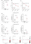
Figure 1
Figure 1. ADGRG1 and its ligands undergo dynamic alterations in expression in the heart during the development of heart failure
(A) Doppler echocardiography was performed 1-week post-TAC surgery to assess peak pressure gradients at the constriction site in C57BL/6 mice. Data are mean ± SEM, ****P <0.0001, two-tailed t-test, n = 8 male mice per group. Serial echocardiography was performed at baseline, 6- and 12-week post-surgery to assess ejection fraction (%EF, B) and fractional shortening (%FS, C). Data are mean ± SEM, *P<0.05, **P<0.01, ****P<0.0001, two-way ANOVA with Tukey’s post-hoc test, n=8 (baseline, 6 weeks) or 4 (12 weeks) for Sham and TAC groups. Hearts were harvested at 6 weeks (D) or 12 weeks (E) post-surgery and LV processed for RNA extraction and RT-qPCR analysis of Adgrg1, Tg2 and Col3a1. Data are mean ± SEM, *P<0.05, ns = not significant, two-tailed t-test, n = 4 male mice per group. (F) To investigate the expression of Adgrg1 in cardiomyocytes specifically following TAC, we analyzed existing single cell RNA-sequencing data (GSE120064) from 2 to 4 male C57BL/6 male mice per timepoint [27]. Dot plots indicate the expression (in TP10K) and % cardiomyocyte expression of Adgrg1 in response to TAC over time (compared with Agtr1a, and with Nppa as a control for TAC-induced changes in transcript expression).
Figure 2
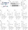
Figure 2. Constitutive cardiomyocyte-specific ADGRG1 deletion
(A) ADGRG1 floxed mice (ADGRG1fl/fl), possessing a loxP site upstream of exon 4 as well as a 3′ loxP site downstream of exon 6, were crossed with transgenic mice expressing an αMHC promoter-driven Cre recombinase (αMHC-Cre) to generate mice with constitutive cardiomyocyte-specific deletion of ADGRG1/GPR56 (CM-ADGRG1-KO). (B) ADGRG1 knockdown was validated in isolated adult mouse cardiomyocytes (AMCM) by Western blot analysis. Data are mean ± SEM, **P<0.01, two-tailed t-test, n = 8 AMCM preparations (from 4 male and 4 female mice) per genotype. Echocardiography was used to assess systolic (C) and diastolic (D) LV volume, and systolic (E) and diastolic (F) internal diameter, ejection fraction (%EF, G), fractional shortening (%FS, H), stroke volume (I) and cardiac output (J) in 8- and 10-week-old CM-ADGRG1-KO (n = 5; 4 female, 1 male) and αMHC-Cre mice (n = 5; 3 female, 2 male). Data are mean ± SEM, **P<0.01, two-way ANOVA with Tukey’s post-hoc test.
Figure 3
Figure 3. Cardiomyocyte-specific ADGRG1 deletion hastens cardiac dysfunction in response to chronic pressure overload
(A) Schematic of TAC study design. Mice were 12 weeks of age at time of TAC surgery. (B) To confirm equivalent surgical constrictions in vivo, doppler echocardiography was performed 1-week post-surgery to assess peak pressure gradient of the constriction site. Data are mean ± SEM, ***P<0.001, ns = not significant; Bartlett’s test associated with initial one-way ANOVA yielded P<0.0001 suggesting unequal variance, thus Brown–Forsythe and Welch’s ANOVA tests with Dunnett’s T3 multiple comparison tests were performed. (C) Echocardiography was performed to assess % ejection fraction (D) and % fractional shortening (E). Data are mean ± SEM, *P<0.05, **P<0.01, ***P<0.001 versus MHC-Cre TAC, ####P<0.0001 versus CM-ADGRG1-KO Sham and ††P<0.01 versus MHC-Cre Sham, two-way ANOVA with Tukey’s post-hoc test. n = 4 (MHC-Cre Sham; 2 males, 2 females), n = 5 (CM-ADGRG1-KO Sham; 1 male, 4 females), n = 6 (MHC-Cre TAC; 3 males, 3 females), n = 7 (CM-ADGRG1-KO TAC; 2 males, 5 females).
Figure 4
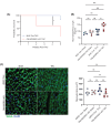
Figure 4. Cardiomyocyte-specific ADGRG1 deletion reduces survival in response to chronic pressure overload
(A) Survival analysis of CM-ADGRG1-KO versus MHC Cre mice following TAC. *P<0.01, Log-rank (Mantel-Cox) test, n = 11 (CM-ADGRG1-KO; 6 males, 5 females) and 6 (MHC Cre; 4 males, 2 females). (B) Gravimetric data was collected from 5-week-post TAC mice and Heart Weight/Tibia Length ratios are indicated. Data are mean ± SEM, *P<0.05, **P<0.01, ns = not significant, one-way ANOVA with Tukey’s post-hoc test. n = 4 (MHC-Cre Sham; 2 males, 2 females), n = 5 (CM-ADGRG1-KO Sham; 1 males, 4 females), n = 6 (MHC-Cre TAC; 3 males, 3 females), n = 6 (CM-ADGRG1-KO TAC; 2 males, 4 females). (C) Wheat germ agglutinin (WGA) staining was performed on cardiac slices from CM-ADGRG1-KO and MHC Cre mice at 5-week post-surgery, with data quantified in histogram. Data are mean ± SEM, ns = not significant, one-way ANOVA with Tukey’s post-hoc test. n = 4 (MHC-Cre Sham; 2 males, 2 females), n = 5 (CM-ADGRG1-KO Sham; 1 male, 4 females), n = 6 (MHC-Cre TAC; 3 males, 3 females), n = 6 (CM-ADGRG1-KO TAC; 2 males, 4 females).
Figure 5
Figure 5. Cardiomyocyte-specific ADGRG1 deletion increases peripheral immune cells recruitment to the heart in response to chronic pressure overload
(A) Masson’s trichrome (MTC) staining was performed on cardiac slices from CM-ADGRG1-KO and MHC Cre mice at 5-week post-surgery, with data quantified in histogram. Data are mean ± SEM, *P<0.05, ns = not significant; Bartlett’s test associated with initial one-way ANOVA yielded P=0.0381 suggesting unequal variance, thus Brown–Forsythe and Welch’s ANOVA tests with Dunnett’s T3 multiple comparison tests were performed. n = 4 (MHC-Cre Sham; 2 males, 2 females), n = 5 (CM-ADGRG1-KO Sham; 1 male, 4 females), n = 6 (MHC-Cre TAC; 3 males, 3 females), n = 6 (CM-ADGRG1-KO TAC; 2 males, 4 females). (B) CD45 (red) and DAPI (blue) staining were performed on cardiac slices from CM-ADGRG1-KO and MHC Cre mice at 5-week post-surgery, with data quantified in histogram. Data are mean ± SEM, **P<0.01, ***P<0.001, ****P<0.0001, ns = not significant, one-way ANOVA with Tukey’s post-hoc test. n = 4 (MHC-Cre Sham; 2 males, 2 females), n=5 (CM-ADGRG1-KO Sham; 1 male, 4 females), n=6 (MHC-Cre TAC; 3 males, 3 females), n=6 (CM-ADGRG1-KO TAC; 2 males, 4 females).

References

    1. Martin S.S., Aday A.W., Almarzooq Z.I., Anderson C.A.M., Arora P., Avery C.L.et al. . (2024) 2024 Heart Disease and Stroke Statistics: a report of US and global data From the American Heart Association. Circulation 149, e347–e913 10.1161/CIR.0000000000001209 - DOI - PubMed
    1. Einspahr J.M. and Tilley D.G. (2022) Pathophysiological impact of the adhesion G protein-coupled receptor family. Am. J. Physiol. Cell Physiol. 323, C640–C647 10.1152/ajpcell.00445.2021 - DOI - PMC - PubMed
    1. Insel P.A., Snead A., Murray F., Zhang L., Yokouchi H., Katakia T.et al. . (2012) GPCR expression in tissues and cells: are the optimal receptors being used as drug targets? Br. J. Pharmacol. 165, 1613–1616 10.1111/j.1476-5381.2011.01434.x - DOI - PMC - PubMed
    1. Wilde C., Mitgau J., Suchy T., Schoneberg T. and Liebscher I. (2022) Translating the force-mechano-sensing GPCRs. Am. J. Physiol. Cell Physiol. 322, C1047–C1060 10.1152/ajpcell.00465.2021 - DOI - PubMed
    1. Liu M., Parker R.M., Darby K., Eyre H.J., Copeland N.G., Crawford J.et al. . (1999) GPR56, a novel secretin-like human G-protein-coupled receptor gene. Genomics 55, 296–305 10.1006/geno.1998.5644 - DOI - PubMed

Publication types

MeSH terms

Substances

LinkOut - more resources