. 2024;44(11):516-527.
doi: 10.1080/10985549.2024.2399358. Epub 2024 Sep 12.

Midnolin, a Genetic Risk Factor for Parkinson's Disease, Promotes Neurite Outgrowth Accompanied by Early Growth Response 1 Activation in PC12 Cells

Affiliations

Midnolin, a Genetic Risk Factor for Parkinson's Disease, Promotes Neurite Outgrowth Accompanied by Early Growth Response 1 Activation in PC12 Cells

Ayano Chiba et al. Mol Cell Biol. 2024.

Abstract

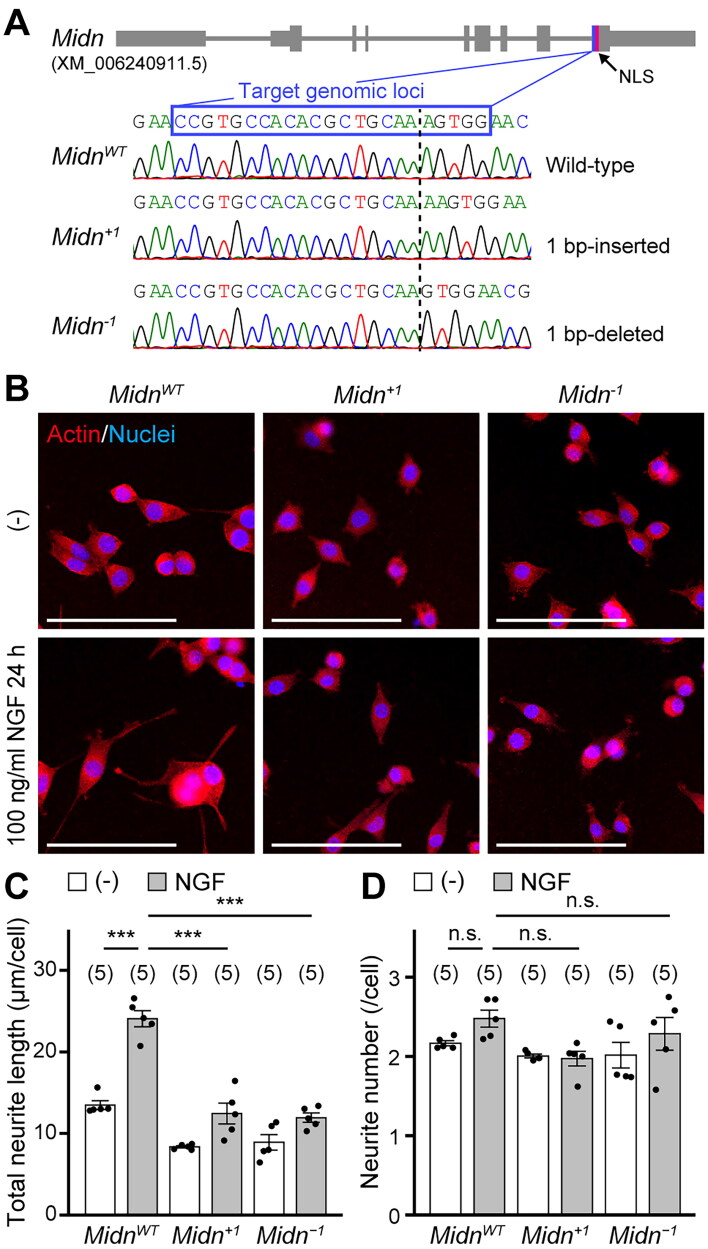
Parkinson's disease (PD) is an age-related progressive neurodegenerative disease. Previously, we identified midnolin (MIDN) as a genetic risk factor for PD. Although MIDN copy number loss increases the risk of PD, the molecular function of MIDN remains unclear. To investigate the role of MIDN in PD, we established monoclonal Midn knockout (KO) PC12 cell models. Midn KO inhibited neurite outgrowth and neurofilament light chain (Nefl) gene expression. Although MIDN is mainly localized in the nucleus, it does not encode DNA-binding domains. We therefore hypothesized that MIDN might bind to certain transcription factors and regulate gene expression. Of the candidate transcription factors, we focused on early growth response 1 (EGR1) because it is required for neurite outgrowth and its target genes are downregulated by Midn KO. An interaction between MIDN and EGR1 was confirmed by immunoprecipitation. Surprisingly, although EGR1 protein levels were significantly increased in Midn KO cells, the binding of EGR1 to the Nefl promoter and resulting transcriptional activity were downregulated as measured by luciferase assay and chromatin immunoprecipitation quantitative real-time polymerase chain reaction. Overall, we identified the MIDN-dependent regulation of EGR1 function. This mechanism may be an underlying reason for the neurite outgrowth defects of Midn KO PC12 cells.

Keywords: PC12 cells; Parkinson’s disease (PD); early growth response 1 (EGR1); neurite outgrowth; neurofilament light chain (NEFL).


Conflict of interest statement

Yoshishiro Koyama is an employee of ThermoFisher Scientific KK (Tokyo, Japan) and contributed to the measurement of neurite outgrowth. Apart from this, the authors report that there are no competing interests to declare.

Figures

Figure 1.
Figure 1.
Midnolin (MIDN) is responsible for neurite outgrowth in PC12 cells. (A) Midn cDNA sequences from wild-type (WT) PC12 cells and monoclonal Midn knockout (KO) PC12 cells established using clustered regularly interspaced short palindromic repeats (CRISPR)/CRISPR-associated protein 9 (Cas9)-mediated genome editing. The gray lines at the top indicate the Midn gene. Bold, medium, and thin lines indicate the coding sequence, untranslated regions, and introns, respectively. The Midn frameshift mutation was homozygously induced near the nuclear localization signal (NLS). Dotted lines indicate inserted or deleted sites. MidnWT, a WT clone; Midn+1, a single nucleotide-inserted clone; Midn−1, a single nucleotide-deleted clone. (B) Representative images of MidnWT, Midn+1, or Midn−1 PC12 cells treated with or without nerve growth factor (NGF, 100 ng/mL, 24 h). Actin fibers and nuclei were stained with rhodamine-phalloidin (5 U/mL) and Hoechst-33258 (1 μg/mL), respectively. NGF-induced neurite outgrowth was reduced in Midn KO PC12 cells. (–), Absence of NGF; scale bars, 100 μm. (C) Quantitative analysis of the total neurite length shown in (B), measured as described in the Materials and Methods. In this and the following graphs, unless otherwise noted, each dot represents a single sample value; the number of samples is indicated at the top. (D) Quantitative analysis of the number of neurites shown in (B), measured as described in the Materials and Methods. n.s., not significant; ***P < 0.001 (Tukey’s test). Data are expressed as the mean ± standard error.
Figure 2.
Figure 2.
MIDN is involved in the expression of neurofilament light chain (NEFL) in PC12 cells. (A) Nefl promoter activity was measured as described in the Materials and Methods. PC12 cells were treated with or without NGF (100 ng/mL, 6 h). Nefl promoter activity was enhanced by NGF (100 ng/mL, 6 h) in MidnWT PC12 cells; this effect was significantly attenuated in Midn KO PC12 cells. (B) Relative expression of Nefl mRNA, analyzed using quantitative real-time polymerase chain reaction (qPCR). PC12 cells were treated with NGF (100 ng/mL, 6 h). NGF-enhanced Nefl mRNA expression was significantly attenuated in Midn KO PC12 cells compared with that in MidnWT PC12 cells. (C) Representative NEFL and glyceraldehyde-3-phosphate dehydrogenase (GAPDH) protein levels, measured by Western blotting. PC12 cells were treated with or without NGF (100 ng/mL, 24 h). NGF-enhanced NEFL protein levels were attenuated in Midn KO PC12 cells compared with those in MidnWT PC12 cells. (D) Representative NEFL, GAPDH, and MIDN-Flag protein levels in PC12 cells transfected with either empty vector (pcDNA3.1[+]) or MIDN-Flag. Cells were treated with or without NGF (100 ng/mL, 24 h). Reduced NEFL expression in Midn KO cells treated with NGF was partially rescued by MIDN-Flag expression. (E) Schematic representation of the MIDN-Flag-dependent rescue of NEFL expression shown in (D). ***P < 0.001 (Tukey’s test). Data are expressed as the mean ± standard error.
Figure 3.
Figure 3.
MIDN (101–200 amino acid [aa]) is required for direct binding to early growth response 1 (EGR1). (A) Representative EGR1 protein levels in PC12 cells co-immunoprecipitated with MIDN-Flag. PC12 cells were treated with or without NGF (100 ng/mL, 2 h). The nuclear fraction of the cells (Input), prepared as described in the Materials and Methods, was immunoprecipitated with sepharose beads (Control) or Flag-agarose beads (Flag-IP). EGR1 was co-immunoprecipitated with MIDN-Flag in PC12 cells. (B) Representative EGR1 protein levels in SH-SY5Y cells co-immunoprecipitated with MIDN-Flag. SH-SY5Y cells were treated with or without insulin (1 μM, 2 h). The nuclear fraction of the cells (Input) was precipitated with Flag-agarose beads (Flag-IP). EGR1 was co-immunoprecipitated with MIDN-Flag in SH-SY5Y cells. (C) Schematic representation of amino-terminal-truncated MIDN mutants, constructed as described in the Materials and Methods. UBL, ubiquitin-like domain; NLS, nuclear localization signal; N-term, amino-terminus; C-term, carboxy-terminus. (D) Representative EGR1 protein levels co-immunoprecipitated with MIDN mutants. PC12 cells were treated with or without NGF (100 ng/mL, 2 h). The nuclear fraction of cells transfected with either empty vector, MIDN-Flag, or Flag-tagged MIDN mutants (Input) was immunoprecipitated with Flag-agarose beads (Flag-IP). EGR1 was co-immunoprecipitated with MIDN-Flag and MIDN-ΔN100-Flag. (E) Representative EGR1 protein levels co-immunoprecipitated with MIDN mutants using in vitro-translated recombinant proteins, prepared as described in the Materials and Methods. MIDN (101–200 aa) was required for the direct binding to EGR1.
Figure 4.
Figure 4.
MIDN is required for EGR1 transcriptional activity. (A) Illustration of the detection of EGR1-dependent transcription using the pGL4-Egr1 (EGR1 reporter) plasmid. (B) EGR1-dependent transcription, measured as described in the Materials and Methods. PC12 cells transfected with either pGL3 empty vector or pGL4-Egr1 were cotransfected with either pcDNA3.1(+) empty vector or HA-tagged EGR1 (HA-EGR1). EGR1-dependent transcription was enhanced in PC12 cells transfected with both pGL4-Egr1 and HA-EGR1. (C) EGR1-dependent transcription that was enhanced by NGF treatment. PC12 cells transfected with pGL4-Egr1 were treated with or without NGF (100 ng/mL, either 6 h or 24 h). Treatment with NGF for 6 h enhanced EGR1-dependent transcription. (D) EGR1-dependent transcription in MidnWT and Midn−1 PC12 cells. PC12 cells transfected with pGL4-Egr1 were cotransfected with either pcDNA3.1(+) empty vector or HA-EGR1. The EGR1-dependent transcription enhanced by HA-EGR1 expression was significantly attenuated in Midn KO PC12 cells compared with that in MidnWT PC12 cells. (E) EGR1-dependent transcription that was enhanced by NGF treatment in MidnWT and Midn−1 PC12 cells. PC12 cells transfected with pGL4-Egr1 were treated with or without NGF (100 ng/mL, 6 h). The EGR1-dependent transcription enhanced by NGF treatment was significantly attenuated in Midn KO PC12 cells compared with that in MidnWT PC12 cells. (F) Schematic representation of ZnEGR1, a dominant-negative form of EGR1, constructed as described in the Materials and Methods. (G) EGR1-dependent transcription in PC12 cells transfected with either pcDNA3.1(+) empty vector or HA-tagged ZnEGR1 (ZnEGR1-HA). PC12 cells were cotransfected with pGL4-Egr1, pcDNA3.1(+) or HA-EGR1, and pcDNA3.1(+) or ZnEGR1-HA. The EGR1-dependent transcription enhanced by HA-EGR1 expression was significantly attenuated by ZnEGR1-HA expression. (H) Nefl promoter activity in PC12 cells transfected with either pcDNA3.1(+) empty vector or ZnEGR1-HA. PC12 cells transfected with Nefl-luciferase reporter were cotransfected with pcDNA3.1(+) or ZnEGR1-HA and treated with or without NGF (100 ng/mL, 6 h). The Nefl promoter activity enhanced by NGF treatment was significantly attenuated in ZnEGR1-expressing PC12 cells. n.s., not significant; *P < 0.05; **P < 0.01; ***P < 0.001 (Tukey’s test). Data are expressed as the mean ± standard error.
Figure 5.
Figure 5.
MIDN is required for EGR1 DNA binding in PC12 cells. (A) Representative EGR1 and GAPDH protein levels measured by Western blotting. PC12 cells were treated with NGF (100 ng/mL, 2 h). EGR1 expression levels normalized to GAPDH are indicated at the bottom. EGR1 protein levels were significantly increased in Midn KO PC12 cells compared with those in MidnWT PC12 cells (n = 3). (B, C) Chromatin immunoprecipitation (ChIP)-qPCR assays using EGR1 antibody or mouse immunoglobulin G (IgG). Primers targeting the reported EGR1 binding sequences in VGF nerve growth factor inducible (Vgf) and tribbles pseudokinase 1 (Trib1) promoter were used as the positive control. (D) ChIP-qPCR primer design. The long line indicates the Nefl gene, and the short lines with P1, P2, and negative control indicate the amplified region by these primer sets used for the ChIP-qPCR analysis. Ex, exon. (E–G) ChIP-qPCR analysis in MidnWT and Midn KO PC12 cells immunoprecipitated with either EGR1 antibody or control IgG. Primers shown in (D) were used. The EGR1 binding to the Nefl promoter region was significantly attenuated in Midn KO PC12 cells. (H) ChIP-qPCR primer design. The long line indicates the cyclin dependent kinase 5 regulatory subunit 1 (Cdk5r1) gene, and the short lines with P3, P4, and negative control indicate the amplified region by these primer sets used for the ChIP-qPCR analysis. (I–K) ChIP-qPCR analysis in MidnWT and Midn KO PC12 cells immunoprecipitated with either EGR1 antibody or control IgG. Primers shown in (H) were used. The EGR1 binding to the Cdk5r1 promoter region was significantly attenuated in Midn KO PC12 cells. P values were determined using Student’s t test (A) or Tukey’s test (B, C, E–G, I–K). n.s., not significant; **P < 0.01; ***P < 0.001. Data are expressed as the mean ± standard error.
Figure 6.
Figure 6.
A putative schematic representation of the MIDN-dependent regulation mechanism of neurite outgrowth in PC12 cells. In MidnWT PC12 cells (top), MIDN directly binds to EGR1 and enhances its DNA binding. As a result, the EGR1-dependent induction of Nefl and Cdk5r1 promotes neurite outgrowth. In Midn KO PC12 cells (bottom), the reduced DNA binding of EGR1 inhibits Nefl and Cdk5r1 expression, thus resulting in defective neurite outgrowth.

Similar articles

Cited by

References

    1. Dorsey ER, Bloem BR.. The Parkinson pandemic—a call to action. JAMA Neurol. 2018;75:9–10. doi:10.1001/jamaneurol.2017.3299. - DOI - PubMed
    1. Dorsey ER, Sherer T, Okun MS, Bloem BR.. The emerging evidence of the Parkinson pandemic. J Parkinsons Dis. 2018;8:S3–S8. doi:10.3233/JPD-181474. - DOI - PMC - PubMed
    1. Lill CM. Genetics of Parkinson’s disease. Mol Cell Probes. 2016;30:386–396. doi:10.1016/j.mcp.2016.11.001. - DOI - PubMed
    1. Billingsley KJ, Bandres-Ciga S, Saez-Atienzar S, Singleton AB.. Genetic risk factors in Parkinson’s disease. Cell Tissue Res. 2018;373:9–20. doi:10.1007/s00441-018-2817-y. - DOI - PMC - PubMed
    1. Blauwendraat C, Makarious MB, Leonard HL, Bandres-Ciga S, Iwaki H, Nalls MA, Noyce AJ, Singleton AB.. A population scale analysis of rare SNCA variation in the UK Biobank. Neurobiol Dis. 2021;148:105182. doi:10.1016/j.nbd.2020.105182. - DOI - PMC - PubMed

Publication types

LinkOut - more resources