. 2024 Sep 12;14(1):83.
doi: 10.1186/s13550-024-01149-8.

Brain perfusion SPECT in the presurgical evaluation of epilepsy: is additional ictal SPECT required in case of high-confidence lateralization of the seizure onset zone by interictal SPECT and vice versa?

Affiliations

Brain perfusion SPECT in the presurgical evaluation of epilepsy: is additional ictal SPECT required in case of high-confidence lateralization of the seizure onset zone by interictal SPECT and vice versa?

Kian Baradaran-Salimi et al. EJNMMI Res. .

Abstract

Background: Ictal brain perfusion SPECT provides higher sensitivity for the identification of the epileptic seizure onset zone (SOZ) than interictal SPECT. However, ictal SPECT is demanding due to the unpredictable waiting period for the next seizure to allow for ictal tracer injection. Thus, starting with an interictal scan and skipping the ictal scan if the interictal scan provides a SOZ candidate with high confidence could be an efficient approach. The current study estimated the rate of high-confidence SOZ candidates and the false lateralization rate among them for interictal and ictal SPECT.

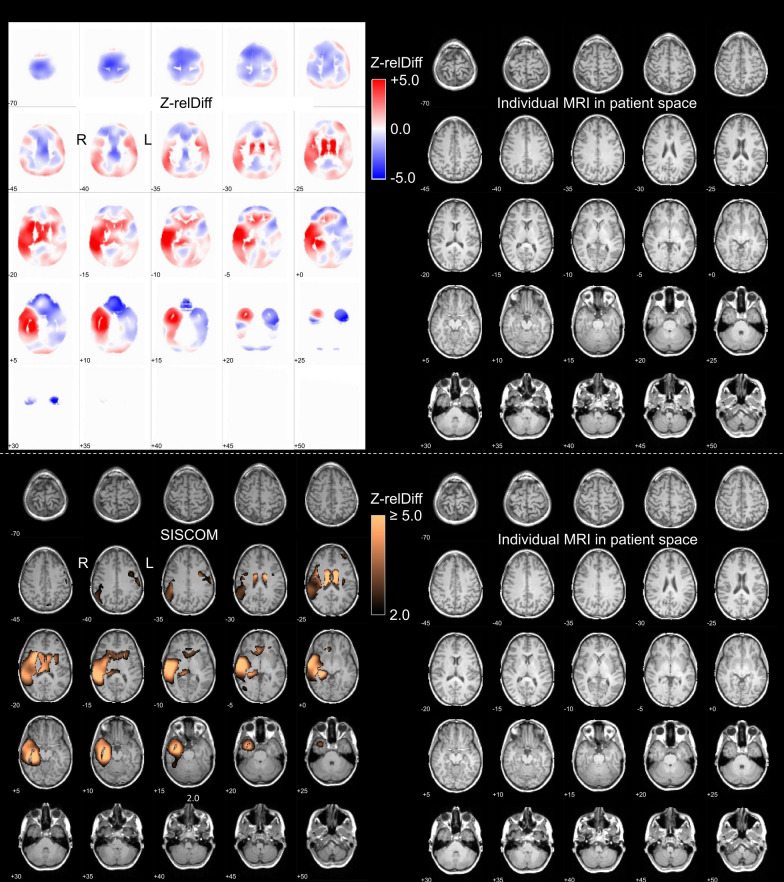
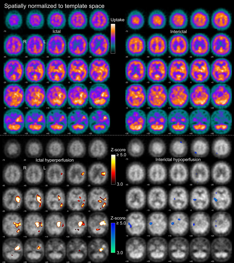
Methods: 177 patients (48% females, median age 38y, interquartile range 27-48y) with ictal and interictal SPECT acquired with 99mTc-HMPAO (n = 141) or -ECD (n = 36) were included retrospectively. The vast majority of the patients was suspected to have temporal lobe epilepsy. Visual interpretation of the SPECT data was performed independently by 3 readers in 3 settings: "interictal only" (interictal SPECT and statistical hypoperfusion map), "ictal only" (ictal SPECT and hyperperfusion map), and "full" setting (side-by-side interpretation of ictal and interictal SPECT including statistical maps and SISCOM analysis). The readers lateralized the SOZ (right, left, none) and characterized their confidence using a 5-score. A case was considered "lateralizing with high confidence" if all readers lateralized to the same hemisphere with at least 4 of 5 confidence points. Lateralization of the SOZ in the "full" setting was used as reference standard.

Results: The proportion of "lateralizing with high confidence" cases was 4.5/31.6/38.4% in the "interictal only"/"ictal only"/"full" setting. One (12.5%) of the 8 cases that were "lateralizing with high confidence" in the "interictal only" setting lateralized to the wrong hemisphere. Among the 56 cases that were "lateralizing with high confidence" in the "ictal only" setting, 54 (96.4%) were also lateralizing in the "full" setting, all to the same hemisphere.

Conclusions: Starting brain perfusion SPECT in the presurgical evaluation of epilepsy with an interictal scan to skip the ictal scan in case of a high-confidence interictal SOZ candidate is not a useful approach. In contrast, starting with an ictal scan to skip the interictal scan in case of a high-confidence ictal SOZ candidate can be recommended.

Keywords: 99mTc-ECD; 99mTc-HMPAO; Cerebral blood flow; Epilepsy; Ictal; Interictal; Perfusion; SISCOM; SPECT; Seizure onset zone.


Conflict of interest statement

The authors declare that they have no competing interests.

Figures

Fig. 1
Fig. 1
Standardized display for visual reading in the “interictal only” setting. The upper and lower parts were provided as page 1 and page 2 of a 2-page pdf-document. On the first page, the left side shows the patient’s interictal perfusion SPECT coregistered to the individual T1-weighted MRI shown on the right. On the second page, the left side shows the patient’s interictal perfusion SPECT after spatial normalization to MNI space, the right side shows the statistical hypoperfusion map thresholded at z ≥ 3.0 and overlaid to the patient’s interictal SPECT. The upper threshold of the color bar for the display of the SPECT images was set to the maximum of the 3-dimensional SPECT image volume, separately for each SPECT scan. The lower threshold of the color bar was set to zero in all cases. The example images were acquired with 99mTc-HMPAO in a 28y old woman with normal MRI
Fig. 2
Fig. 2
Standardized display for visual reading in the “ictal only” setting. The upper and lower parts were provided as page 1 and page 2 of a 2-page pdf-document. On the first page, the left side shows the patient’s ictal perfusion SPECT coregistered to the individual T1-weighted MRI shown on the right. On the second page, the left side shows the patient’s ictal perfusion SPECT after spatial normalization to MNI space, the right side shows the statistical hyperperfusion map thresholded at z ≥ 3.0 and overlaid to the patient’s ictal SPECT. The example images are from the same patient as in Fig. 1. Ictal SPECT was performed with 700 MBq 99mTc-HMPAO injected 36 s after electrical seizure onset. The seizure continued for 70 s after the tracer injection
Fig. 3
Fig. 3
Standardized display for visual reading of SISCOM results in the “full” setting. The upper and lower parts were provided as page 7 and page 8 of an 8-page pdf-document. On page 7, the left side shows the full (non-thresholded) z-relDiff map coregistered to the individual T1-weighted MRI shown on the right. The z-relDiff map is displayed with a split color table to support the discrimination between positive (ictal > interictal) and negative (interictal > ictal) z-scores. On page 8, the left side shows the SISCOM map thresholded at z ≥ 2.0, the right side again shows the individual T1-weighted MRI. The example images are from the same patient as in Figs. 1, 2
Fig. 4
Fig. 4
Between-settings differences of the lateralizing 3-score. A Proportion of “non-lateralizing” (Non-Lat), “lateralizing with low confidence” (Low-Conf) and “lateralizing with high confidence” (High-Conf) cases in each of the three settings (“interictal only”, “ictal only”, “full”). BD Cross-tables of the lateralizing 3-score for each pair of settings. The numbers represent the percentages of cases relative to the whole patient sample (n = 177)
Fig. 5
Fig. 5
Concordant and discordant lateralization relative to the “full” setting. Frequency (number of cases) of concordant lateralization (same hemisphere) and discordant lateralization (different hemispheres) compared with the “full” setting, separately for the “interictal only” setting (A, B) and the “ictal only” setting (C, D), and separately for “lateralization with high confidence” (A, C) and “lateralization with low confidence” (B, D). In each of the subplots (AD), the cases are sorted according to the confidence regarding the lateralization in the “full” setting (left: high confidence, High-Conf; right: low confidence, Low-Conf). Cases that were not lateralizing in the “full” setting are excluded (Fig. 4C, D). The scaling of the vertical axis differs between the “interictal only” setting (A, B: 0–24) and the “ictal only” setting (C, D: 0–40) to account for the lower number of lateralizing cases in the “interictal only” setting
Fig. 6
Fig. 6
False lateralization by interictal SPECT. Ictal SPECT and interictal SPECT with.99mT-HMPAO in a 29 y old man with therapy refractory epilepsy and unclear lesion in the left temporal lobe (ictal SPECT: injection of 569 MBq 33 s after electrical seizure onset, post-injection seizure duration 40 s, start of SPECT acquisition 91 min after injection; interictal SPECT: injection of 534 MBq, start of SPECT acquisition 93 min after injection). In the ictal SPECT, the SOZ was lateralized to the left hemisphere with highest possible confidence (confidence score = 5 by all readers). In the interictal SPECT, the SOZ was lateralized to the right hemisphere with high confidence (confidence score = 4/5 by 1/2 readers). In the “full” setting, the SOZ was lateralized to the left hemisphere with low confidence (confidence score = 3/5 by 1/2 readers)
Fig. 7
Fig. 7
Lateralization accuracy relative to favorable surgical outcome. Frequency (number of cases) of correct lateralization (to the operated hemisphere, green) and false lateralization (contralateral to the operated hemisphere, orange) in 31 patients with favorable seizure outcome (Engel I or II) 12 months after temporal lobe surgery. The frequencies are shown separately for each of the 3 considered settings (“interictal only”, “ictal only”, “full” setting) and separately for “lateralization with high confidence” and “lateralization with low confidence” (left: high confidence, High-Conf; right: low confidence, Low-Conf). Non-lateralizing cases were excluded, separately for each setting

References

    1. Leveille J, Demonceau G, De Roo M, Rigo P, Taillefer R, Morgan RA, et al. Characterization of technetium-99m-L, L-ECD for brain perfusion imaging, Part 2: biodistribution and brain imaging in humans. J Nucl Med. 1989;30:1902–10. - PubMed
    1. Walovitch RC, Hill TC, Garrity ST, Cheesman EH, Burgess BA, O’Leary DH, et al. Characterization of technetium-99m-L, L-ECD for brain perfusion imaging, Part 1: pharmacology of technetium-99m ECD in nonhuman primates. J Nucl Med. 1989;30:1892–901. - PubMed
    1. Vallabhajosula S, Zimmerman RE, Picard M, Stritzke P, Mena I, Hellman RS, et al. Technetium-99m ECD: a new brain imaging agent: in vivo kinetics and biodistribution studies in normal human subjects. J Nucl Med. 1989;30:599–604. - PubMed
    1. Sharp PF, Smith FW, Gemmell HG, Lyall D, Evans NT, Gvozdanovic D, et al. Technetium-99m HM-PAO stereoisomers as potential agents for imaging regional cerebral blood flow: human volunteer studies. J Nucl Med. 1986;27:171–7. - PubMed
    1. Neirinckx RD, Canning LR, Piper IM, Nowotnik DP, Pickett RD, Holmes RA, et al. Technetium-99m d, l-HM-PAO: a new radiopharmaceutical for SPECT imaging of regional cerebral blood perfusion. J Nucl Med. 1987;28:191–202. - PubMed

LinkOut - more resources