Case Reports
. 2024 Sep 12;45(1):1.
doi: 10.1007/s10875-024-01799-2.

Non-Skewed X-inactivation Results in NF-κB Essential Modulator (NEMO) Δ-exon 5-autoinflammatory Syndrome (NEMO-NDAS) in a Female with Incontinentia Pigmenti

Affiliations
Case Reports

Non-Skewed X-inactivation Results in NF-κB Essential Modulator (NEMO) Δ-exon 5-autoinflammatory Syndrome (NEMO-NDAS) in a Female with Incontinentia Pigmenti

Jessica Eigemann et al. J Clin Immunol. .

Abstract

Purpose: Genetic hypomorphic defects in X chromosomal IKBKG coding for the NF-κB essential modulator (NEMO) lead to ectodermal dysplasia and immunodeficiency in males and the skin disorder incontinentia pigmenti (IP) in females, respectively. NF-κB essential modulator (NEMO) Δ-exon 5-autoinflammatory syndrome (NEMO-NDAS) is a systemic autoinflammatory disease caused by alternative splicing and increased proportion of NEMO-Δex5. We investigated a female carrier presenting with IP and NEMO-NDAS due to non-skewed X-inactivation.

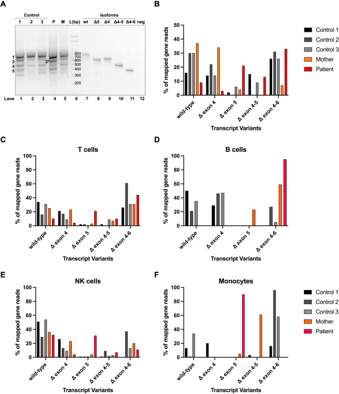
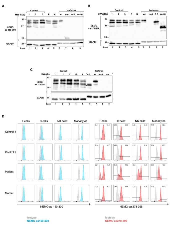
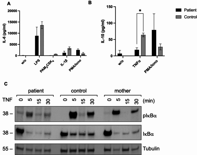
Methods: IKBKG transcripts were quantified in peripheral blood mononuclear cells isolated from the patient, her mother, and healthy controls using RT-PCR and nanopore sequencing. Corresponding proteins were analyzed by western blotting and flow cytometry. Besides toll-like receptor (TLR) and tumor necrosis factor (TNF) signaling, the interferon signature, cytokine production and X-inactivation status were investigated.

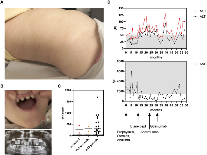
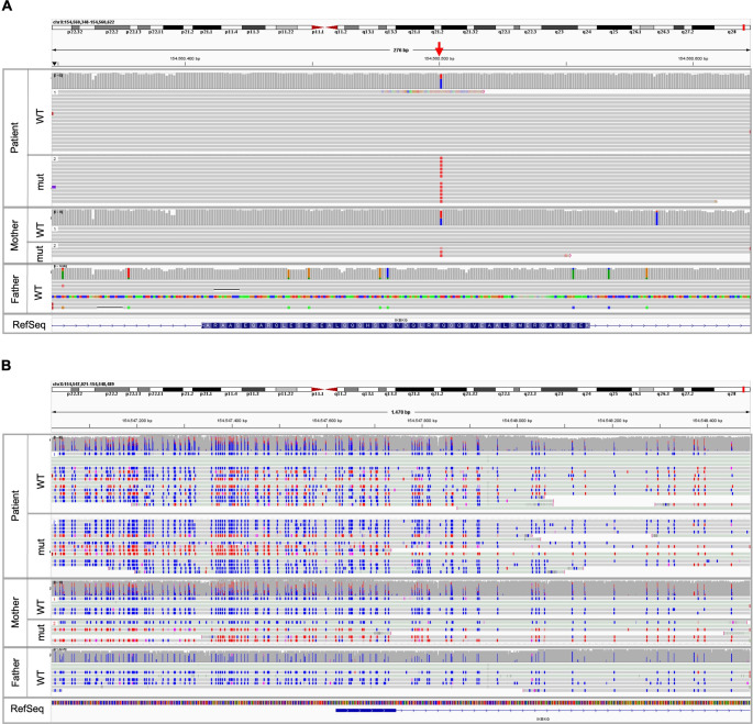
Results: IP and autoinflammation with recurrent fever, oral ulcers, hepatitis, and neutropenia, but no immunodeficiency was observed in a female patient. Besides moderately reduced NEMO signaling function, type I interferonopathy, and elevated IL-18 and CXCL10 were found. She and her mother both carried the heterozygous variant c.613 C > T p.(Gln205*) in exon 5 of IKBKG previously reported in NEMO-deficient patients. However, X-inactivation was skewed in the mother, but not in the patient. Alternative splicing led to increased ratios of NEMO-Dex5 over full-length protein in peripheral blood cell subsets causing autoinflammation. Clinical symptoms partially resolved under treatment with TNF inhibitors.

Conclusion: Non-skewed X-inactivation can lead to NEMO-NDAS in females with IP carrying hypomorphic IKBKG variants due to alternative splicing and increased proportions of NEMO-∆ex5.

Keywords: Autoinflammation; Immunodeficiency; Incontinentia pigmenti; NEMO; Non-skewed X-inactivation.


Conflict of interest statement

The authors declare no competing interests.

Figures

Fig. 1
Fig. 1
Clinical presentation of incontinentia pigmenti (IP) and NEMO-NDAS. At the age of 4y, the patient presented with ectodermal dysplasia characterized by hyperpigmentation along the Blashko lines (A) and conical teeth (B) (top: teeth at the age of 4y, bottom: dental X-ray at the age of 9y). Type I IFN-scores were calculated based on expression of IFN-stimulated genes analyzed in peripheral blood cells before start of treatment and under treatment with TNF inhibitors (C). IFN-scores of a cohort of 20 patients with typical interferonopathy (Aicardi-Goutières syndrome; AGS) are shown as control. Median values are displayed by horizontal bars. The cut-off for pathologic levels is indicated by the dotted line. Under treatment with TNF inhibitors as indicated, the patient developed mild hepatitis shown by elevated liver enzymes (ALT: alanine aminotransferase and AST: aspartate aminotransferase), and neutropenia (ANC: absolute neutrophil count) (D). Normal ranges are marked by grey-shaded areas
Fig. 2
Fig. 2
The variant c.613 C > T leads to expression of IKBKG-∆ex5 transcripts and NEMO-∆ex5 proteins. RNA was isolated from peripheral blood obtained from the patient, her mother and three healthy controls. IKBKG transcripts were amplified from cDNA and separated by electrophoresis (A). Full-length IKBKG transcripts (wt) and transcript variants ∆ex4 (∆4), ∆ex5 (∆5), ∆ex4-5 (∆4–5), ∆ex4-6 (∆4–6) were cloned into the pcDNA6 vector and used as size markers. In addition, bands obtained were excised and sequenced by Sanger sequencing: 1: wt (780 bp), 2: ∆4 (661 bp), 3 (*only observed in patient cells): ∆5 (627 bp), 4: ∆4–5 (508 bp), 5: ∆4–6 (411 bp). Transcript variants were further analyzed by nanopore sequencing and mapped gene reads were quantified using a library of all detected IKBKG transcripts. Shown are the frequencies of the most abundant variants ∆ex4, ∆ex5, ∆ex4-5, and ∆ex4-6 as % of all mapped gene reads obtained from whole PBMCs of patient, mother and controls (B). IKBKG transcripts were amplified from cDNA of T, B, NK lymphocytes, and monocytes and analyzed by nanopore sequencing (C-F). Shown are the frequencies of the most abundant variants Δex4, Δex5, Δex4-5, and Δex4-6 as % of all mapped gene reads. Of note, Δex4-5 could not be detected in B cells (D)
Fig. 3
Fig. 3
NEMO-∆ex5 is expressed in patient peripheral blood cells. Protein lysates obtained from PBMCs isolated from patient, mother and 3 controls were separated by SDS-PAGE and blotted on a PVDF membrane by western blotting. Proteins synthesized in-vitro from cloned full-length (wt), full-length with c.613 C > T variant in exon 5 (mut), ∆ex5 (∆5), and ∆ex4-6 (∆4–6) IKBKG transcripts were used as size markers in addition to a 10-250 kDa ladder. NEMO proteins were detected using primary antibodies binding to amino acids (aa) 150–300 (NEMO150–300) (A) and aa278-396 (NEMO278–396) (B, C).Besides the wt protein (48 kDa), several cross-reactional bands were detected by both antibodies, as described by the manufacturer. No protein could be detected from Δex4 and ∆ex4-5 transcripts due to disruption of the reading frame. The NEMO-∆ex5 protein could be observed in patient lysates following a longer period of separation (C). GAPDH was used as loading control, whereas bands resulting from non-specified proteins contained in the wheat germ extract were detected for in-vitro synthesized NEMO isoforms. Shown are representative western blots obtained from 3 experiments. In addition, NEMO expression was investigated in T, B, NK lymphocytes and monocytes by flow cytometry using abNEMO150–300 (left panel, blue histograms) and abNEMO278–396(right panel, red histograms) combined with anti-human CD3, CD19, CD56, and CD14 surface antibodies (D). Alexa fluor 488 anti-rabbit, and anti-mouse, respectively, were used as secondary antibodies and background controls (isotypes, grey histograms). In contrast to the controls, two peaks of intensity were observed in peripheral blood cell subsets obtained from the patient and her mother, which were quantified by gating
Fig. 4
Fig. 4
NEMO signaling function is partially impaired. Functional studies were performed on whole blood obtained from the patient and compared to shipping controls. Expression of IL-6 was measured in the supernatant following stimulation with LPS, PAM2CSK2, IL-1β and PMA/ionomycin (A). Levels of IL-10 were investigated after stimulation with TNF and PMA/ionomycin (B). Shown are the mean results with standard deviations obtained from two independent investigations. Statistics were calculated using unpaired student’s t test. PBMCs isolated from the patient and a healthy control were stimulated with TNF (200ng/ml) for 0, 5, 15, and 30 min. Levels of pIκBα and IκBα were investigated by western blotting (C). Protein sizes are marked on the left-hand side. Tubulin served as loading control
Fig. 5
Fig. 5
NEMO-NDAS results in elevation of cytokines and chemokines. Levels of 36 cytokines were investigated in serum obtained from the patient, her mother, and control 1 and 2 using the Human Proteome Profiler Human Cytokine Array Kit (R&D Systems). Pixel densities were analyzed by optical densitometry and normalized on included background proteins. Shown are mean densities of 13 detectable cytokines. Statistics were analyzed using two-way Anova (* p ≤ 0,05, ** p ≤ 0,01, *** p ≤ 0,001, **** p ≤ 0,0001)
Fig. 6
Fig. 6
The DNA methylation status of the IKBKG transcription start / promoter region is skewed to one X allele in the mother, but non-skewed in the patient (A) Exon 5 of IKBKG (chrX:154,560,348 − 154,560,622 in hg38) in patient, mother and father was analyzed by Oxford Nanopore Technologies (ONT) sequencing and results are displayed in the Integrative Genomics Viewer (IGV). The IKBKG variant c.613 C > T p.(Gln205*) is indicated by the red arrow and was found in the mother and the patient. (B) The DNA methylation pattern of patient, mother and father in the transcription start / promotor region of IKBKG (chrX:154,547,021–154,548,489 in hg38) was analyzed by ONT sequencing and is displayed in the IGV viewer. Wildtype (WT) allele and mutated (mut) allele of the patient (top panel) show similar DNA methylation patterns, whereas the WT allele from the mother is predominantly unmethylated (blue, middle panel). The mutated allele from the mother shows a DNA methylation of ~ 60%. The WT allele from the father (lower panel) is completely unmethylated (blue). No methylation = blue, methylation = red

Similar articles

Cited by

References

    1. Yamaoka S, Courtois G, Bessia C, Whiteside ST, Weil R, Agou F, et al. Complementation cloning of NEMO, a component of the IkappaB kinase complex essential for NF-kappaB activation. Cell. 1998;93(7):1231–40. 10.1016/S0092-8674(00)81466-X - DOI - PubMed
    1. Sen R, Baltimore D. Inducibility of kappa immunoglobulin enhancer-binding protein Nf-kappa B by a posttranslational mechanism. Cell. 1986;47(6):921–8. 10.1016/0092-8674(86)90807-X - DOI - PubMed
    1. Picard C, Casanova JL, Puel A. Infectious diseases in patients with IRAK-4, MyD88, NEMO, or IkappaBalpha deficiency. Clin Microbiol Rev. 2011;24(3):490–7. 10.1128/CMR.00001-11 - DOI - PMC - PubMed
    1. Mercurio F, Murray BW, Shevchenko A, Bennett BL, Young DB, Li JW, et al. IkappaB kinase (IKK)-associated protein 1, a common component of the heterogeneous IKK complex. Mol Cell Biol. 1999;19(2):1526–38. 10.1128/MCB.19.2.1526 - DOI - PMC - PubMed
    1. Rothwarf DM, Zandi E, Natoli G, Karin M. IKK-gamma is an essential regulatory subunit of the IkappaB kinase complex. Nature. 1998;395(6699):297–300. 10.1038/26261 - DOI - PubMed

Publication types

LinkOut - more resources