. 2024 Dec 16;84(24):4264-4282.
doi: 10.1158/0008-5472.CAN-24-0800.

Simultaneous Targeting of NQO1 and SOD1 Eradicates Breast Cancer Stem Cells via Mitochondrial Futile Redox Cycling

Affiliations

Simultaneous Targeting of NQO1 and SOD1 Eradicates Breast Cancer Stem Cells via Mitochondrial Futile Redox Cycling

Ming Luo et al. Cancer Res. .

Abstract

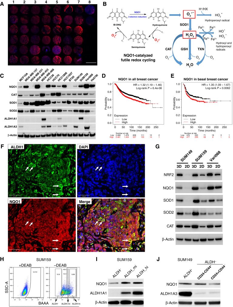
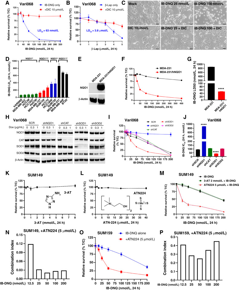
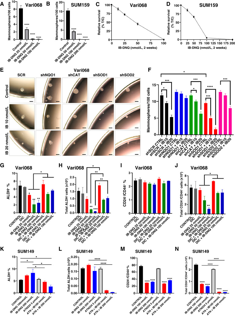
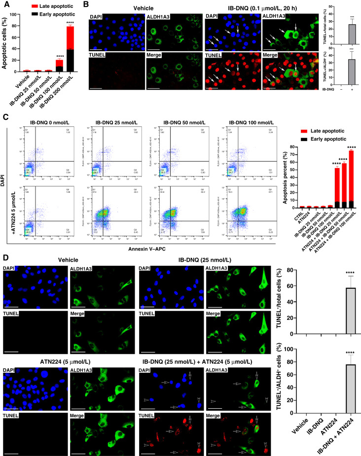
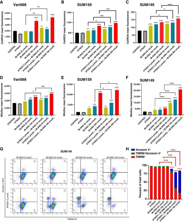
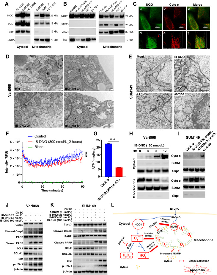
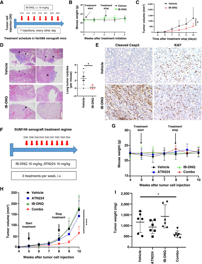
Triple-negative breast cancer (TNBC) contains the highest proportion of cancer stem-like cells (CSC), which display intrinsic resistance to currently available cancer therapies. This therapeutic resistance is partially mediated by an antioxidant defense coordinated by the transcription factor NRF2 and its downstream targets that include NAD(P)H quinone oxidoreductase 1 (NQO1). In this study, we identified the antioxidant enzymes NQO1 and superoxide dismutase 1 (SOD1) as therapeutic vulnerabilities of ALDH+ epithelial-like CSCs and CD24-/loCD44+/hi mesenchymal-like CSCs in TNBC. Effective targeting of these CSC states was achieved by using isobutyl-deoxynyboquinone (IB-DNQ), a potent and specific NQO1-bioactivatable futile redox cycling molecule, which generated large amounts of reactive oxygen species including superoxide and hydrogen peroxide. Furthermore, the CSC killing effect was specifically enhanced by genetic or pharmacologic inhibition of SOD1, a copper-containing superoxide dismutase highly expressed in TNBC. Mechanistically, a significant portion of NQO1 resides in the mitochondrial intermembrane space, catalyzing futile redox cycling from IB-DNQ to generate high levels of mitochondrial superoxide, and SOD1 inhibition markedly potentiated this effect, resulting in mitochondrial oxidative injury, cytochrome c release, and activation of the caspase-3-mediated apoptotic pathway. Treatment with IB-DNQ alone or together with SOD1 inhibition effectively suppressed tumor growth, metastasis, and tumor-initiating potential in xenograft models of TNBC expressing different levels of NQO1. This futile oxidant-generating strategy, which targets CSCs across the epithelial-mesenchymal continuum, could be a promising therapeutic approach for treating patients with TNBC. Significance: Combining NQO1-bioactivatable futile oxidant generators with SOD1 inhibition eliminates breast cancer stem cells, providing a therapeutic strategy that may have wide applicability, as NQO1 and SOD1 are overexpressed in several cancers.


Conflict of interest statement

P.J. Hergenrother reports personal fees from Vanquish Oncology, Systems Oncology, Oncoteq, and Arrepath outside the submitted work, as well as US patent 9233960 issued. No disclosures were reported by the other authors.

Figures

Figure 1.
Figure 1.
NQO1 and related antioxidant enzyme expression in TNBC tissues, cell lines, and CSC populations. A, Immunofluorescent labeling of NQO1 in a human TNBC tissue array (columns 1 to 7) vs. two normal mammary tissues (column 8). Scale bar, 2 mm. B, NQO1-catalyzed futile redox cycle from IB-DNQ. GSH, glutathione; TXN, thioredoxin. C, NQO1 and related antioxidant enzyme expression in 13 TNBC cell lines vs. MCF10A. D and E, NQO1 expression predicts poor recurrence-free survival in a patient cohort containing all breast cancer subtypes (D) and basal breast cancer (E). F, Double labeling of TNBC tissues using specific antibodies against NQO1 and ALDH1. Bar, 50 µm. G, NQO1 and its related antioxidant enzyme expression in tumorspheres vs. two-dimensional adherent cells. H and I, ALDHhi, ALDHmed, and ALDH bulk tumor cells were sorted based on ALDEFLUOR assay (H) and examined by immunoblotting with ALDH1A1 and NQO1 antibodies (I). J, NQO1 expression in ALDH+ and ALDHCD24CD44+ CSCs vs. bulk tumor cells of SUM149.
Figure 2.
Figure 2.
IB-DNQ is more potent and specific than β-Lap in killing TNBC cells, and genetic or pharmacologic inhibition of SOD1 synergistically enhances IB-DNQ–elicited lethality. A and B, LD50 of IB-DNQ (A) vs. β-Lap (B) in Vari068 breast cancer cells treated with or without DIC (n = 6 wells). C, Vari068 cells treated with Mock, DIC (10 µmol/L), IB-DNQ (25 and 100 nmol/L), or IB-DNQ plus DIC (10 µmol/L) for 24 hours and examined by light microscopy. Scale bar, 50 µm. D, IB-DNQ LD50 in 13 TNBC cells (n = 2). E–G, NQO1 expression in NQO MDA-MB-231 (E) renders these cells sensitive to IB-DNQ (n = 6 wells; F) with LD50 <50 nmol/L (G). ****, P < 0.0001 vs. NQO cells (n = 2, unpaired Student t test). H–J, Dox-inducible KD of NQO1, CAT, SOD1, and SOD2 in Vari068 validated by immunoblotting with respective antibodies (H), and relative survival curve (n = 6 wells; I) and IC50 (J) of Vari068 cells with KD of NQO1, CAT, SOD1, or SOD2 vs. SCR cells following IB-DNQ treatment. ****, P < 0.0001 vs. shSCR (n = 2, unpaired Student t test). K and L, Relative survival of SUM149 treated with 3-AT (K) or ATN224 (L) at different doses for 24 hours (n = 6 wells). M and N, Relative survival of SUM149 cells treated with IB-DNQ alone or together with ATN224 or 3-AT (n = 6 wells; M) and combination index of ATN224 (5 µmol/L) with IB-DNQ from 12.5 to 200 nmol/L in SUM149 (N). O and P, Relative survival of SUM159 cells treated with IB-DNQ alone or with ATN224 (n = 6 wells; O) and combination index of ATN224 with IB-DNQ from 12.5 to 200 nmol/L in SUM159 (P).
Figure 3.
Figure 3.
IB-DNQ treatment preferentially inhibits CSC activity in TNBC cells expressing NQO1. A–D, Tumorsphere formation of Vari068 (A) and SUM159 (C) breast cancer cells in medium containing control (DMSO) or IB-DNQ of various doses, and relative survival of Vari068 (B) and SUM159 (D) cells grown in adherent culture with IB-DNQ for 14 days, n = 6 wells. E and F, Sphere formation of Vari068 cells subjected to Dox-induced KD of NQO1, CAT, SOD1, or SOD2 and IB-DNQ treatment (E) and the impact of KD on IB-DNQ suppression of sphere-forming capacity (F). G–N, Vari068 (G–J) and SUM149 (K–N) cells were treated with indicated compounds for 20 hours and examined for the content and absolute number of ALDH+ and CD24CD44+ CSCs. *, P < 0.05; **, P < 0.01; ***, P < 0.001; ****, P < 0.0001 vs. control or indicated by brackets. n = 3, one-way ANOVA for A, C, F, G–J, and K–N.
Figure 4.
Figure 4.
IB-DNQ induces apoptosis of Vari068 ALDH+ CSCs, and ATN224 enhances IB-DNQ–mediated apoptosis in SUM149 breast cancer cells expressing low/modest levels of NQO1. A and B, Vari068 cells treated with IB-DNQ for 20 hours were examined by flow cytometry to assess early and late apoptotic cells (A), or fixed and examined by ALDH1A3 and TUNEL dual labeling fluorescent microscopy (B) and percentage of TUNEL+ over total or ALDH1A3+ cells was scored in three different areas. C and D, SUM149 cells treated with IB-DNQ alone or together with ATN224 (5 µmol/L) for 20 hours were examined by flow cytometry to detect early and late apoptotic cells (C), or fixed and examined by ALDH1A3 and TUNEL dual labeling (D), and percentage of TUNEL+ over total or ALDH1A3+ cells was scored in three different areas. ***, P < 0.001; ****, P < 0.0001 vs. vehicle. n = 3, one-way ANOVA for A and C and unpaired Student t test for B and D. Scale bar, 50 µm.
Figure 5.
Figure 5.
IB-DNQ in combination with ATN224 synergistically enhances total ROS and MitoROS production, resulting in the loss of mitochondrial membrane potential. A–F, Vari068, SUM159, and SUM149 cells were treated with control (DMSO), ATN224 (5 µmol/L), IB-DNQ alone, or IB-DNQ plus ATN224 for 20 hours and examined by CellROX (A–C) or MitoSOX (D–F) labeling, followed by flow cytometry. G and H, SUM149 cells treated with IB-DNQ (0–100 nmol/L) alone or together with 5 µmol/L of ATN224 for 20 hours were stained with Annexin V–APC plus TMRM-PE and examined by flow cytometry (G), and percentage of TMRM+, TMRM+Annexin V+ and Annexin V+ cells was plotted (H). *, P < 0.05; **, P < 0.01; ***, P < 0.001; ****, P < 0.0001 vs. control or indicated by brackets; n = 3, one-way ANOVA for A–F and H.
Figure 6.
Figure 6.
NQO1 residing in the mitochondrial IMS drives IB-DNQ redox cycling, promoting mitochondrial oxidative damage, Cyto c release, and activation of Casp3-mediated mitochondrial apoptotic pathway. A and B, NQO1med and NQO1lo (A) and NQO1hi (B) TNBC cells were examined for NQO1, SOD1, succinate dehydrogenase complex subunit A (SDHA), and S-phase kinase-associated protein-1 (Skp1) expression in the mitochondrial and cytosol fraction. C, SUM159 cells labeled with specific antibodies against NQO1 and Cyto c were examined by confocal microscopy. Scale bar, 120 µm. D and E, Vari068 and SUM149 cells treated with 100 nmol/L of IB-DNQ for 0 to 8 hours (D) or with vehicle, IB-DNQ (100 nmol/L), ATN224 (5 µmol/L), IB-DNQ plus ATN224 for 16 hours (E) were processed and examined by transmission electron microscopy. Bars, 200 (D) and 500 (E) nm. F, SUM159 cells were treated with vehicle or IB-DNQ for 2 hours and measured for oxygen consumption based on fluorescent intensity (RFU) of an oxygen-bleaching fluorescent dye in the media covered by mineral oil at 1.5 minutes intervals for 90 minutes. ****, P < 0.0001 (n = 3, two-way ANOVA). G, ATP levels in the lysates of SUM159 cells treated with vehicle or IB-DNQ for 2 hours were measured and normalized by protein concentrations. ****, P < 0.0001 with unpaired Student t test (n = 3). H and I, Vari068 and SUM149 cells treated with IB-DNQ (100 nmol/L) for 0 to 12 hours (H) or IB-DNQ and ATN224 alone or in combination for 16 hours (I) were used to isolate mitochondrial and cytosol fractions and examine Cyto c, SDHA, and Skp1 expression. J and K, Lysates of Vari068 (J) and SUM149 (K) cells treated with IB-DNQ alone or IB-DNQ plus ATN224 for 20 hours were subjected to immunoblotting to examine the expression of apoptosis-related proteins. L, A model illustrating the mechanisms of action for IB-DNQ–mediated prooxidant therapy targeting TNBC cells including CSCs. MOMP, mitochondrial outer membrane permeability.
Figure 7.
Figure 7.
IB-DNQ alone or together with SOD1 inhibition suppresses tumor growth, metastasis, and tumor-initiating potential of TNBC xenografts. A–C, Vari068 mammary tumor xenograft mice were randomized and treated with vehicle or IB-DNQ as indicated in A. B and C, Mouse body weight was examined weekly for 4 weeks after treatment initiation (B) and mammary tumor growth following the last treatment was monitored for 2 weeks (C). *, P < 0.05 (n = 6). D, Lung histologic sections from each group of mice were harvested to examine metastatic tumor nodule formation. *, P < 0.05. E, staining of tumor sections treated with IB-DNQ vs. vehicle using antibodies against cleaved Casp3 or Ki67. F and H, SUM149 mammary tumor xenograft mice were randomized into four groups and treated with the regime indicated in F, and mouse body weight (G) and mammary tumor growth (H) were examined weekly for 6 weeks. ****, P < 0.0001 (vs. vehicle or IB-DNQ, n = 6). I, Tumor weight for each group of mice at the end of tumor monitoring. *, P < 0.05 (vehicle vs. combo, n = 6). C and H, Two-way ANOVA; D and I, two-tailed Student t test.

Similar articles

Cited by

References

    1. Garrido-Castro AC, Lin NU, Polyak K. Insights into molecular classifications of triple-negative breast cancer: improving patient selection for treatment. Cancer Discov 2019;9:176–98. - PMC - PubMed
    1. Loizides S, Constantinidou A. Triple negative breast cancer: immunogenicity, tumor microenvironment, and immunotherapy. Front Genet 2022;13:1095839. - PMC - PubMed
    1. Charafe-Jauffret E, Ginestier C, Iovino F, Wicinski J, Cervera N, Finetti P, et al. . Breast cancer cell lines contain functional cancer stem cells with metastatic capacity and a distinct molecular signature. Cancer Res 2009;69:1302–13. - PMC - PubMed
    1. Abdoli Shadbad M, Hosseinkhani N, Asadzadeh Z, Derakhshani A, Karim Ahangar N, Hemmat N, et al. . A systematic review to clarify the prognostic values of CD44 and CD44+CD24− phenotype in triple-negative breast cancer patients: lessons learned and the road ahead. Front Oncol 2021;11:689839. - PMC - PubMed
    1. Olsson M, Larsson P, Johansson J, Sah VR, Parris TZ. Cancer stem cells are prevalent in the basal-like 2 and mesenchymal triple-negative breast cancer subtypes in vitro. Front Cell Dev Biol 2023;11:1237673. - PMC - PubMed

MeSH terms