. 2024 Sep 12;20(9):e1011402.
doi: 10.1371/journal.pgen.1011402. eCollection 2024 Sep.

Subfunctionalization of NRC3 altered the genetic structure of the Nicotiana NRC network

Affiliations

Subfunctionalization of NRC3 altered the genetic structure of the Nicotiana NRC network

Ching-Yi Huang et al. PLoS Genet. .

Abstract

Nucleotide-binding domain and leucine-rich repeat (NLR) proteins play crucial roles in immunity against pathogens in both animals and plants. In solanaceous plants, activation of several sensor NLRs triggers their helper NLRs, known as NLR-required for cell death (NRC), to form resistosome complexes to initiate immune responses. While the sensor NLRs and downstream NRC helpers display diverse genetic compatibility, molecular evolutionary events leading to the complex network architecture remained elusive. Here, we showed that solanaceous NRC3 variants underwent subfunctionalization after the divergence of Solanum and Nicotiana, altering the genetic architecture of the NRC network in Nicotiana. Natural solanaceous NRC3 variants form three allelic groups displaying distinct compatibilities with the sensor NLR Rpi-blb2. Ancestral sequence reconstruction and analyses of natural and chimeric variants identified six key amino acids involved in sensor-helper compatibility. These residues are positioned on multiple surfaces of the resting NRC3 homodimer, collectively contributing to their compatibility with Rpi-blb2. Upon activation, Rpi-blb2-compatible NRC3 variants form membrane-associated punctate and high molecular weight complexes, and confer resistance to the late blight pathogen Phytophthora infestans. Our findings revealed how mutations in NRC alleles lead to subfunctionalization, altering sensor-helper compatibility and contributing to the increased complexity of the NRC network.


Conflict of interest statement

I have read the journal’s policy and the authors of this manuscript have the following competing interests: JK received funding from industry on NLR biology at the time of the study. JK has filed patents on NLR biology. Other authors have declared that no competing interests exist.

Figures

Fig 1
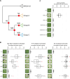
Fig 1. Rpi-blb2 signals through NRC3a but not NRC3b or NRC3c.
(A) Cell death assays of Rpi-blb2 with different NRCs. Rpi-blb2 and AVRblb2 were co-expressed with indicated NRCs cloned from tomato and N. benthamiana in nrc2/3/4_KO N. benthamiana. Cell death intensity and phenotypes were recorded at 6 dpi. The line in the boxplots represents the medium, the box edges represent the 25th and 75th percentiles, and the whiskers extend to the most extreme data points no more than 1.5x of the interquartile range. Statistical differences between the negative control (EV) and tested groups were examined by paired Wilcoxon signed rank test (* = p < 0.0001). (B) Assays of Rpi-blb2-mediated cell death rescued by NRC variants. Rpi-blb2 and AVRblb2 were co-expressed with NRCs as indicated in both WT or nrc2/3/4_KO N. benthamiana. (C) Phylogenetic analysis of NRC3 natural variants identified from tomato, tobacco potato, pepper, and eggplant. Sequence alignment of the NB-ARC domain was used to generate the phylogenetic tree using the Maximum likelihood method with 1000 bootstrap tests. SlNRC1 and SlNRC2 were selected as outgroups. The scale bars indicate the evolutionary distance in amino acid substitution per site. The orange, green, and blue boxes indicate allelic groups A, B, and C, respectively. (D) Cell death assays of different sensor NLRs with NRC3 natural variants. The cloned NRC3 natural variants were co-expressed with Rpi-blb2/AVRblb2, Pto/AvrPto, or Rx/CP in nrc2/3/4_KO N. benthamiana. (E) Protein accumulation of NRC3 natural variants. NRC3 natural variants were transiently expressed in WT N. benthamiana. The proteins were extracted from leaf samples at 2 dpi and the NRC3 protein accumulations were detected by α-myc antibody. SimplyBlue SafeStain-staining of Rubisco was used as the loading control. The dot plots represent cell death intensity quantified by UVP ChemStudio PLUS at 6 dpi. The line in the boxplots represents the medium, the box edges represent the 25th and 75th percentiles, and the whiskers extend to the most extreme data points no more than 1.5x of the interquartile range. Statistical differences between the negative control (EV) and tested groups were examined by paired Wilcoxon signed rank test (* = p<0.05, ** = p < 0.0001).
Fig 2
Fig 2. Subfunctionalization contributes to the evolution of NRC3c.
(A) Phylogenetic tree of NRC3 natural variants. Orange, green, and blue boxes represent allelic groups A, B, and C, respectively. Red dots indicate the nodes of reconstructed ancestral NRC3 variants. (B) Cell death assays of ancestral NRC3 variants. The ancestral variants were co-expressed with Rpi-blb2/AVRblb2, Pto/AvrPto, or Rx/CP in nrc2/3/4_KO N. benthamiana. (C) Cell death analysis of NRC3b_DV, N88_DV and N95_DV. The NRC3_DV variants carry a D to V mutation in the MHD motif. These variants were expressed alone in WT N. benthamiana. The dot plots represent cell death intensity quantified by UVP ChemStudio PLUS at 6 dpi. The line in the boxplots represents the medium, the box edges represent the 25th and 75th percentiles, and the whiskers extend to the most extreme data points no more than 1.5x of the interquartile range. Statistical differences between the negative control (EV) and tested groups were examined by paired Wilcoxon signed rank test (* = p < 0.0001).
Fig 3
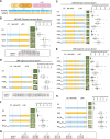
Fig 3. Changing six residues in NbNRC3 enabled it to function with Rpi-blb2.
(A) Sequence identity between NbNRC3 (NNN) and SlNRC3 (SSS) in CC, NB-ARC, and LRR domains. Cell death assays of chimeric NRC3 variants designed to investigate polymorphisms in (B) the NB-ARC domain, (C) the LRR domain, (D) LRR domain region 2, and (E) LRR domain region 5 that contribute to the sensor-helper compatibility. (F) Cell death assays of chimeric NRC3 variants that carry polymorphisms identified in (B-E). (G) Polymorphisms of NbNRC3 and SlNRC3 at the seven positions identified (highlighted in red). (H) Cell death assays of chimeric NRC3 variants NNPKKNTHKI and NNPKKNTHK. Cell death assays were performed by co-expressing chimeric NRC3 variants with Rpi-blb2/AVRblb2 in nrc2/3/4_KO N. benthamiana. In the schematic representations of the NRC3 variants, the blue color indicates the regions/residues from NbNRC3 (N), and the orange color indicates the regions/residues from SlNRC3 (S). The dot plots represent cell death intensity quantified by UVP ChemStudio PLUS at 6 dpi. The line in the boxplots represents the medium, the box edges represent the 25th and 75th percentiles, and the whiskers extend to the most extreme data points no more than 1.5x of the interquartile range. Statistical differences were examined using Dunn’s test (p < 0.05).
Fig 4
Fig 4. Two K to N mutations in the NB-ARC and LRR domains play major roles in NRC3 subfuntionalization.
(A) Left panel, the polymorphisms in the NRC3 natural variants at the 6 positions identified. Right panel, cell death assay testing these polymorphisms in NbNRC3c background. Introducing PKKTHK or PKKTRK enabled it to function with Rpi-blb2. (B) Left panel, the polymorphisms in the ancestral NRC3 variants at the 6 positions identified. Right panel, cell death assay testing two lysine-asparagine changes in both N88 and N89 backgrounds. Swapping two K to N in N88/N89 changed the compatibility to Rpi-blb2. The dot plots in (A) and (B) represent cell death intensity quantified by UVP ChemStudio PLUS at 6 dpi. The line in the boxplots represents the medium, the box edges represent the 25th and 75th percentiles, and the whiskers extend to the most extreme data points no more than 1.5x of the interquartile range. Statistical differences were examined using Dunn’s test (p < 0.05) (C) The entropy analysis of natural NRC3 variants. The protein sequences of NRC3s were aligned using MAFFT and the Shannon entropy was calculated. The positions of the two K to N changes were highlighted in green. (D) dN-dS calculation of natural NRC3 variants by using SLAC analysis. The positions of K to N changes were highlighted in green. The two residues were under neutral selection based on FEL analysis.
Fig 5
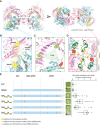
Fig 5. Multiple protein surface contribue to the sensor-helper compatibility determination.
(A) Structure of NbNRCc homodimer shown in two orthogonal views. The NB-ARC domains of two protomers are shown in light blue and the LRR domains are shown in pink. The 6 residues involved in sensor-helper compatibility are highlighted in yellow, green, and red. (B) Details of the exposed surface between S202(P)/T203(K) and N832(K). (C) Details of the cavity in between the NB-ARC domain and the LRR domain. This cavity is surrounded by I642(T), C824(H), and N221(K). (D) Details of the interface between two NbNRC3c protomers where the N221(K) is located. The interfaces are highlighted based on the resting NbNRC2 homodimer. (E) Cell death assays of NRC3 variants carrying identified residues from SlNRC3 in only two of the surfaces. The residues on the exposed surface, on the concave surface of the LRR domain, and on the interface between NRC3 protomers are highlighted in yellow, green, and red, respectively. Cell death assays were performed by co-expressing chimeric NRC3 variants with Rpi-blb2/AVRblb2 in nrc2/3/4_KO N. benthamiana. The dot plots represent cell death intensity quantified by UVP ChemStudio PLUS at 6 dpi. The line in the boxplots represents the medium, the box edges represent the 25th and 75th percentiles, and the whiskers extend to the most extreme data points no more than 1.5x of the interquartile range. Statistical differences were examined using Dunn’s test (p < 0.05).
Fig 6
Fig 6. Introducing the 6 mutations into NbNRC3c enabled it to form resistosomes upon activation, and to confer resistance against P. infestans.
(A) Compatible NRC3 variants form membrane-associated punctate upon immune activation. NRC3 variants fused with GFP were co-expressed with Rpi-blb2/AVRblb2 or Pto/AvrPto. Samples were examined at 3 dpi. Scale bars represent 10μm. SOBIR1-mcherry was used as a plasma membrane marker. (B) Quantification of punctate of NRC3 variants in (A). (C) BN-PAGE analyzing the complex size of NRC3 variants. The NRC3 variants were co-expressed with Rpi-blb2 in the presence or absence of AVRblb2 in nrc2/3/4_KO N. benthamiana. Leaf samples were collected at 2 dpi. (D) Infection assay of P. infestans in nrc2/3/4_KO N. benthamiana transiently expressing Rpi-blb2 and NRC variants. The dot plots represent the lesion sizes observed at 5 days post-infection quantified by UVP ChemStudio PLUS. The line in the boxplots represents the medium, the box edges represent the 25th and 75th percentiles, and the whiskers extend to the most extreme data points no more than 1.5x of the interquartile range. Statistical differences were examined using Tukey’s HSD test (p < 0.05).

Similar articles

Cited by

References

    1. Jones JD, Vance RE, Dangl JL. Intracellular innate immune surveillance devices in plants and animals. Science. 2016/12/10 ed. 2016;354. doi: 10.1126/science.aaf6395 - DOI - PubMed
    1. Duxbury Z, Wu CH, Ding P. A Comparative Overview of the Intracellular Guardians of Plants and Animals: NLRs in Innate Immunity and Beyond. Annu Rev Plant Biol. 2021/03/11 ed. 2021;72: 155–184. doi: 10.1146/annurev-arplant-080620-104948 - DOI - PubMed
    1. Wang G, Roux B, Feng F, Guy E, Li L, Li N, et al.. The Decoy Substrate of a Pathogen Effector and a Pseudokinase Specify Pathogen-Induced Modified-Self Recognition and Immunity in Plants. Cell Host & Microbe. 2015;18: 285–295. doi: 10.1016/j.chom.2015.08.004 - DOI - PubMed
    1. Wang J, Hu M, Wang J, Qi J, Han Z, Wang G, et al.. Reconstitution and structure of a plant NLR resistosome conferring immunity. Science. 2019/04/06 ed. 2019;364. doi: 10.1126/science.aav5870 - DOI - PubMed
    1. Bi G, Su M, Li N, Liang Y, Dang S, Xu J, et al.. The ZAR1 resistosome is a calcium-permeable channel triggering plant immune signaling. Cell. 2021/05/14 ed. 2021;184: 3528–3541 e12. doi: 10.1016/j.cell.2021.05.003 - DOI - PubMed

LinkOut - more resources