. 2024 Sep 12;18(9):e0012502.
doi: 10.1371/journal.pntd.0012502. eCollection 2024 Sep.

Development of a Lipid-encapsulated TGFβRI-siRNA Drug for Liver Fibrosis Induced by Schistosoma mansoni

Affiliations

Development of a Lipid-encapsulated TGFβRI-siRNA Drug for Liver Fibrosis Induced by Schistosoma mansoni

Ying-Chou Chen et al. PLoS Negl Trop Dis. .

Abstract

Schistosoma mansoni infection leads to chronic schistosomiasis and severe hepatic fibrosis. We designed a liver-targeted lipid nanoparticle (LNP) carrying siRNA against type I TGF-β receptor (TGFβRI) mRNA to treat schistosomiasis-induced liver fibrosis in BALB/c mice. Knockdown of TGFβRI by LNP-siTGFβRI reduced LX-2 cell activation in vitro and alleviated liver fibrosis in S. mansoni-infected mice. αSMA and Col1a1 fibrotic markers in the liver tissues of infected mice were significantly suppressed in the treatment groups. In the serum of the LNP-siTGFβRI-treated groups, cytokines IFNγ, IL-1α, IL-6, IL-12, RANTES (CCL5), and TNFα increased, while GM-CSF, IL-2, IL-4, IL-10, IL-13, and KC (CXCL1) decreased compared to the control. Cell proportions were significantly altered in S. mansoni-infected mice, with increased CD56d NK cells and decreased CD19+ B cells and CD4+ T cells compared to naïve mice. Following LNP-siTGFβRI treatment, CD56d NK cells were downregulated, while B and memory Th cell populations were upregulated. The density of fibrotic regions significantly decreased with LNP-siTGFβRI treatment in a dose-dependent manner, and no systemic toxicity was observed in the major organs. This targeted siRNA delivery strategy effectively reduced granulomatous lesions in schistosomiasis-induced liver fibrosis without detectable side effects.


Conflict of interest statement

The authors have declared that no competing interests exist.

Figures

Fig 1
Fig 1. Characterization of LNP-siTGFβRI.
(A) Schematic of LNP production by mixing lipid mixtures and siRNA aqueous solution via a microfluidic system. (B) Particle size distribution analysis and zeta potential intensity plots of LNP-siTGFβRI. (C) Transmission electron microscopy (TEM) image of LNP-siTGFβRI (indicated by arrows). LNP, lipid nanoparticle.
Fig 2
Fig 2. Evaluation of in vitro activity of LNP-siRNA on LX-2 cells stimulated by 2 ng/mL of TGF-β.
(A) RT-PCR analysis of TgfbrI mRNA expression. (B) RT-PCR analysis of αSMA and Col1a1 mRNA expression. (C) Western blotting (WB) analysis. (D) protein expression levels of TGFβRI, αSMA, and collagen 1 in the LX-2 cells stimulated by 2 ng/mL of TGF-β and treated with either 100 ng/mL of LNP-scrambled RNA (Scr) or LNP-siTGFβRI for 48 hours (RT-PCR) or 72 hours (WB). * p<0.05, ** p<0.01, *** p<0.001, **** p<0.0001 compared with control. # p<0.05, ## p<0.01, #### p<0.0001 indicate comparisons between two indicated groups.
Fig 3
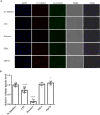
Fig 3. Cellular uptake properties of LNP-siRNA by LX-2 cells.
(A) Fluorescence microscopy images of LX-2 cells incubated with LNP-Cy3 labeled siRNA (red), LysoTracker (green), and DAPI (blue) in presence of endocytic inhibitors CPZ, Dynasore, EIPA, and MβCD. (B) Analysis of the endocytosis mechanisms of LNP-siRNA by LX-2 cells in presence of various endocytic inhibitors. **** p<0.0001 compared with untreated control. LNP, lipid nanoparticle.
Fig 4
Fig 4. In vivo specific delivery and biodistribution of LNP-Fluc mRNA.
(A) Bioluminescent image of BALB/c mice administrated with LNP-Fluc mRNA (particle size = 79.04 ± 3.77 nm) via intravenous injection. (B) Ex vivo images of major organs excised from mice 6 hours post injection. LNP, lipid nanoparticle.
Fig 5
Fig 5. Effect of intravenous injection of LNP-siTGFβRI on Schistosoma mansoni-induced liver fibrosis in mice.
(A) Scheme illustrating the experimental design for the in vivo study. (B) Macroscopic appearances of hepatic and splenic tissues from naïve and infected mice treated with PBS, LNP-Scr, and LNP-siTGFβRI at the end of the experiment after 10 weeks of infection. (C) H&E staining of liver sections, shown at 100× magnifications. (D) Masson’s trichrome staining of liver sections, shown at 100× magnifications. (E) Granuloma size measured from trichrome staining of liver sections. (F) ALT and AST levels in serum. (G) Real-time PCR analysis of mRNA levels of αSMA, Col1a1, and TgfbrI in liver tissues. (H) Western blotting analysis of protein levels of αSMA, collagen 1, and TGFβRI in liver tissues. Data are expressed as the mean ± standard deviation (SD). The arrows indicate the worm egg locations. * p<0.05, *** p<0.001, **** p<0.0001 compared with naïve mice. # p<0.05, ## p<0.01, ### p<0.001, #### p<0.0001 indicate comparisons between two indicated groups. LNP, lipid nanoparticle; Scr, scrambled RNA.
Fig 6
Fig 6. Serum cytokine concentrations and venous immune cell profiles in naïve and S. mansoni-infected mice at 10 weeks post-infection.
(A) Concentrations of Th1/Th2 cytokines in the serum. (B) Stacked bar charts showing cell frequencies as a proportion of total live cells at 10 weeks post-infection. Data are presented with averages calculated from experiments. (C) Cumulative charts showing differences in cell frequencies among naïve, PBS-treated, and LNP-siTGFβRI-treated mice. (D) The frequencies of Th1, Th2 and CD56dNK cells in different groups were calculated, respectively. Data are expressed as the mean ± standard deviation (SD). * p<0.05, ** p<0.01 compared with naïve mice. # p<0.05, ## p<0.01, ### p<0.001 indicate comparisons between two indicated groups.
Fig 7
Fig 7
Histopathological examination (H&E) of the (A) brain, (B) heart, (C) lung, and (D) kidney collected at 48 h after the last administration of LNP-siTGFβRI (1 mg/kg/dose) injected twice a week for 4 weeks, or PBS. Images were taken with a 10× objective lens.
Fig 8
Fig 8. Proposed mechanism of LNP-siTGFβRI action on schistosomiasis-induced liver fibrosis.
S. mansoni eggs release SEA, which promotes TGF-β production from AAMφ. TGF-β significantly activates hepatic stellate cells and increases collagen production. LNP-siTGFβRI inhibits the TGF-β/SMAD signaling pathway, reducing extracellular matrix levels. This process also influences the infiltration of CD4+ T cells and CD8+ T cells, shifting the immune response from Th2 to Th1 polarization, thereby aiding in egg elimination.

References

    1. Kołodziej P, Szostakowska B, Lass A, Sulima M, Sikorska K, Kocki J, et al. Chronic intestinal schistosomiasis caused by co-infection with Schistosoma intercalatum and Schistosoma mansoni. The Lancet Infectious Diseases. 2023. doi: 10.1016/S1473-3099(23)00486-3 - DOI - PubMed
    1. Lima RRM, Lima JVA, Ribeiro JFF, Nascimento JB, Oliveira WF, Cabral Filho PE, et al. Emerging biomedical tools for biomarkers detection and diagnostics in schistosomiasis. Talanta. 2023;265:124900. Epub 2023/07/10. doi: 10.1016/j.talanta.2023.124900 . - DOI - PubMed
    1. Chitsulo L, Engels D, Montresor A, Savioli L. The global status of schistosomiasis and its control. Acta Trop. 2000;77(1):41–51. doi: 10.1016/s0001-706x(00)00122-4 . - DOI - PMC - PubMed
    1. Gryseels B, Polman K, Clerinx J, Kestens L. Human schistosomiasis. Lancet. 2006;368(9541):1106–18. doi: 10.1016/S0140-6736(06)69440-3 . - DOI - PubMed
    1. Zhou YB, Zheng HM, Jiang QW. A diagnostic challenge for Schistosomiasis japonica in China: consequences on praziquantel-based morbidity control. Parasit Vectors. 2011;4:194. Epub 2011/10/11. doi: 10.1186/1756-3305-4-194 ; PubMed Central PMCID: PMC3195757. - DOI - PMC - PubMed