. 2024 Nov 1;327(5):F775-F787.
doi: 10.1152/ajprenal.00172.2024. Epub 2024 Sep 12.

Spns1 is an iron transporter essential for megalin-dependent endocytosis

Affiliations

Spns1 is an iron transporter essential for megalin-dependent endocytosis

Andrew Beenken et al. Am J Physiol Renal Physiol. .

Abstract

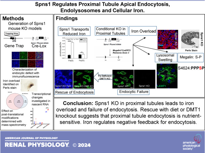
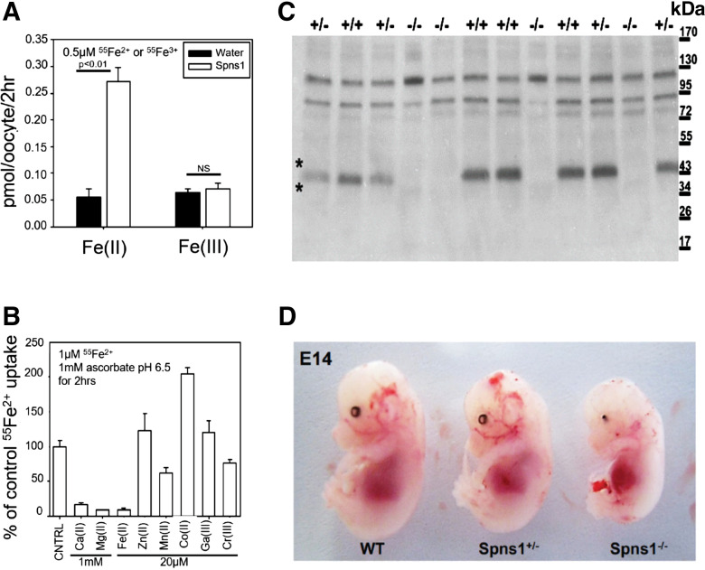
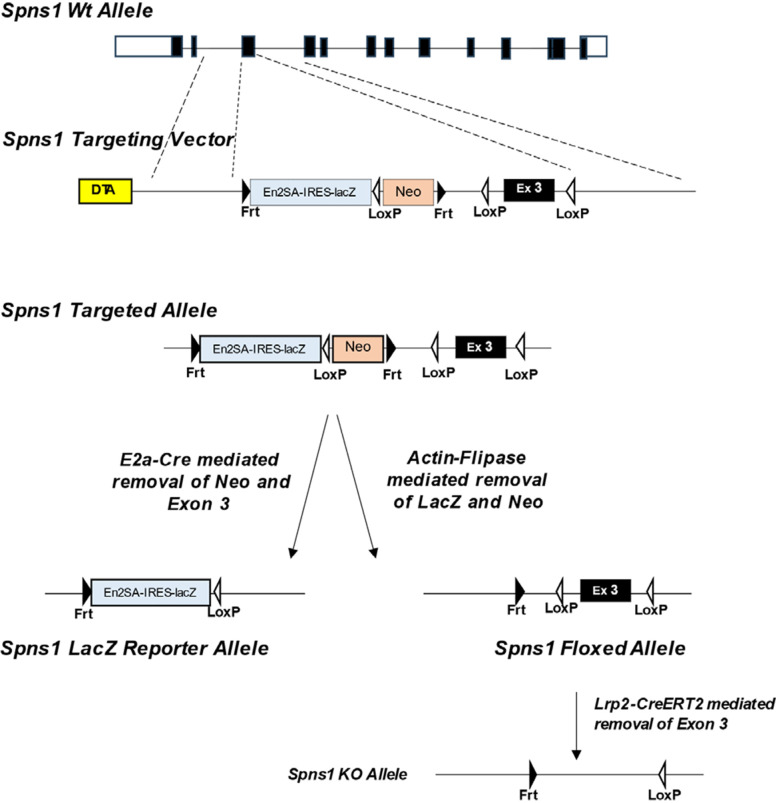
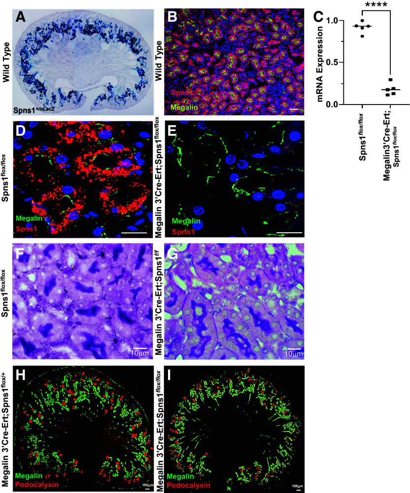
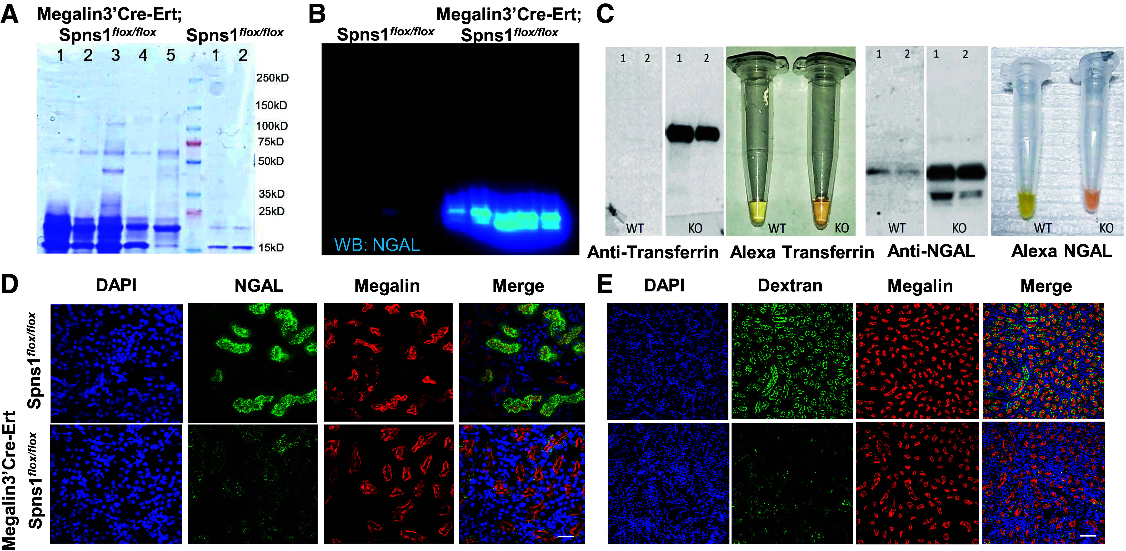
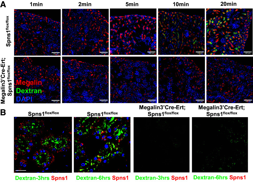
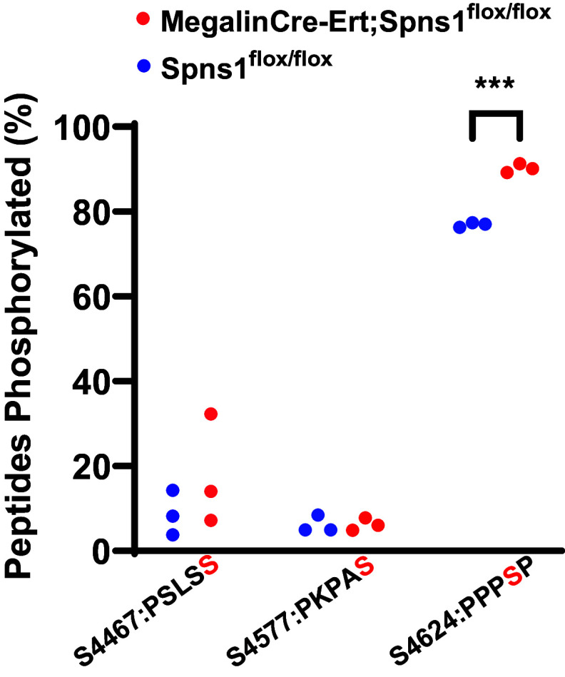
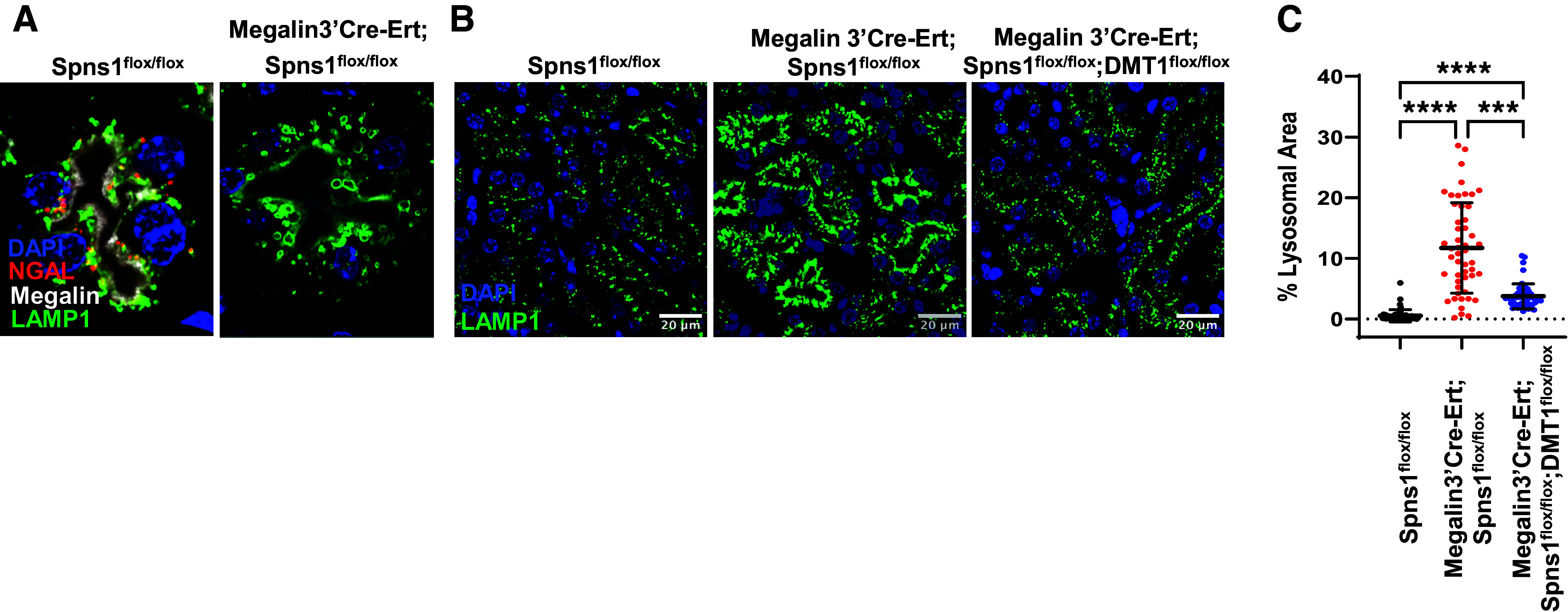
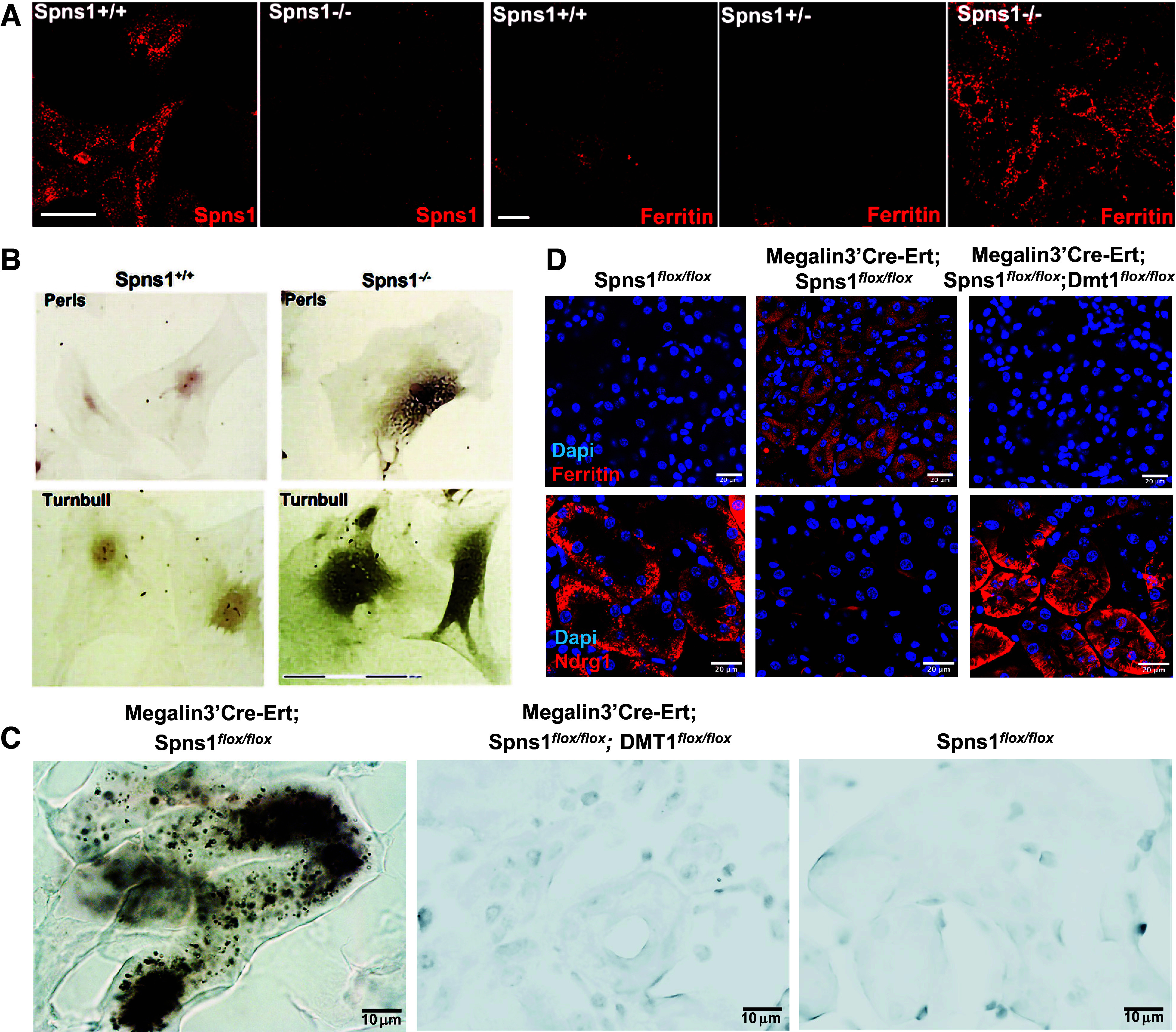
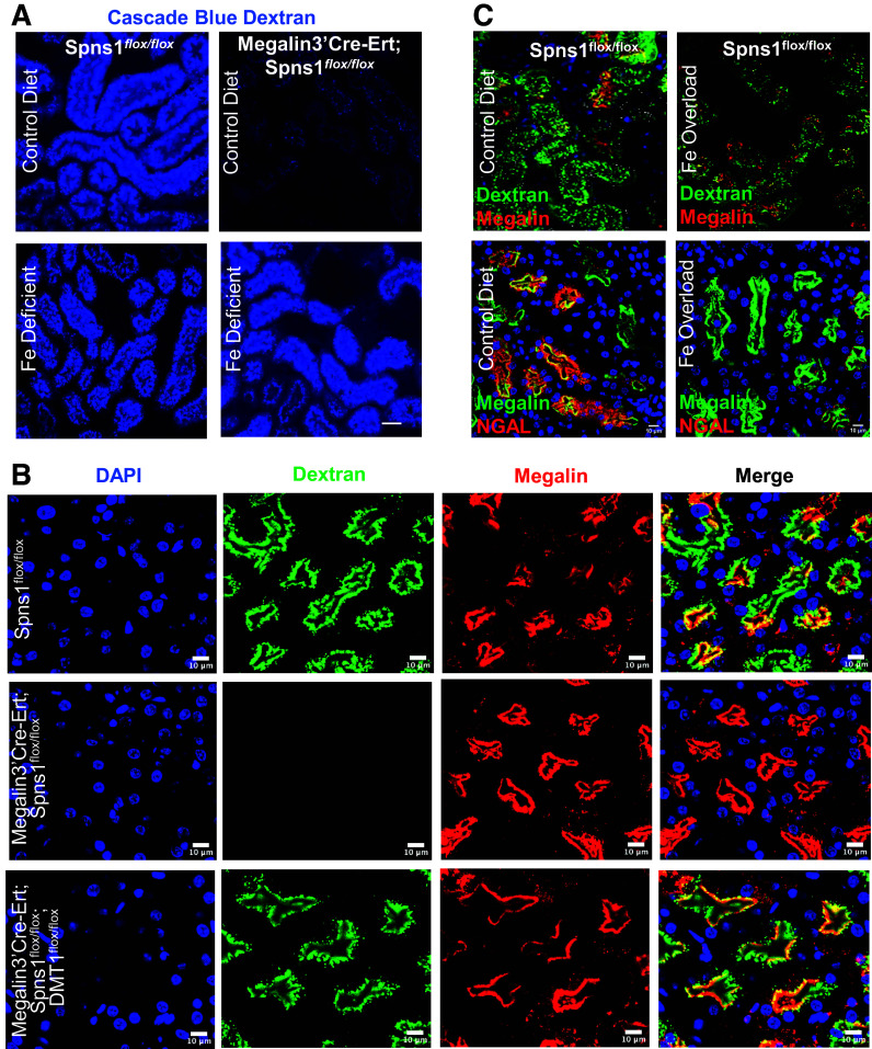
Proximal tubule endocytosis is essential to produce protein-free urine as well as to regulate system-wide metabolic pathways, such as the activation of Vitamin D. We have determined that the proximal tubule expresses an endolysosomal membrane protein, protein spinster homolog1 (Spns1), which engenders a novel iron conductance that is indispensable during embryonic development. Conditional knockout of Spns1 with a novel Cre-LoxP construct specific to megalin-expressing cells led to the arrest of megalin receptor-mediated endocytosis as well as dextran pinocytosis in proximal tubules. The endocytic defect was accompanied by changes in megalin phosphorylation as well as enlargement of lysosomes, confirming previous findings in Drosophila and Zebrafish. The endocytic defect was also accompanied by iron overload in proximal tubules. Remarkably, iron levels regulated the Spns1 phenotypes because feeding an iron-deficient diet or mating Spns1 knockout with divalent metal transporter1 knockout rescued the phenotypes. Conversely, iron-loading wild-type mice reproduced the endocytic defect. These data demonstrate a reversible, negative feedback for apical endocytosis and raise the possibility that regulation of endocytosis, pinocytosis, megalin activation, and organellar size and function is nutrient-responsive.NEW & NOTEWORTHY Spns1 mediates a novel iron conductance essential during embryogenesis. Spns1 knockout leads to endocytic and lysosomal defects, accompanied by iron overload in the kidney. Reversal of iron overload by restricting dietary iron or by concurrent knockout of the iron transporter, DMT1 rescued the endocytic and organellar defects and reverted markers of iron overload. These data suggest feedback between iron and proximal tubule endocytosis.

Keywords: Spns1; endocytosis; iron; megalin; proteinuria.


Conflict of interest statement

No conflicts of interest, financial or otherwise, are declared by the authors.

Figures

None
Graphical abstract
Figure 1.
Figure 1.
Spns1 is an iron transporter essential for organogenesis. A: oocytes inoculated with Spns1 message transport 55Fe2+ but not Fe3+ (7–10 oocytes/assay; two-tailed t test with Welch’s correction). B: Fe2+, Ca2+, and Mg2+ competed for 55Fe2+ capture (7–10 oocytes/assay; P < 0.01). C: loss of SPNS1 protein in Spns1−/− mice. In the immunoblot, the ∼50 kDa SPNS1 band is bracketed by asterisks (*). D: failure of organogenesis in gene-trapped Spns1 knockout mice. Eye development and dorsal cranial structures were most affected.
Figure 2.
Figure 2.
Generation of Spns1 LacZ reporter and conditional knockout. We obtained Spns1-targeted ES cells from https://www.informatics.jax.org/allele/MGI:4432137. Heterozygotes were bred with E2a-Cre to generate Spns1LacZ reporter mice or bred with Actin-Flipase to generate Spns1flox/+ mice, which, in turn, were bred with MegalinCre-Ert2 mice to obtain Spns1 deletion specifically in the proximal tubule.
Figure 3.
Figure 3.
Conditional knockout of Spns1. A: whole-mount Spns1+/Lacz in proximal tubules (E13.5). B: expression of Spns1 (red) in megalin+ (yellow) proximal tubules. C: deletion of Spns1 expression in Megalin3’Cre-Ert; Spns1flox/flox kidneys (P = 1.7 × 10⁻⁷, two-tailed t test with Welch’s correction). D and E: kidneys from littermates showing with immunofluorescence depicting loss of SPNS1 (red stain) but not megalin (green stain) in Megalin3’Cre-Ert;Spns1flox/flox knockout proximal tubules (right); Spns1flox/flox is shown as a control (left). F and G: kidneys from littermates with. periodic acid-Schiff staining demonstrating intact apical brush borders in both Spns1flox/flox controls and Megalin3’Cre-Ert;Spns1flox/flox knockout proximal tubules. H and I: kidneys from littermates with immunofluorescence depicting grossly similar levels and distributions of podocalyxin (red stain) and megalin (green stain) in Megalin3’Cre-Ert;Spns1flox/flox knockout proximal tubules (right) and Megalin3’Cre-Ert;Spns1flox/+ control (left) proximal tubules. Bars = 50 µm in B, 20 µm in D and E, 10 µm in F and G, and 100 µm in H and I.
Figure 4.
Figure 4.
Conditional knockout of Spns1 impairs proximal tubule endocytosis. A: low molecular weight proteinuria in Spns1 knockouts. Coomassie staining is shown. B: urinary NGAL excreted in Spns1 knockouts. C: exogenous Alexa568-transferrin and -NGAL excreted in Spns1 knockout. Urine immunoblots are shown. D and E: failure to capture FITC-NGAL and FITC-dextran despite expression of megalin in Megalin3’Cre-Ert;Spns1flox/flox knockout proximal tubules. Bars = 20 µm in D and 80 µm in E.
Figure 5.
Figure 5.
Conditional knockout of Spns1 impairs proximal tubule endocytosis: time course. A: wild-type (Spns1flox/llox) and Spns1-deleted proximal tubules (Megalin3’Cre-Ert;Spns1flox/llox) could be distinguished within 10 min of infusion of FITC-dextran. B: dextran was captured by endosomes at the luminal surface and then subsequently located in SPNS1+ late endosomes and lysosomes deep in the cytoplasm. No labeling was found in Spns1 knockouts. Bars = 100 µm in A and 20 µM in B.
Figure 6.
Figure 6.
Conditional knockout of Spns1 regulates phospho-megalin. Megalin3’Cre-Ert;Spns1flox/flox mice have a significant increase in phosphorylation at the PPPSP motif compared with Spns1flox/flox (***P = 2.79 × 10⁻⁴ by an unpaired two-tailed t test with Welch's correction; n = 3 mice each). Results from experiments in three separate mice yielded statistical significance. Phosphopeptides: S4467: KLPSLSSLAKPSENGNGVTFRS; S4577: R.SIDPSEIVPEPKPASPGADETQGTK.W; and S4624: K.EAVAVAPPPSPSLPAKA.
Figure 7.
Figure 7.
Conditional knockout of Spns1 leads to endolysosomal swelling. AC: LAMP1+ lysosomes doubled in size in Megalin3’Cre-Ert;Spns1flox/flox proximal tubules and occupied 22 times more area than lysosomes in Spns1flox/flox proximal tubules. The size of the lysosomes reverted to control size in Megalin3’Cre-Ert;Spns1flox/flox;DMT1flox/flox double knockout proximal tubules. C quantification of cytoplasmic area occupied by LAMP1+ lysosomes. Kruskal–Wallis with Dunn’s correction for multiple comparisons was used. ****WT vs. Spns1 KO, Padj = 1.7 × 10⁻²³; ****WT vs. Spns1KO;DMT1 KO, Padj = 2.5 × 10⁻⁹; and ***Spns1KO vs. Spns1KO; DMT1 KO, Padj = 2.2 × 10⁻⁴. Bars = 10 µm in B.
Figure 8.
Figure 8.
Conditional knockout of Spns1 leads to iron overload. A: deletion of SPNS1 protein and accumulation of ferritin immunoreactivity in Spns1−/− deleted MEFs. B: Spns1−/− MEFs stain with Perls-DAB (top row) and Turnbull stains (bottom row) depicting Fe3+ or Fe2+ deposits, respectively. C: Perls-DAB staining of kidney sections. Increased staining in Megalin3’Cre-Ert;Spns1flox/flox kidneys was prevented by the additional deletion of DMT1. D: increased ferritin and decreased NDRG1 proteins in Megalin3’Cre-Ert;Spns1flox/flox knockout; there was reversion in Megalin3’Cre-Ert;Spns1flox/flox;DMT1flox/flox double knockouts. Bars = 20 µm in A, 150 µm in B, 10 µm in C, and 20 µm in D.
Figure 9.
Figure 9.
Iron-deficient chow or DMT1 deletion rescues endocytosis. A: iron-deficient chow (bottom row) rescued the endocytosis of blue-dextran. B: likewise, double knockout Megalin3’Cre-Ert;Spns1flox/flox;DMT1flox/flox mice rescued endocytosis of FITC-dextran. C: conversely, iron overload of the kidney suppressed dextran and protein NGAL capture in wild-type kidneys. Bars = 25 µm in A and 10 µm in B and C.

References

    1. Nielsen R, Christensen EI, Birn H. Megalin and cubilin in proximal tubule protein reabsorption: from experimental models to human disease. Kidney Int 89: 58–67, 2016. doi:10.1016/j.kint.2015.11.007. - DOI - PubMed
    1. Lieu PT, Heiskala M, Peterson PA, Yang Y. The roles of iron in health and disease. Mol Aspects Med 22: 1–87, 2001. doi:10.1016/s0098-2997(00)00006-6. - DOI - PubMed
    1. Ponka P. Cellular iron metabolism. Kidney Int Suppl 69: S2–S11, 1999. doi:10.1046/j.1523-1755.1999.055suppl.69002.x. - DOI - PubMed
    1. Ponka P, Lok CN. The transferrin receptor: role in health and disease. Int J Biochem Cell Biol 31: 1111–1137, 1999. doi:10.1016/s1357-2725(99)00070-9. - DOI - PubMed
    1. Aisen P. Transferrin receptor 1. Int J Biochem Cell Biol 36: 2137–2143, 2004. doi:10.1016/j.biocel.2004.02.007. - DOI - PubMed

MeSH terms

Substances