. 2024;16(1):451-469.
doi: 10.1159/000540744. Epub 2024 Sep 12.

Inhibition of WNK Kinases in NK Cells Disrupts Cellular Osmoregulation and Control of Tumor Metastasis

Affiliations

Inhibition of WNK Kinases in NK Cells Disrupts Cellular Osmoregulation and Control of Tumor Metastasis

Ji Sung Kim et al. J Innate Immun. 2024.

Abstract

Introduction: The serine/threonine with-no-lysine (WNK) kinase family function in blood pressure control, electrolyte homeostasis, and cellular osmoregulation. These kinases and their downstream effectors are considered promising therapeutic targets in hypertension and stroke. However, the role of WNK kinases in immune cells remains poorly understood.

Methods: Using the small-molecule WNK kinase inhibitors WNK463 and WNK-IN-11, we investigated how WNK kinase inhibition affects natural killer (NK) cell physiology.

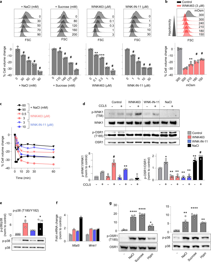
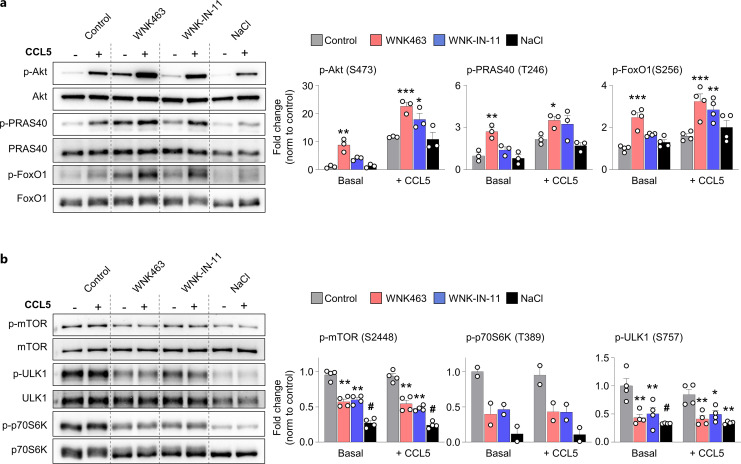
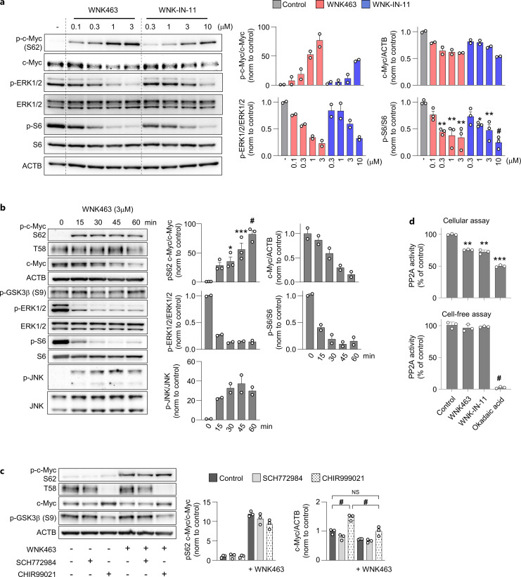
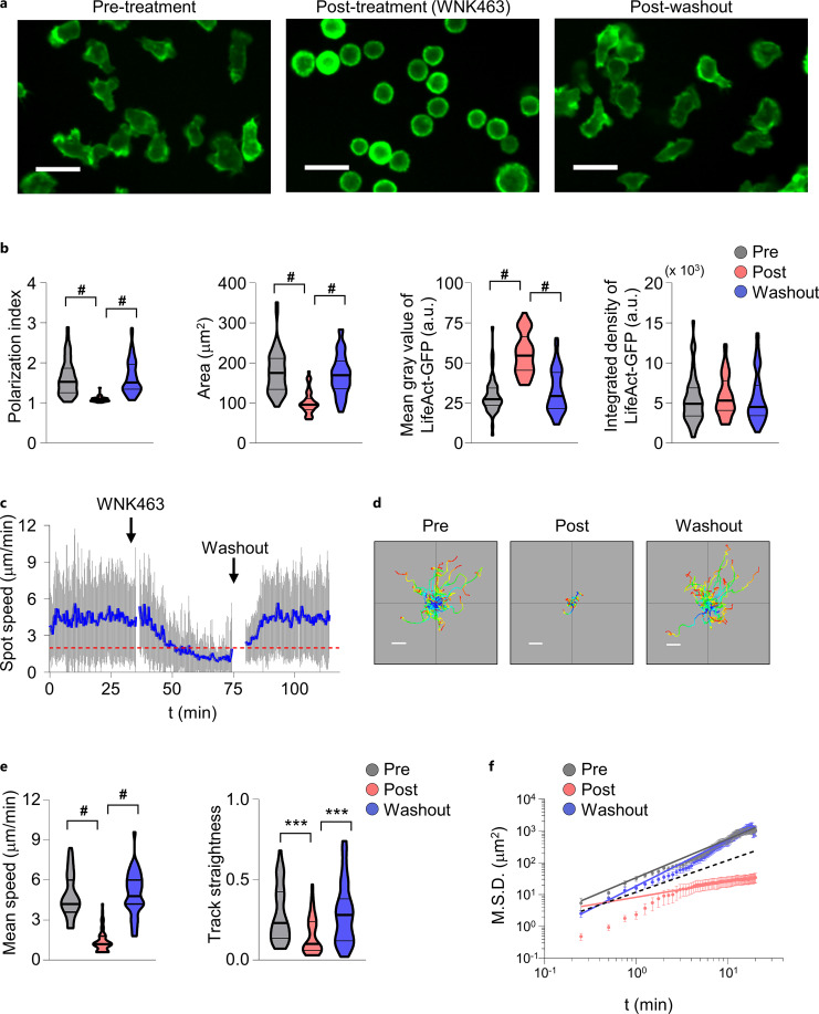
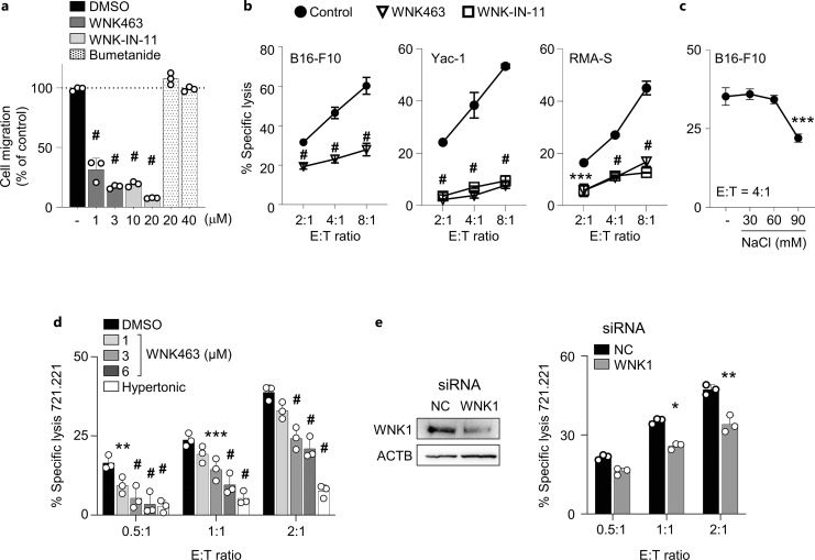
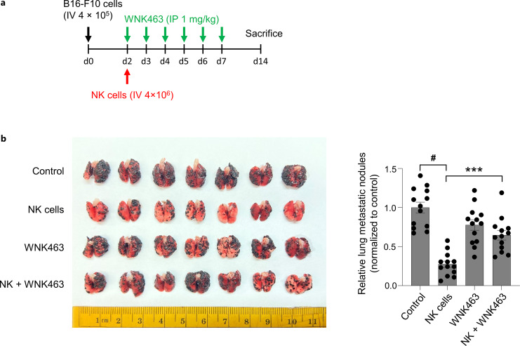
Results: WNK kinase inhibition with WNK463 or WNK-IN-11 significantly decreased IL-2-activated NK cell volume, motility, and cytolytic activity. Treatment of NK cells with these inhibitors induced autophagy by activating AMPK and inhibiting mTOR signaling. Moreover, WNK kinase inhibition increased phosphorylation of Akt and c-Myc by misaligning activity of activating kinases and inhibitory phosphatases. Treatment of tumor-bearing mice with WNK463 impaired tumor metastasis control by adoptively transferred NK cells.

Conclusion: The catalytic activity of WNK kinases has a critical role of multiple aspects of NK cell physiology and their pharmacologic inhibition negatively impacts NK cell function.

Keywords: Autophagy; Cytotoxicity; Natural killer; Osmoregulation; WNK kinases.


Conflict of interest statement

The authors declare that the research was conducted in the absence of any commercial or financial relationships that could be construed as a potential conflict of interest. The content is solely the responsibility of the authors and does not necessarily represent the official views of the National Institutes of Health.

Figures

Fig. 1.
Fig. 1.
WNK inhibition decreases NK cell volume. a Cell volume changes in primary IL-2-activated mouse NK cells, which were exposed to various concentration of extra NaCl, WNK463, or WNK-IN-11 for 1 h. Data pooled from three independent experiments. % Cell volume change is shown relative to the untreated cells. b Cell volume changes at various hypoosmotic pressure in the presence of 3 µM WNK463. NK cells were first treated with 3 µM WNK463 for 30 min. The cells were then exposed to a hypotonic medium, prepared by diluting with water while maintaining 3 µM WNK463, for an additional 30 min. Data pooled from two independent experiments. % Cell volume change is shown relative to the untreated cells. c Cell volume changes overtime after exposure to WNK463, WNK-IN-11, or NaCl. Representative of two independent experiments with two replications. d Representative immunoblots of phosphorylation of WNK1 at T58 residue and OSR1 at T185 residue in NK cells treated with WNK463 (3 µM), WNK-IN-11 (10 µM), or NaCl (extra 90 mm) for 1 h and then stimulated by CCL5 for 1 min. Phosphoproteins were quantified from three independent experiments. ns, nonspecific band. e Representative immunoblots of phospho-p38 in NK cells treated with WNK463, WNK-IN-11, or NaCl for 1 h. Phosphoprotein was quantified from three independent experiments. f mRNA expression levels of NFAT5 and WNK1 in NK cells treated with WNK463, WNK-IN-11, or NaCl for 6 h. Expression was quantified from two independent experiments with three replications. g Representative immunoblots of phospho-OSR1 and p38 in NK cells treated with NaCl, sucrose, or hypotonic medium prepared by dilution with water. Phosphoproteins was quantified from four independent experiments. All phosphorylated proteins were quantified by their total proteins and normalized by fold changes to the control. Fold mRNA change is normalized to GAPDH and shown relative to the control. One-way ANOVA Tukey tests were used for analysis, with *p < 0.05, **p < 0.01, ***p < 0.005, and #p < 0.0001.
Fig. 2.
Fig. 2.
WNK inhibition increases autophagic flux and suppresses mTORC1 signaling. a Representative immunoblots of LC3-II and p62 expression in primary IL-2-activated mouse NK cells. Cell lysates were prepared from NK cells treated with WNK463 (3 µM), WNK-IN-11 (10 µM), NaCl (extra 90 mm), or DMSO as a control at the presence or absence bafilomycin A1 for an hour. Autophagic flux was calculated by references and normalized to control. p62 expression was normalized to control. Autophagic flux and p62 expression were quantified from three, or four independent experiments. b, c Immunoblots analysis of various phosphorylated proteins in NK cells treated for an hour with WNK463 (3 µM), WNK-IN-11 (10 µM), NaCl (extra 90 mm), or DMSO as a control. b p-LKB1 (S428), LKB1; pAMPKα (T172), AMPKα; p-TSC2 (S1387), TSC2. Phosphoproteins were quantified from two or five independent experiments. c p-mTOR (S2448), mTOR; pS6 (S235/S236), S6; p-4E-BP1 (T37/T46), 4E-BP1; p-4E-BP1 (S65), 4E-BP1. Phosphoproteins were quantified from three or six independent experiments. All phosphorylated proteins were quantified by their total proteins and normalized by fold changes to the control. One-way ANOVA Tukey tests, or non-parametric Kruskal-Wallis tests (LKB1 S428 (b) and 4E-BP1 S65 (c)) were used for analysis, with *p < 0.05, **p < 0.01, and ***p < 0.005.
Fig. 3.
Fig. 3.
WNK inhibition increases Akt phosphorylation. a, b Akt or mTOR signaling pathway in primary IL-2-activated mouse NK cells treated with WNK463 (3 µM), WNK-IN-11 (10 µM), NaCl (extra 90 mm), or DMSO control for 1 h and then stimulated by CCL5 for 1 min. a Representative immunoblots of phosphorylated or total Akt, PRAS40, FoxO1, and their quantification. b Representative immunoblots of phosphorylated or total mTOR, ULK1, or p70S6K and their quantification. All phosphorylated proteins from two or four independent experiments were quantified by their total proteins and normalized by fold changes to the basal control. One-way ANOVA Tukey tests were used for analysis, with *p < 0.05, **p < 0.01, ***p < 0.005, and #p < 0.0001.
Fig. 4.
Fig. 4.
WNK inhibition impairs c-Myc expression. a Representative immunoblots of phosphorylated c-Myc, ERK, or S6 in primary IL-2-activated mouse NK cells treated indicated concentration of WNK463, or WNK-IN-11 and quantification of phosphorylated proteins in NK cells from two or three independent experiments. pS62 c-Myc (upper left), c-Myc (upper right), pERK1/2 (lower left), or p-S6 (lower right) is normalized to c-Myc, actin, ERK1/2, or S6, respectively. b Representative immunoblots of phosphorylated c-Myc, ERK, S6, and JNK in NK cells treated with WNK463 for indicated time points and quantification of phosphorylated proteins from two, or three independent experiments. pS62 c-Myc (top left), c-Myc (top right), pERK1/2 (middle left), p-S6 (middle right), or p-JNK (bottom left) is normalized to c-Myc, actin, ERK1/2, S6, or JNK, respectively. All phosphorylated proteins were quantified by their total proteins and normalized by fold changes to the control. c Representative immunoblots of phospho-c-Myc, total c-Myc, and actin in NK cells treated with SCH772984 or CHIR99021 at the presence or absence of WNK463 for 20 min from three independent experiments and quantification of pS62 c-Myc and c-Myc, which were normalized to c-Myc, or actin, respectively. d PP2A activity of cells or cell lysates treated with WNK463 (3 µM), WNK-IN-11 (10 µM), or okadaic acid (100 nM) for an hour. Repeated twice with similar results. % of control is shown relative to the control. One-way ANOVA Tukey tests were used for analysis, with *p < 0.05, **p < 0.01, ***p < 0.005, and #p < 0.0001.
Fig. 5.
Fig. 5.
WNK inhibition induces cell shrinkage and arrests NK cell motility. a Snapshots from spinning disk confocal live-cell images of migrating LifeAct NK cells treated with WNK463 pre-, posttreatment, or after washout. Scale bar: 20 µm. b Analysis of morphological changes in NK cells by WNK463. From the left: polarization index, cell area, mean gray value of LifeAct-GFP, and integrated density of LifeAct-GFP. n = 52, 47, or 58 for pre, post, or washout, respectively. c Spot speed changes in NK cells by WNK463. A blue line indicated average spot speed. Spot speed less than 2 µm/min (red dash line) is considered that cells were stopped, defined by estimated the diffusion coefficient. d Trajectories of cell migration. Scale bar: 20 μm. e Mean speeds of NK cells with WNK463 pre-, posttreatment, or after washout are 4.85, 1.39, or 4.98, respectively. Track straightness. n = 37, 54, or 43 for pre, post, or washout, respectively. f The mean-squared displacement (M.S.D.) on a log-log plot, where α = 1 (dash line, Brownian motion). Slopes are 1.21 for pretreatment, 0.48 for posttreatment, and 1.45 for after washout. Repeated twice with similar results. Non-parametric Kruskal-Wallis tests were used for analysis, with ***p < 0.005, and #p < 0.0001.
Fig. 6.
Fig. 6.
WNK inhibition impairs NK cell migration and their cytolytic activity. a Effect of WNK463, WNK-IN-11, and bumetanide on primary IL-2-activated mouse NK cell chemotaxis toward CCL5 was measured by transwell migration assay. Each inhibitor remained for the duration of the assay. b Effect of WNK inhibitors on cytolytic activity of NK cells. Various ratio of NK cells was cocultured with B16-F10, Yac-1, or RMA-S for 2 h at the presence or absence of WNK463 (3 µM), or WNK-IN-11 (10 µM). c Effect of hypertonicity on cytolytic activity of NK cells. NK cells were cocultured with B16-F10 in various hypertonicity by adding indicated extra NaCl. d Cytolytic activity of YTS human NK cell line against 721.221 target cells with indicated concentration of WNK463, or NaCl (extra 90 mm). e WNK1 in YTS cells was knocked down by siRNA electroporation. Representative immunoblots of WNK1 and actin (left). Cytolytic activity of YTS cells transfected with WNK1 siRNA or negative control siRNA against 721.221 cells was determined over indicated E:T ratios (right). Error bars represent standard deviation of three replicates. The data are representative of two independent experiments with similar results. One- or two-way ANOVA Tukey tests were used for analysis, with *p < 0.05, **p < 0.01, ***p < 0.005, and #p < 0.0001.
Fig. 7.
Fig. 7.
WNK inhibition impairs NK cell-mediated control of lung metastasis. a Schedule of mouse treatment. Mice were intravenously injected with B16-F10 melanoma cells on day 0. The mice were treated with IL-2-activated NK cells on day 2 and/or WNK463 daily from day 2 to day 7. Mice were sacrificed for analysis on day 14 after tumor injection. b Representative lung images (left) and quantified metastatic foci in the lung (right) (n = 13 per group). The data are pooled and normalized from two independent experiments. One-way ANOVA Tukey tests were used for analysis, with ***p < 0.001, and #p < 0.0001.

Similar articles

Cited by

References

    1. Shekarabi M, Zhang J, Khanna AR, Ellison DH, Delpire E, Kahle KT. WNK kinase signaling in ion homeostasis and human disease. Cell Metab. 2017;25(2):285–99. - PubMed
    1. Verissimo F, Jordan P. WNK kinases, a novel protein kinase subfamily in multi-cellular organisms. Oncogene. 2001;20(39):5562–9. - PubMed
    1. Moriguchi T, Urushiyama S, Hisamoto N, Iemura S, Uchida S, Natsume T, et al. . WNK1 regulates phosphorylation of cation-chloride-coupled cotransporters via the STE20-related kinases, SPAK and OSR1. J Biol Chem. 2005;280(52):42685–93. - PubMed
    1. Vidal-Petiot E, Elvira-Matelot E, Mutig K, Soukaseum C, Baudrie V, Wu S, et al. . WNK1-related Familial Hyperkalemic Hypertension results from an increased expression of L-WNK1 specifically in the distal nephron. Proc Natl Acad Sci U S A. 2013;110(35):14366–71. - PMC - PubMed
    1. Lalioti MD, Zhang J, Volkman HM, Kahle KT, Hoffmann KE, Toka HR, et al. . Wnk4 controls blood pressure and potassium homeostasis via regulation of mass and activity of the distal convoluted tubule. Nat Genet. 2006;38(10):1124–32. - PubMed

Publication types

Substances

LinkOut - more resources