. 2024 Nov 7;32(11):1963-1972.e3.
doi: 10.1016/j.str.2024.08.011. Epub 2024 Sep 11.

Structural organization of pyruvate: ferredoxin oxidoreductase from the methanogenic archaeon Methanosarcina acetivorans

Affiliations

Structural organization of pyruvate: ferredoxin oxidoreductase from the methanogenic archaeon Methanosarcina acetivorans

Matteo Cossu et al. Structure. .

Abstract

Enzymes of the 2-oxoacid:ferredoxin oxidoreductase (OFOR) superfamily catalyze the reversible oxidation of 2-oxoacids to acyl-coenzyme A esters and carbon dioxide (CO2)using ferredoxin or flavodoxin as the redox partner. Although members of the family share primary sequence identity, a variety of domain and subunit arrangements are known. Here, we characterize the structure of a four-subunit family member: the pyruvate:ferredoxin oxidoreductase (PFOR) from the methane producing archaeon Methanosarcina acetivorans (MaPFOR). The 1.92 Å resolution crystal structure of MaPFOR shows a protein fold like those of single- or two-subunit PFORs that function in 2-oxoacid oxidation, including the location of the requisite thiamine pyrophosphate (TPP), and three [4Fe-4S] clusters. Of note, MaPFOR typically functions in the CO2 reductive direction, and structural comparisons to the pyruvate oxidizing PFORs show subtle differences in several regions of catalytical relevance. These studies provide a framework that may shed light on the biochemical mechanisms used to facilitate reductive pyruvate synthesis.


Conflict of interest statement

Declaration of interests The authors declare no competing interests.

Figures
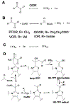
Figure 1.
Figure 1.. Reaction schemes for members of the OFOR superfamily.
(A) The CoA-independent OOR reaction. (B) The CoA-dependent reactions catalyzed by PFOR, OGOR, VOR and IOR. (C) Shuttling of the low potential electrons generated above to the cellular ferredoxin pool for subsequent usages. (D) Proposed reaction mechanism for pyruvate oxidation by members of the PFOR class. The CO2 fixation reaction runs in the reverse direction.
Figure 2.
Figure 2.. Characterization of MaPFOR.
(A) Gene organization of the por locus in the M. acetivorans genome showing the composition of the individual subunits that constitute MaPFOR. (B) Homologous expression and affinity purification of MaPFOR is enabled by attaching an affinity tag to the δ subunit. Abbreviations: M: molecular weight markers, with mass in kDa listed to the left; lane 1: unbound protein; lanes 2-7: wash fractions; Lanes 8-13: elution fractions. (C, D) Kinetic analysis of anoxically purified MaPFOR. Pyruvate-dependent reduction of benzyl viologen was assayed spectrophotometrically as described. The data were fit to standard Michaelis-Menten kinetics to generate the kinetic parameters shown. Units (U) are defined as mmol*min-1*mg. All experiments were carried out in triplicate and errors were calculated based in standard regression analysis.
Figure 3.
Figure 3.. Structural organization of MaPFOR and its orthologs.
(A) The overall structure of the MaPFOR (αβγδ)2 assembly showing the arrangement of the individual subunits color coded as described. Only of the αβγδ subunit assembly is shown in color and the other is shown in gray. (B) Close-up view showing the organization of the TPP co-factor between the α and β subunits, the proximal [4Fe-4S] cluster in the β subunit, and the medial and distal clusters in the δ subunit. (C) Relationship of the subunit arrangements in MaPFOR and the domain arrangements of other structurally characterize OFORs.
Figure 4.
Figure 4.. Comparison of OFOR active sites.
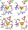
(A) Stereo figure showing the superposition of the structures of MtOOR in the absence of substrate (tan; PDB 5C4I) with that of the COOM-TPP bound form (blue; PDB 5EXD). The active site of the CoA-independent MtOOR employs a ‘switch loop’ to enable 2-oxoacid decarboxylation, and a similar switching mechanism has also been proposed for MmOGOR. (B) Close up stereo view of the active site of MaPFOR shows that residues in the α subunit form a hydrophobic environment that is suited for the binding of CO2. The structure of MaPFOR does not contain a ‘switch loop’ nor a negatively charged residue as these would disrupt the hydrophobic pocket necessary for CO2 engagement.
Figure 5.
Figure 5.. Comparison of OFOR proximal clusters.
(A) The proximal cluster of MaPFOR is composed of small or polar residues and a charge Arg42 is provided by the γ subunit. (B) The proximal cluster of MmOGOR is shielded from solvent and is surrounded by hydrophobic residues.
Figure 6.
Figure 6.. Interactions of the MaPFOR δ subunit.
The ferredoxin-like δ subunit is tethered to the complex through numerous interactions including those formed by residues in the unique N-terminal region and those in the β and γ subunits.
Figure 7.
Figure 7.. Comparison of OFOR CoA binding domains.
(A) The γ subunit of MaPFOR (equivalent to the CoA-binding domain III is structurally divergent from other characterized OFORs and would require significant conformational reorganization to form a catalytically competent structure. The P loop (motif 1) is colored in magenta (B) Structural superposition of the MaPFOR γ subunit (pink) with domain III of MtPFOR (gray) showing the structural differences between the two. (C) Binding of CoA to MtPFOR results in the movement of two helices to situate CoA-binding residues. The MaPFOR γ subunit would require significantly more dramatic changes to bind CoA. (D) View of MaPFOR with CoA modelled showing that several unique residues may contribute to CoA binding.

Similar articles

Cited by

References

    1. Charon MH, Volbeda A, Chabriere E, Pieulle L, and Fontecilla-Camps JC (1999). Structure and electron transfer mechanism of pyruvate:ferredoxin oxidoreductase. Curr Opin Struct Biol 9, 663–669. - PubMed
    1. Gibson MI, Chen PY, and Drennan CL (2016). A structural phylogeny for understanding 2-oxoacid oxidoreductase function. Curr Opin Struct Biol 41, 54–61. - PMC - PubMed
    1. Ragsdale SW (2003). Pyruvate ferredoxin oxidoreductase and its radical intermediate. Chem Rev 103, 2333–2346. - PubMed
    1. Ragsdale SW (2008). Enzymology of the wood-Ljungdahl pathway of acetogenesis. Ann N Y Acad Sci 1125, 129–136. - PMC - PubMed
    1. Yun NR, Arai H, Ishii M, and Igarashi Y (2001). The genes for anabolic 2-oxoglutarate: ferredoxin oxidoreductase from Hydrogenobacter thermophilus TK-6. Biochem Biophys Res Commun 282, 589–594. - PubMed

Publication types

MeSH terms

LinkOut - more resources