. 2024 Sep 11;12(9):e009147.
doi: 10.1136/jitc-2024-009147.

Regression of renal cell carcinoma by T cell receptor-engineered T cells targeting a human endogenous retrovirus

Affiliations

Regression of renal cell carcinoma by T cell receptor-engineered T cells targeting a human endogenous retrovirus

Stefan Barisic et al. J Immunother Cancer. .

Abstract

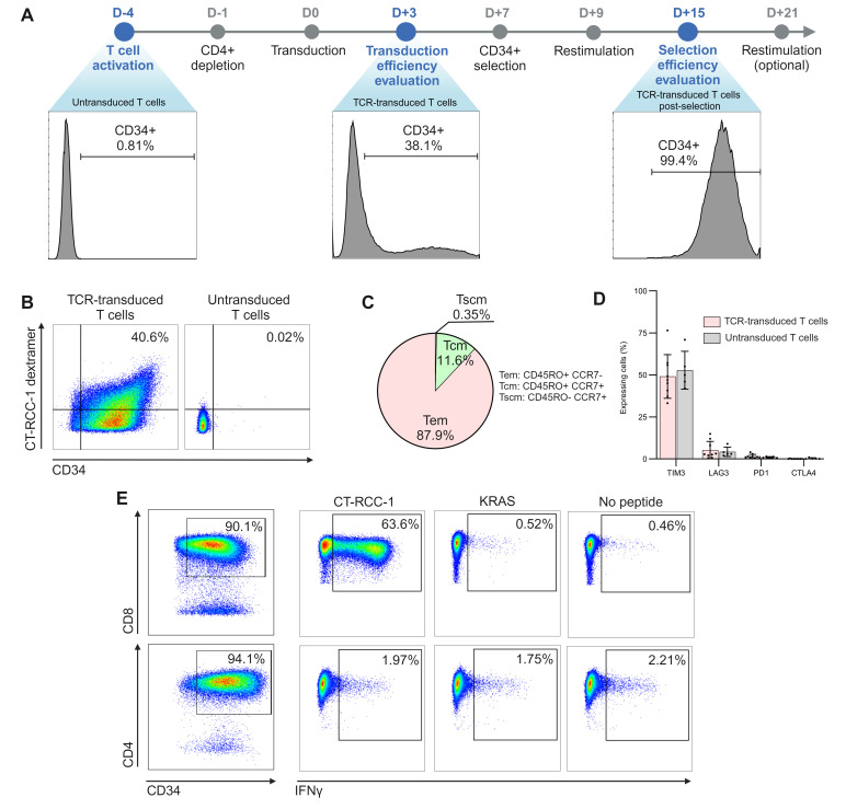
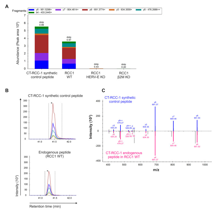
Background: We discovered a novel human endogenous retrovirus (CT-RCC HERV-E) that was selectively expressed in most clear cell renal cell carcinomas (ccRCC) and served as a source of antigens for T cell-mediated killing. Here, we described the cloning of a novel T cell receptor (TCR) targeting a CT-RCC HERV-E-derived antigen specific to ccRCC and characterized antitumor activity of HERV-E TCR-transduced T cells (HERV-E T cells).

Methods: We isolated a CD8+ T cell clone from a patient with immune-mediated regression of ccRCC post-allogeneic stem cell transplant that recognized the CT-RCC-1 HERV-E-derived peptide in an HLA-A11-restricted manner. We used 5'Rapid Amplification of cDNA Ends (RACE) to clone the full length HERV-E TCR and generated retrovirus encoding this TCR for transduction of T cells. We characterized HERV-E T cells for phenotype and function in vitro and in a murine xenograft model. Lastly, we implemented a good manufacturing practice-compliant method for scalable production of HERV-E T cells.

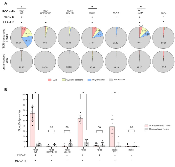
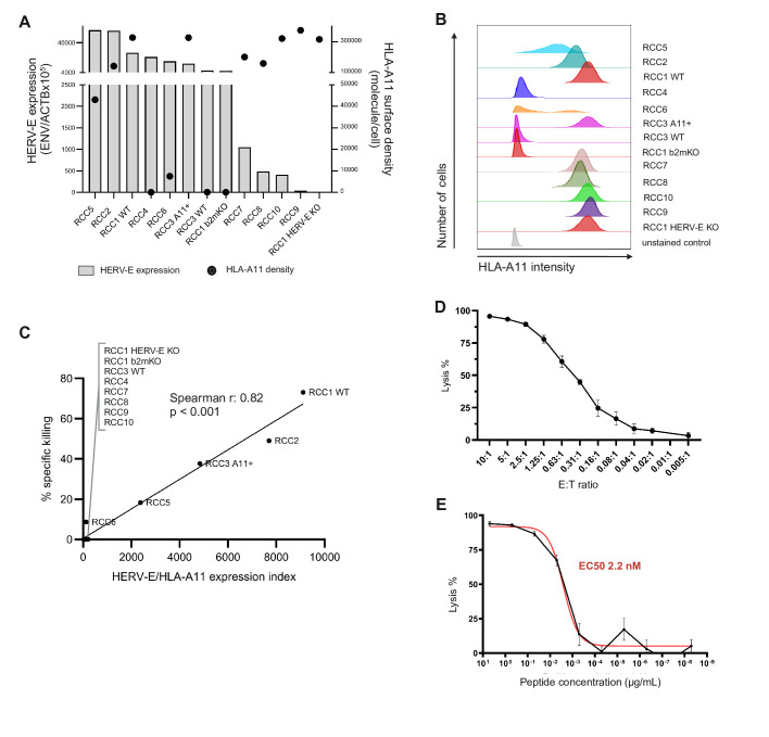
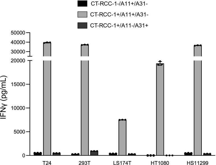
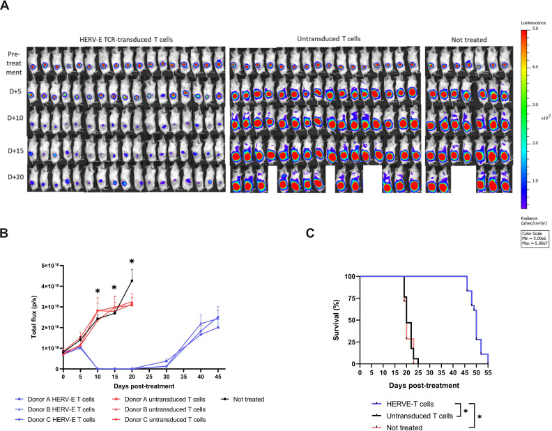
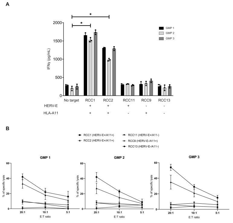
Results: The HLA-A11-restricted HERV-E-reactive TCR exhibited a CD8-dependent phenotype and demonstrated specific recognition of the CT-RCC-1 peptide. CD8+ T cells modified to express HERV-E TCR displayed potent antitumor activity against HLA-A11+ ccRCC cells expressing CT-RCC HERV-E compared with unmodified T cells. Killing by HERV-E T cells was lost when cocultured against HERV-E knockout ccRCC cells. HERV-E T cells induced regression of established ccRCC tumors in a murine model and improved survival of tumor-bearing mice. Large-scale production of HERV-E T cells under good manufacturing practice conditions generated from healthy donors retained specific antigen recognition and cytotoxicity against ccRCC.

Conclusions: This is the first report showing that human ccRCC cells can be selectively recognized and killed by TCR-engineered T cells targeting a HERV-derived antigen. These preclinical findings provided the foundation for evaluating HERV-E TCR-transduced T cell infusions in patients with metastatic ccRCC in a clinical trial (NCT03354390).

Keywords: T cell receptor - TCR; adoptive cell therapy - ACT; kidney cancer.


Conflict of interest statement

Competing interests: The authors (EC, MIN and RWC) declare a filed patent WO2018006054A1, licensed by T-Cure BioScience, represented by GZ and GP.

Figures

Figure 1
Figure 1. Characterization of HERV-E TCR-transduced T cells. (A) The preclinical method used to generate and expand HERV-E TCR-transduced CD8+ T cells. Histograms show a representative example of transduction efficiency (n=7 donors) before and after CD34 enrichment as determined by CD34 surface expression by flow cytometry. (B) Representative plot of flow cytometry analysis of HLA-A11:01/CT-RCC-1 dextramer binding of HERV-E TCR-transduced T cells (n=7 donors). Untransduced T cells, which underwent the same treatment as retroviral-transduced T cells, were used as a negative control for dextramer binding. (C) A pie chart depicting the T cell subpopulation composition of TCR-transduced T cells (n=7 donors, values shown are median). (D) Surface expression of select inhibitory markers on HERV-E TCR-transduced T cells 2 weeks post-transduction (mean±SD, n=7 donors). (E) Intracellular staining for IFNγ shows HERV-E TCR-transduced CD8+ T cells but not HERV-E TCR-transduced CD4+ T cells recognize T2-A11+ cells pulsed with the CT-RCC-1 HERV-E peptide. T2-A11+ cells were peptide-pulsed with either CT-RCC-1 HERV-E peptide, a mock peptide (HLA-A11-restricted KRAS G12D7-16), or were not pulsed. HERV-E, human endogenous retrovirus type E; RCC, renal cell carcinoma.
Figure 2
Figure 2. Characterization of the HERV-E T cell function versus ccRCC cells in vitro. (A) The lytic and cytokine-producing ability of HERV-E T cells and untransduced T cells from the same donors are shown when cocultured with ccRCC cells with different HLA-A11 and CT-RCC HERV-E expression profiles. The target cell name and the expression of CT-RCC HERV-E and HLA-A11 are indicated above each pie chart group. Pie chart percentages are median values (n=3 donors), represented as CD107a+/IFNγ-/TNFα- (lytic), CD107a-/IFNγ+/TNFα+ (cytokine secreting) and CD107a+/IFNγ+/TNFα+ (both lytic and cytokine secreting, polyfunctional). Each experiment was done in technical triplicates. *p<0.001. (B) HERV-E T cell-mediated cytolysis of ccRCC tumor lines in vitro, as determined by Celigo Imaging Cytometry (E:T 10:1). The target cell name and the expression of CT-RCC HERV-E and HLA-A11 are indicated under each cell line name. The data displayed are representative of three healthy donors. Each experiment was done in technical triplicates. *p<0.001, ns, not significant. ccRCC, clear cell renal cell carcinoma; E:T, effector to target; HERV-E, human endogenous retrovirus type E.
Figure 3
Figure 3. The cytotoxic potential of HERV-E T cells is dependent on HLA-A11 surface density and CT-RCC HERV-E mRNA expression levels in ccRCC tumor cells. (A) HLA-A11 surface density and CT-RCC HERV-E mRNA expression levels relative to the ACTB house-keeping gene in 13 different ccRCC cell lines used in cytotoxicity experiments. HLA-A11 surface density was determined by quantitative flow cytometry and CT-RCC HERV-E mRNA expression by qRT-PCR. Each assay was done in triplicate. (B) HLA-A11 surface expression flow cytometry profiles of the cell lines used in this experiment. (C) The cytotoxic activity of HERV-E T cells expressed as % specific killing correlates with the HERV-E/HLA-A11 index (r=0.82, p<0.001). HERV-E/HLA-A11 index is a multiplicative measure incorporating both HERV-E expression and HLA-A11 surface density as one value. Data show HERV-E T cells generated from two healthy donors targeted against 13 different ccRCC tumors (all assays were done in triplicates, E:T 10:1). (D) HERV-E T cells cocultured with an RCC1 WT (HLA-A11+/CT-RCC HERV-E+) ccRCC tumor at different E:T ratios show specific lysis of target cells even at low E:T ratios. (E) The avidity of HERV-E T cells for CT-RCC-1 antigen. A functional avidity assay with the % target cell lysis in a 4-hour coculture as readout. The target cells are T2-A11+ cells pulsed with CT-RCC-1 peptide at different concentrations. Error bars represent the mean of three technical replicates. ccRCC, clear cell renal cell carcinoma; E:T, effector to target; HERV-E, human endogenous retrovirus type E.
Figure 4
Figure 4. Testing the specificity of HERV-E T cells. Verification of HLA-A11 dependence of CT-RCC-1 peptide recognition by HERV-E T cells. Non-RCC cell lines T24 (bladder cancer), 293T (embryonic kidney), LS174T (colon cancer), HT1080 (sarcoma), HS1299 (lung cancer) encoding HLA-A11 or HLA-A31 either alone or in combination with plasmids encoding the CT-RCC-1 peptide were used as targets in an IFNγ ELISA assay. Transduced T cells only recognized target cells expressing CT-RCC-1 peptide and HLA-A11. Cells expressing either HLA-A11 or HLA-A31 alone failed to activate transduced T cells, as was the case for cells expressing HLA-A31 and the CT-RCC-1 epitope. The assay was done in triplicates. HERV-E, human endogenous retrovirus type E; RCC, renal cell carcinoma.
Figure 5
Figure 5. CT-RCC-1 peptide is exclusively derived from the CT-RCC HERV-E region. (A) Targeted PRM MS analysis for the presence of the CT-RCC-1 peptide among peptides presented by HLA class I molecules showed a distinct CT-RCC-1 peptide fingerprint to be present in RCC1 WT (HLA-A11+/CT-RCC HERV-E+) ccRCC tumors but not in its subclones (RCC1 HERV-E KO and RCC1 β2m KO) that had CT-RCC HERV-E or β2m knocked out using CRISPR. (B) Elution profiles of synthetic and endogenous CT-RCC-1 peptide. (C) The MS/MS fragmentation pattern further confirms the presence of the endogenous peptide. HERV-E, human endogenous retrovirus type E; KO, knockout; MS, mass spectrometry; PRM, parallel reaction monitoring; ccRCC, clear cell renal cell carcinoma.
Figure 6
Figure 6. HERV-E T cells mediate regression of ccRCC tumors in vivo in a tumor-bearing mouse model. NSG mice with established (14 days) luciferase expressing subcutaneous RCC1 WT tumors were treated with a single intravenous injection of either HERV-E T cells, untransduced T cells, or did not receive T cells. Tumor burden was evaluated by serial BLI at the indicated time points. (A) Bioluminescence signal shows tumor burden in each treatment group at indicated time points after the i.v. T cell injection. (B) Bioluminescent quantification of ccRCC tumor burden in mice. Error bars represent the SEM (n=17 in the group treated with HERV-E T cells, n=16 in the group treated with untransduced T cells, and n=7 in the no T cell treated group (Not treated). *p<0.05 (C) Kaplan-Meyer survival curves show that mice treated with HERV-E T cells had significantly prolonged survival (median survival: 50 days, p<0.001) compared with mice that received untransduced T cells from the same donor (median survival: 20 days, p<0.001) or mice that did not receive T cells (median survival: 20 days, p<0.001). Mice that developed tumor ulceration were euthanized as per animal use committee standards. *p<0.001. ccRCC, clear cell renal cell carcinoma; HERV-E, human endogenous retrovirus type E.
Figure 7
Figure 7. Antigen specificity of clinical-grade HERV-E TCR-transduced T cells manufactured from three healthy donors. (A) IFNγ secretion was determined by ELISA after coculturing HERV-E TCR-transduced T cells with different ccRCC targets. Compared with cultures without targets, IFNγ secretion levels were significantly higher only in cocultures with HLA-A11+/CT-RCC HERV-E+ tumor cells (p<0.05) and not in cocultures with other cell lines. The assay was done in triplicates. (B) Dose-dependent cytotoxic activity of the three clinical-grade HERV-E TCR-transduced T cell products after coculturing with different ccRCC cell lines at 20:1, 10:1, and 5:1 effector to target (E:T) ratios, as determined by an LDH release cytotoxicity assay. The assay was done in triplicates. ccRCC, clear cell renal cell carcinoma; HERV-E, human endogenous retrovirus type E; LDH, lactate dehydrogenase.

References

    1. Kotecha RR, Motzer RJ, Voss MH. Towards individualized therapy for metastatic renal cell carcinoma. Nat Rev Clin Oncol. 2019;16:621–33. doi: 10.1038/s41571-019-0209-1. - DOI - PubMed
    1. Janiszewska AD, Poletajew S, Wasiutyński A. Spontaneous regression of renal cell carcinoma. Contemp Oncol (Pozn) 2013;17:123–7. doi: 10.5114/wo.2013.34613. - DOI - PMC - PubMed
    1. Klapper JA, Downey SG, Smith FO, et al. High-dose interleukin-2 for the treatment of metastatic renal cell carcinoma : A retrospective analysis of response and survival in patients treated in the surgery branch at the National Cancer Institute between 1986 and 2006. Cancer. 2008;113:293–301. doi: 10.1002/cncr.23552. - DOI - PMC - PubMed
    1. Motzer RJ, Tannir NM, McDermott DF, et al. Nivolumab plus Ipilimumab versus Sunitinib in Advanced Renal-Cell Carcinoma. N Engl J Med. 2018;378:1277–90. doi: 10.1056/NEJMoa1712126. - DOI - PMC - PubMed
    1. Barisic S, Childs RW. Graft-versus-solid-tumor effect: from hematopoietic stem cell transplantation to adoptive cell therapies. Stem Cells. 2022;40:556–63. doi: 10.1093/stmcls/sxac021. - DOI - PMC - PubMed

Substances

Associated data

LinkOut - more resources