. 2024 Sep 19;38(15-16):772-783.
doi: 10.1101/gad.351907.124.

Developmental regulation of dermal adipose tissue by BCL11b

Affiliations

Developmental regulation of dermal adipose tissue by BCL11b

Sarah Traynor et al. Genes Dev. .

Abstract

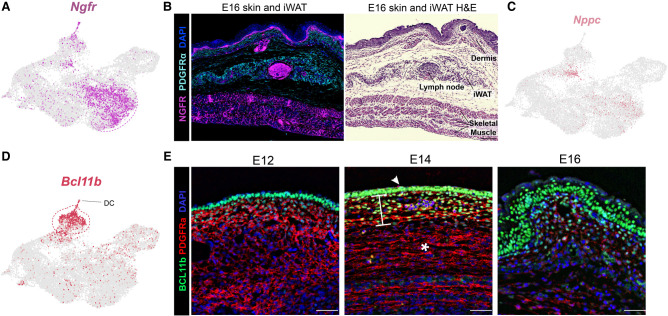
The distinct anatomic environment in which adipose tissues arise during organogenesis is a principle determinant of their adult expansion capacity. Metabolic disease results from a deficiency in hyperplastic adipose expansion within the dermal/subcutaneous depot; thus, understanding the embryonic origins of dermal adipose is imperative. Using single-cell transcriptomics throughout murine embryogenesis, we characterized cell populations, including Bcl11b + cells, that regulate the development of dermal white adipose tissue (dWAT). We discovered that BCL11b expression modulates the Wnt signaling microenvironment to enable adipogenic differentiation in the dermal compartment. Subcutaneous and visceral adipose arises from a distinct population of Nefl + cells during embryonic organogenesis, whereas Pi16 + /Dpp4 + fibroadipogenic progenitors support obesity-stimulated hypertrophic expansion in the adult. Together, these results highlight the unique regulatory pathways used by anatomically distinct adipose depots, with important implications for human metabolic disease.

Keywords: Bcl11b; FAP; Nefl; Ngfr; Pi16; adipose development; adipose lineage; dermal adipose; single cell.


Figures

Figure 1.
Figure 1.
Diversity of embryonic mesenchymal progenitors at single-cell resolution. (A) Histologic time course of embryonic dermal and subcutaneous organogenesis. Scale bar, 100 μm. (B) Single-cell transcriptomics of surgically isolated mesenchymal tissues (indicated by blue shading in Supplemental Fig. S1A) from E12–E16 embryos (36,665 cells). (C) Subgroup analysis of Pdgfrα+ cells in relative transcriptional proximity (dashed line in B) to committed preadipocytes (15,543 cells). (D) Temporal analysis of Pdgfrα+ mesenchymal progenitor subset single-cell data from E12 to E16.
Figure 2.
Figure 2.
Pi16+/Dpp4+ progenitors contribute to adult hyperplasic adipogenesis. (A) Single-cell expression of Pi16. (B) Lineage tracing of Pi16+ progenitor progeny chased to P12, demonstrating no contribution to subcutaneous/dermal embryonic adipose organogenesis. Scale bar, 50 μm. (C, top panels) “Chow”: Pi16 lineage tracing to P24 demonstrating no contribution to subcutaneous or visceral adipose organogenesis in lean adults. Scale bar, 50 μm. (Bottom panels) “High-fat diet”: Pi16 lineage tracing in adults following 4 weeks of HFD feeding. Scale bar, 100 μm.
Figure 3.
Figure 3.
Nefl+ cells contribute to subcutaneous and visceral adipose organogenesis. (A) Single-cell expression of Nefl. (B) Single-cell expression of Nefl+ cell marker genes Fst and Maf. (C) Lineage tracing of Nefl+ cells clustered near the developing iWAT depot (asterisk) at E14. (D) Lineage tracing of Nefl+ progeny to P12–P14 showing numerous labeled adipocytes in the iWAT (left) but none in the dWAT (right). Scale bar, 100 μm.
Figure 4.
Figure 4.
Ngfr+ cells are concentrated in skeletal muscle, and Bcl11b+ cells are located in the skin. (A) Single-cell expression of Ngfr. (B) IHC staining of E16 embryos showing NGFR+ spindle-shaped cells intercalated within body wall skeletal muscle. Scale bar, 100 μm. (C) Single-cell expression of Nppc. (D) Single-cell expression of Bcl11b. (E) IHC staining of E12–E16 embryos showing BCL11b expression (green; nuclear) in the epidermis (arrowhead), as well as PDGFRa+ cells (red) in the underlying dermis (bracket) and a lack of expression in PDGFRa+ cells of the developing iWAT (asterisk). Scale bar, 50 μm.
Figure 5.
Figure 5.
Bcl11b is required for dWAT adipogenesis. (A) In vitro adipogenic differentiation assay of WT versus Bcl11b-KO preadipocytes isolated from the dWAT versus the iWAT. Equal cell numbers were plated at high density to eliminate expansion artifacts. Representative images of n = 3 biological replicates. Scale bar, 0 μm. (B) Quantification of adipogenesis in A using the adipogenic index (Merrick et al. 2019). (C) IHC imaging of dermal and iWAT tissues isolated from WT versus Bcl11b-KO mice. Representative images of n = 7–9 biological replicates. Scale bar, 100 μm. (D) Quantification of adipocyte development by measurement of the perilipin area per micrometer of tissue length. (E) Flow quantification of the relative DPP4+ versus ICAM1+ population, represented as a percentage of PDGFRa+ progenitors.
Figure 6.
Figure 6.
BCL11b modulates Wnt regulatory proteins. (A) Representative BCL11b ChIP-seq tracks showing identified peaks near select Wnt constituent genes. (B) GO analysis of Bcl11b peak-associated genes. (C) RNA-seq analysis of gene expression changes in ICAM1+ preadipocytes from the dermis of WT versus Bcl11b-KO mice. n = 3 biological replicates. (D) Proposed model of BCL11b function in modulating the local Wnt microenvironment to enable dermal adipogenesis.

References

    1. Alexander CM, Kasza I, Yen CL, Reeder SB, Hernando D, Gallo RL, Jahoda CA, Horsley V, MacDougald OA. 2015. Dermal white adipose tissue: a new component of the thermogenic response. J Lipid Res 56: 2061–2069. 10.1194/jlr.R062893 - DOI - PMC - PubMed
    1. Angueira AR, Sakers AP, Holman CD, Cheng L, Arbocco MN, Shamsi F, Lynes MD, Shrestha R, Okada C, Batmanov K, et al. 2021. Defining the lineage of thermogenic perivascular adipose tissue. Nat Metab 3: 469–484. 10.1038/s42255-021-00380-0 - DOI - PMC - PubMed
    1. Ataca D, Aouad P, Constantin C, Laszlo C, Beleut M, Shamseddin M, Rajaram RD, Jeitziner R, Mead TJ, Caikovski M, et al. 2020. The secreted protease Adamts18 links hormone action to activation of the mammary stem cell niche. Nat Commun 11: 1571. 10.1038/s41467-020-15357-y - DOI - PMC - PubMed
    1. Belligoli A, Compagnin C, Sanna M, Favaretto F, Fabris R, Busetto L, Foletto M, Dal Prà C, Serra R, Prevedello L, et al. 2019. Characterization of subcutaneous and omental adipose tissue in patients with obesity and with different degrees of glucose impairment. Sci Rep 9: 11333. 10.1038/s41598-019-47719-y - DOI - PMC - PubMed
    1. Bhattacharya S, Wheeler H, Leid M, Ganguli-Indra G, Indra AK. 2015. Transcription factor CTIP2 maintains hair follicle stem cell pool and contributes to altered expression of LHX2 and NFATC1. J Invest Dermatol 135: 2593–2602. 10.1038/jid.2015.281 - DOI - PMC - PubMed

MeSH terms

LinkOut - more resources