. 2024 Sep 12;15(1):7998.
doi: 10.1038/s41467-024-52396-1.

Clearance and transport of amyloid β by peripheral monocytes correlate with Alzheimer's disease progression

Affiliations

Clearance and transport of amyloid β by peripheral monocytes correlate with Alzheimer's disease progression

Xin Huang et al. Nat Commun. .

Abstract

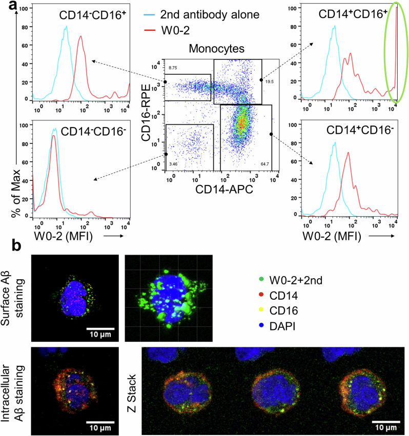
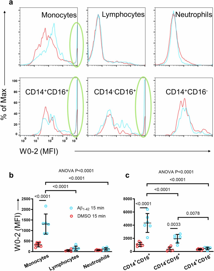
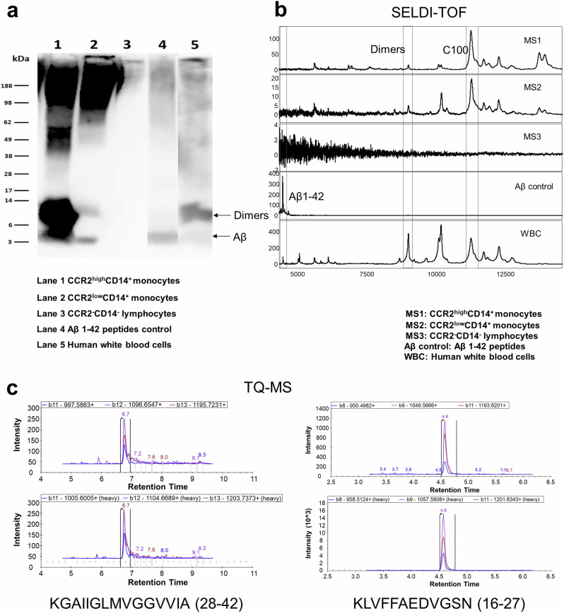
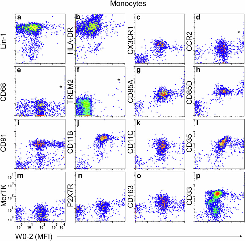
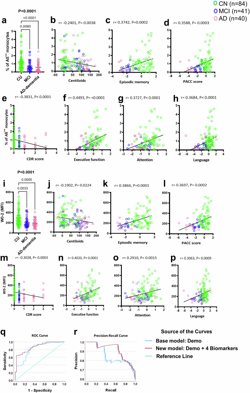
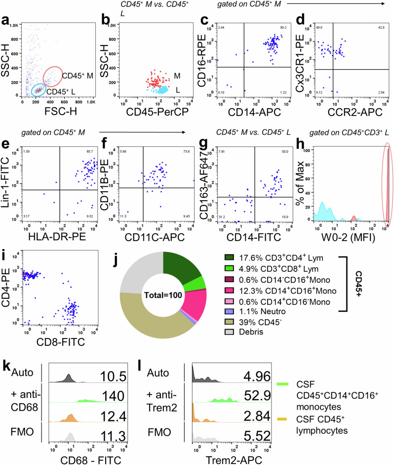
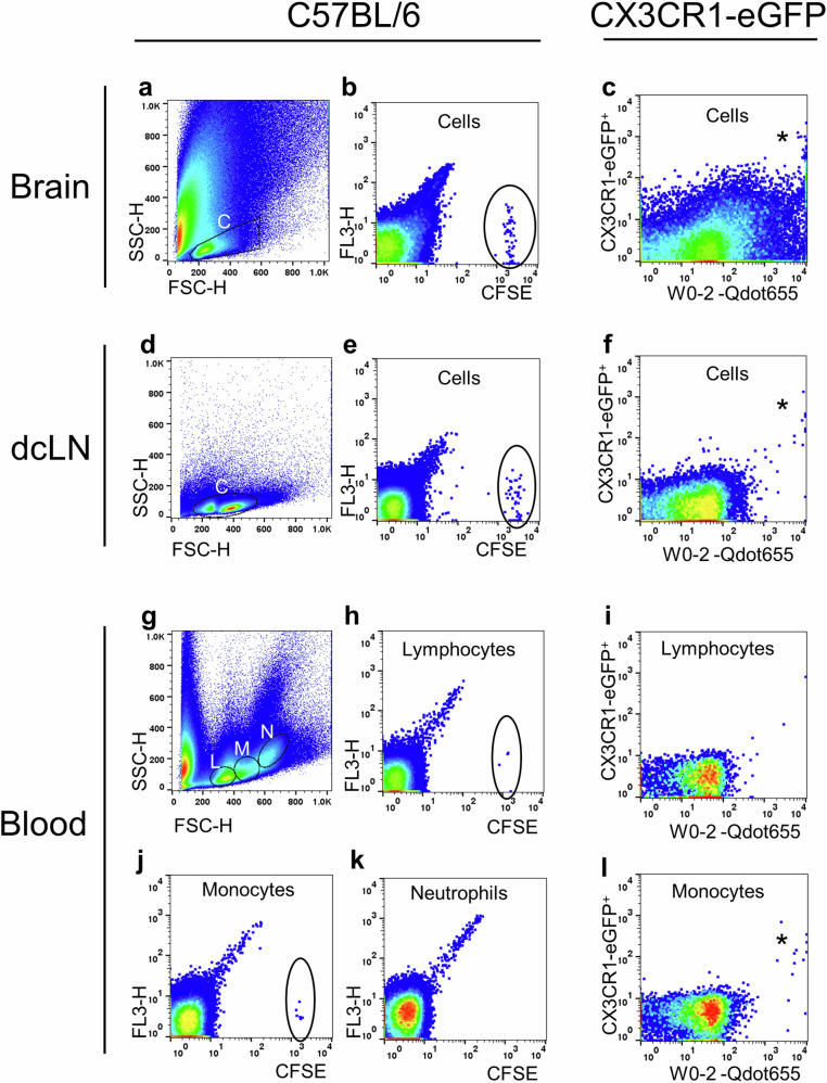
Impaired clearance of amyloid β (Aβ) in late-onset Alzheimer's disease (AD) affects disease progression. The role of peripheral monocytes in Aβ clearance from the central nervous system (CNS) is unclear. We use a flow cytometry assay to identify Aβ-binding monocytes in blood, validated by confocal microscopy, Western blotting, and mass spectrometry. Flow cytometry immunophenotyping and correlation with AD biomarkers are studied in 150 participants from the AIBL study. We also examine monocytes in human cerebrospinal fluid (CSF) and their migration in an APP/PS1 mouse model. The assay reveals macrophage-like Aβ-binding monocytes with high phagocytic potential in both the periphery and CNS. We find lower surface Aβ levels in mild cognitive impairment (MCI) and AD-dementia patients compared to cognitively unimpaired individuals. Monocyte infiltration from blood to CSF and migration from CNS to peripheral lymph nodes and blood are observed. Here we show that Aβ-binding monocytes may play a role in CNS Aβ clearance, suggesting their potential as a biomarker for AD diagnosis and monitoring.


Conflict of interest statement

The authors declare no competing interests.

Figures

Fig. 1
Fig. 1. The flow cytometry-based assay.
a Flow cytometry dot plot outlined monocyte subsets based on CD14 and CD16 expression, displaying surface Aβ detection on monocytes via W0-2 and secondary antibodies in the histograms. b Confocal microscopy displayed Aβ peptides on the surface and cytoplasm of intermediate monocytes, identified by CD14 and CD16 (n = 3). Intracellular Aβ was observed using the 6E10 mAb and captured through Z-stack imaging (n = 2). Nuclei were counter-stained with DAPI. MFI: The mean fluorescence intensity.
Fig. 2
Fig. 2. The affinity of monocytes to synthetic Aβ1-42 peptides.
Human blood samples (n = 6, all CU) were incubated with synthetic Aβ1-42 peptides to validate the assay’s specificity. a Monocytes showed significant binding compared to the DMSO (circled). Lymphocytes and neutrophils did not show comparable binding. Among monocyte subsets, CD14+CD16+ and CD14CD16+ monocytes exhibited significant Aβ1-42 binding (circled). CD14+CD16 monocytes did not show binding. b, c These findings confirm the specific binding of monocytes to Aβ1-42 peptides, particularly CD14+CD16+ and CD14CD16+ monocytes. The column dot plots represent mean ± standard deviation. All datasets were normally distributed. Group comparison was determined by one-way ordinary ANOVA, followed by Tukey’s multiple comparisons test (solid zig-zag line).
Fig. 3
Fig. 3. Confirmation of Aβ1-42 presence on Aβ-binding monocytes in blood.
PBMCs from CU individuals were FACS sorted based on CCR2 and CD14 expression. a Western blots (n = 2) showed substantial Aβ1-42 on CCR2++CD14+ monocytes, smaller amounts on CCR2+CD14+ monocytes, and no signal on CCR2-CD14- lymphocytes. b SELDI-TOF mass spectrometry (n = 2) confirmed diverse Aβ1-42 species, including dimers, in CCR2++CD14+ and CCR2+CD14+ monocytes, but not in CCR2CD14 lymphocytes. c TQ-MS (n = 3) identified Aβ-specific fragments (28–42) on various cell types, including CCR2++CD14+ and CCR2+CD14+ monocytes, and CCR2CD14 lymphocytes, with fragment (16–27) also detected abundantly.
Fig. 4
Fig. 4. Characterization of Aβ-binding monocytes.
Peripheral Aβ-binding monocytes (n ≥ 3, with a total of 22 blood samples analyzed) were analyzed using four-color immunophenotypic analysis. Aβ-binding monocytes exhibited a LIN-1+ and HLA-DR+ (MHC-II) phenotype (a, b), indicating differentiation from dendritic cells, and expressed CX3CR1 and CCR2 chemotactic receptors (c, d), suggesting strong migratory capabilities. They also showed CD68+ macrophage-like (e) and TREM2+ microglia-like phenotypes (f). Aβ-binding monocytes expressed Aβ-binding receptors (CD85A, CD85D, CD91) (gi), complement receptors (CD11b, CD11c, CD35) (jl), and phagocytosis-associated receptors (MerTK, P2X7R, CD163, CD33) (mp). It is worth noting that in the four-color panel for TREM2 (f), what matches TREM2, CD14, and CD16 was Qdot525 conjugated W0-2, unlike in the other panels where W0-2 was used in conjunction with a secondary antibody.
Fig. 5
Fig. 5. Peripheral monocyte surface Aβ as a potential biomarker for AD.
Quantifying Aβ on peripheral monocytes may serve as a non-invasive biomarker for AD. Our study included 150 participants (78 CU, 36 MCI, 36 AD-dementia). ap A decrease in the percentage of Aβ++ monocytes in MCI and AD-dementia compared to CU. Fluorescence intensity also showed a decrease in MCI and AD-dementia. A negative correlation was observed between the percentage of Aβ++ monocytes and brain Aβ-PET burden. Similarly, fluorescence intensity is weakly correlated with the Aβ-PET burden. Correlations between the percentage of Aβ++ monocytes, fluorescence intensity, and cognitive function were also observed, showing moderate correlations. The datasets were not normally distributed, and P-values were determined using the Kruskal-Wallis test, followed by Dunn’s multiple comparisons test (solid zig-zag line). The column dot plots represent mean ± standard deviation. Correlation coefficients (r) and P-values were calculated using Spearman correlation analysis. q, r A multivariate model revealed the potential of monocyte surface Aβ in predicting brain Aβ-PET burden, with a panel of %Aβ++ monocytes, %Aβ+ NK cells, %Aβ++ classical monocytes, and %classic monocytes performing best (AUC = 0.871). The new panel significantly improved AD discrimination, with a sensitivity of 0.65 and specificity of 0.976.
Fig. 6
Fig. 6. Investigating monocyte infiltration into CSF.
To demonstrate the presence of peripheral monocytes entering the CSF, CSF pellet cell samples from 15 participants (2 AD-dementia, 2 MCI, and 11 CU) were investigated. a, b Presence of peripheral lymphocytes and monocytes, identified by CD45, a pan-peripheral leukocyte surface marker, in the CSF occurred in all participants, regardless of clinical classification. ch CSF monocytes expressed CD14, CD16, CX3CR1, Lin-1, HLA-DR, CD11b, CD11c, and CD163.In addition, they showed surface Aβ binding compared to lymphocytes. i Within CSF CD3+ lymphocytes, the presence of CD4+ or CD8+ T cells was identified. j A visual representation of the CSF cellular component was illustrated in a pie chart. k, l Furthermore, most CSF monocytes expressed CD68 and TREM2. Auto: Autofluorescence. Numbers within the histogram represent the mean fluorescence intensity (MFI). FMO = fluorescence minus one.
Fig. 7
Fig. 7. Detection of Adoptive PBMCs in Blood and Deep Cervical Lymph Nodes.
PBMCs were sourced from donor C57BL/6 mice and labeled with CFSE (n = 13) or from CX3CR1-eGFP mice without CFSE labeling (n = 5). Both sets of PBMCs were injected into the lateral ventricles of APP/PS1 mice between 15 and 80 weeks of age. Two days later, recipients’ brains, deep cervical lymph nodes (dcLN), and blood were examined using fluorescent flow cytometry. a, d, and g display typical flow cytometry light scatter dot plots of the brain, dcLN, and blood, respectively. The presence of donors’ CFSE+ cells in the recipients’ brains confirmed successful delivery (circled in b). Likewise, CFSE+ cells in recipients’ dcLN (circled in e) and blood (circled in h, j) validated peripheral migration. No CFSE+ cells were found in the recipients’ blood neutrophil gating (k). Notably, the donors’ CX3CR1-eGFP+ monocytes were identified as carrying substantial amounts of Aβ in the recipients’ brain (asterisked in c), dcLN (asterisked in f) and blood (asterisked in l). A few CX3CR1-eGFP+ monocytes were found in the recipients’ blood lymphocyte gating (i) due to an overlap between lymphocytes and monocytes. This flow cytometry analysis demonstrates the migration and Aβ-carrying capabilities of adoptive PBMCs in different tissues. C: Cells; L: Lymphocytes; M: Monocytes; and N: Neutrophils.

References

    1. Masters, C. L., Bateman, R., Blennow, K., Rowe, C. C., Sperling, R. A. & Cummings, J. L. Alzheimer’s disease. Nat. Rev. Dis. Prim.1, 15056 (2015). 10.1038/nrdp.2015.56 - DOI - PubMed
    1. Villemagne, V. L. et al. Amyloid beta deposition, neurodegeneration, and cognitive decline in sporadic Alzheimer’s disease: a prospective cohort study. Lancet Neurol.12, 357–367 (2013). 10.1016/S1474-4422(13)70044-9 - DOI - PubMed
    1. Mawuenyega, K. G. et al. Decreased clearance of CNS beta-amyloid in Alzheimer’s disease. Science330, 1774 (2010). 10.1126/science.1197623 - DOI - PMC - PubMed
    1. Masters, C. L. et al. Neuronal origin of a cerebral amyloid: neurofibrillary tangles of Alzheimer’s disease contain the same protein as the amyloid of plaque cores and blood vessels. EMBO J.4, 2757–2763 (1985). 10.1002/j.1460-2075.1985.tb04000.x - DOI - PMC - PubMed
    1. Lewis, J. et al. Enhanced neurofibrillary degeneration in transgenic mice expressing mutant tau and APP. Science293, 1487–1491 (2001). 10.1126/science.1058189 - DOI - PubMed

Publication types

MeSH terms

LinkOut - more resources